VIEWS AND REVIEWS OPEN ACCESS
Antisense oligonucleotides
A primer
Daniel R. Scoles, PhD, Eric V. Minikel, MS, and Stefan M. Pulst, MD, Dr med
Neurol Genet2019;5:e323. doi:10.1212/NXG.0000000000000323
Correspondence Dr. Scoles
[email protected] or Dr. Pulst
Abstract
There are few disease-modifying therapeutics for neurodegenerative diseases, but successes on the development of antisense oligonucleotide (ASO) therapeutics for spinal muscular atrophy and Duchenne muscular dystrophy predict a robust future for ASOs in medicine. Indeed, existing pipelines for the development of ASO therapies for spinocerebellar ataxias, Huntington disease, Alzheimer disease, amyotrophic lateral sclerosis, Parkinson disease, and others, and increased focus by the pharmaceutical industry on ASO development, strengthen the outlook for using ASOs for neurodegenerative diseases. Perhaps the most significant advantage to ASO therapeutics over other small molecule approaches is that acquisition of the target sequence provides immediate knowledge of putative complementary oligonucleotide therapeutics. In this review, we describe the various types of ASOs, how they are used therapeutically, and the present efforts to develop new ASO therapies that will contribute to a forthcoming toolkit for treating multiple neurodegenerative diseases.
From the Department of Neurology (D.R.S., S.M.P.), University of Utah, Salt Lake City, UT; and Center for the Science of Therapeutics (E.V.M.), Broad Institute of MIT and Harvard, Cambridge, MA.
Funding information and disclosures are provided at the end of the article. Full disclosure form information provided by the authors is available with the full text of this article at Neurology.org/NG.
The Article Processing Charge was waived at the discretion of the Editor.
The genetic revolution led to the identification of many neu-rologic disease genes. The initial hope thatfinding the mutated protein and placing it into a known cellular pathway would lead to rapid development of therapies has remained largely un-fulfilled. The ability to target the disease gene or its encoded messenger RNAs (mRNAs) has opened new opportunities for therapy development. Of the many ways to target the expres-sion of RNA, this review will focus on the use of antisense oligonucleotides (ASOs) for therapy of neurologic diseases.
Therapeutic ASOs range from 18 to 30 base pairs (bp) in length. They modify expression of a target mRNA, by either altering splicing or by recruiting RNase H leading to target degradation. RNase H is a ubiquitous cellular enzyme that recognizes DNA:RNA hybrids and cleaves the RNA in the hybrid. The mode of ASO action depends on the target se-quence and the precise ASO chemistry used in the design. An illustration of ASO action depending on target type and ASO chemistry is provided infigure 1.
Leading the way currently in medical use is the ASO drug nusinersen that is approved for treating multiple forms of spinal muscular atrophy (SMA).1 Other ASO therapeutics that are Food and Drug Administration (FDA) approved include eteplirsen for Duchene muscular dystrophy (DMD)2 and inotersen for familial amyloid polyneuropathy (FAP).3
ASOs targetingHTTfor Huntington disease (HD),4SOD1and
C9ORF72for amyotrophic lateral sclerosis (ALS),5,6andMAPT
(TAU) for Alzheimer disease (AD)7are in early-phase clinical trials. Most current ASO therapeutics do not cross the blood-brain barrier and therefore for targets in the CNS have to be delivered by intraventricular injection in mice or lumbar puncture in humans. Eteplirsen and inotersen have targets that are not in the CNS and are delivered by intravenous or subcutaneous in-jection, respectively. On the other hand, systemic side effects are limited. Uptake into the CNS is an active process and is not uniform for all cell types or neurons. Chemical modifications can dramatically increase the half-life of ASOs and minimize toxicity.
Oligonucleotide chemistry
Natural, or unmodified, nucleic acids are susceptible to nu-clease degradation and have poor protein binding and thus inefficient tissue uptake precluding their use as drugs.8,9 Multiple types of modifications made to nucleotides, and their
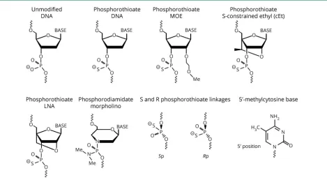
linkages can improve various properties increasing ASO suitability as drugs. Most of these modifications alter phar-macokinetics (improved nuclease resistance resulting in a longer half-life), pharmacodynamics (superior affinity for the target RNA), or endocytic uptake, which is controlled by specific sets of cell surface proteins.10,11But, with the excep-tion of the phosphorothioate (PS) modification to the ASO backbone, most also preclude cleavage by RNase H, which is the desired mechanism of action for many ASOs. Thus, many RNase H ASOs are designed as chimeras, where different bases are a mix of different chemistries, or as gapmers, where some modifications are placed on the “wings”and not the central bases. Yet, for ASOs intended to alter mRNA splicing or translation, chemical compositions that do not support RNase H can be optimal. Thus, ASOs are highly versatile, customizable therapeutic tools. Some of the different ASO compositions and their resultant effects are discussed here, and the relevant structures are presented infigure 2.
Oligonucleotide phosphate linkage modifications
Modifications to the oligonucleotide phosphate linkages predominantly assist in nuclease avoidance. In the phos-phorodiamidate morpholino (PMO), the phosphodiester (PO) linkages in the oligonucleotide backbone are replaced with nonionic phosphorodiamidate linkages leading to re-sistance to PO.12Other ASO types have PS modifications that result in resistance to a broad spectrum of nucleases, support RNase H activity, and increase protein binding, which also improves tissue uptake.10,11
Morpholinos
Morpholinos are oligonucleotides with unique modifications to the ribose sugar that lead to greater target affinity and facilitate nuclease avoidance.12 This modification reduces oligonucleotide-protein interactions.12 Eteplirsen, a 30-bp morpholino-based ASO drug for the treatment of Duchenne muscular dystrophy (DMD), represents the only FDA-approved morpholino therapy for a neurodegenerative (or neuromuscular) disease.
Methoxyethyl oligonucleotides
A chemical modification included in most so-called second-generation ASOs is 29O-methoxyethyl (MOE). MOE ASOs have an MOE modification at the 29-position of the ribose sugar. This change enables enhanced binding affinity to the target mRNA and is considerably less toxic than the 29-O
Glossary
methyl (OMe) modification. In addition, MOE is sufficiently nuclease resistant that some MOE nucleotides can be syn-thesized with normal PO linkages so that a mix of PO and PS linkages can be used tofine tune the pharmacokinetics of the ASO. This can facilitate more rapid distribution into tissue while keeping the terminal elimination rate slow.13 MOE modifications also reduce plasma protein binding, which seem to shift ASOs away from hepatic metabolism and toward the kidney for excretion in urine.1329-MOE PS ASOs delivered to the CSF can have biological half-lives exceeding 6 months.50,58
Constrained nucleic acids
Nucleotides that are covalently modified to limit conforma-tion are referred to as constrained or locked. Nucleic acids are considered “locked” when they have a methylene bridge connection made between 29-oxygen and the 49-carbon of the
ribose sugar molecule (figure 2). This bond effectively locks the base into a conformation predominantly characterizing the RNA ribose sugar and prevents the conformation char-acteristic of the deoxyribose sugar.14The benefit of locked nucleic acids (LNAs) is that they can produce both increased target specificity and reduced recognition by nucleases. LNAs can hybridize to both DNAs and RNAs forming highly stable double-helix duplexes.14 LNAs can be incorporated into siRNAs15and gapmer ASOs supporting RNAse H activity.16 LNA ASOs can be more potent in vivo than their 29-MOE analogs.17
LNAs have been associated with increased liver toxicity18but can be used in combination with unmodified bases to reduce toxicity while improving ASO efficacy.19,20 To get around hepatotoxicities associated with LNA chemistry, chemists have created a sort of LNA/MOE hybrid by adding a
methyl-Figure 1ASO functions
bridge characteristic of LNAs to MOE oligonucleotides. The result is ASOs with reduced liver toxicity and increased po-tency.21There are multiple types of constrained ethyl (cET) and MOE oligonucleotides (S-cEt, R-cEt, S-cMOE, and R-cMOE), where S and R refer to the left and right chiral structures, respectively. ASOs with these structures hybridize to target RNAs with affinities like their corresponding LNAs and have improved liver toxicity and increased resistance to nuclease degradation compared with the LNA chemistry.
Stereopure PS ASOs
PS ASOs are usually stereorandom with regard to chiral PS centers, each of which has 2 distinct stereochemical confi g-urations, making 219stereoisoforms possible for a 20mer ASO with 19 linkages. Although it is recognized that the 2 stereo-isomers (Rp and Sp) differ in their binding affinity and sus-ceptibility to degradation,22–24there are tradeoffs between the two, and it is still debated whether stereopure isomers can be more potent than stereorandom ASOs. Studies in cell culture have failed to show any potency differences between stereopure and stereorandom ASOs. On the other hand, 1 study reported that ASOs with repeated left-left-right (or SSR) chiral PS centers optimized ASO recognition by RNAse H and resulted in greater potency in vivo.22 Therapeutic development of stereopure ASOs is the strategy used at WAVE Life Sciences.
Peptide nucleic acid
The peptide nucleic acid (PNA) has a peptide in the position of the ribose sugar. PNAs have typically been used for modulating transcription and other applications not supporting RNase H activity.25 But, PNA gapmers have been shown to support
RNase H cleavage of target mRNAs.26However, a PNA ASO targeting exon skipping in theDMDgene was poorly effective compared with an ASO with the 29-OMe modification.27
59-methylcytosine modification
Methylation of cytosines at the 59 position is one way to increase ASO specificity. For example, all cytosines in an ASO againstSOD1included the 59-methylcytosine modification.5 This inclusion can enhance base pairing by modifying the hydrophobic nature of the ASO. On the other hand, 59 -methylcytosine-thymidine repeats can increase cytotoxicity,28 and CpG motifs can stimulate immunoreactivity that can be at least partially alleviated by including 59-methylcytosine.29,30
Gapmers, mixed chemistry, and target fate
Gapmer ASOs have“wings”on either side of LNA or MOE modified basesflanking a tract of unmodified bases with PS linkages usually throughout the ASO backbone. Mixed chemistry is intended to maximize resistance to nucleases and minimize toxicity while supporting RNAse H activity.31The target mRNAs cleaved by RNAse H will be degraded in either the nucleus or the cytoplasm by the exosome complex and XRN exonucleases32 (figure 1). Lead optimization may in-clude a screen for optimal structure activity relationship. For PS ASOs, this may combine cET, LNA, MOE, cMOE, and 59 -methylcytosine chemistries. Tracts of 4 guanosines should be avoided because they can result in complex ASO structures.30 For gapmer ASOs, this may include unmodified bases. Splice-switching oligonucleotides (SSOs) will exclude a gap feature as RNase H activity that could lead to target degradation is unwanted.
Figure 2Structural elements commonly used in ASOs
Development of ASO therapeutics for
neurodegenerative diseases
At the time of writing this review, only 3 ASO therapeutics have been approved by the FDA for neurodegenerative or muscular dystrophy diseases. These are nusinersen for SMA, eteplirsen for Duchenne muscular dystrophy (DMD), and inotersen for FAP. A handful of others are in clinical trials or in preclinical development (table).
Spinocerebellar ataxias
Three Spinocerebellar ataxias (SCAs), all caused by DNA CAG repeat expansion encoding a polyglutamine (polyQ) repeat, have been investigated as diseases targetable by ASO treatment in mouse models. SCA2 is caused by CAG repeat expansion in exon 1 of theATXN2gene.33The phenotypes in 2 SCA2 mouse models have been well characterized at the morphologic, physiologic, and transcriptome levels.34–36
ATXN2mutation is also associated with a substantial rise in the expression of the stress granule protein Staufen1 (STAU1) and production of ATXN2/STAU1/TIA1– positive stress granules.37After screening for ASOs targeting
ATXN2in a human cell line, we took the best ASOs reducing expression to in vivo testing by injection into the lateral ventricle of the mouse. These ASOs had a gapmer design. Several ASOs induced a glial or microglial response, a com-mon off-target effect of some ASOs, and were not further evaluated.
The top lead ASO was designated ASO7 and loweredATXN2
expression in SCA2 mouse cerebella by >60% for up to 13 weeks, without activating markers of gliosis. When injected
into symptomatic mice, ASO7 improved motor function and restored proteomic and physiologic Purkinje cell abnormalities.36,38 The study provided proof of concept of lowering of ATXN2 for treatment of ATXN2-related diseases and the impetus for identifying more efficacious SCA2 ASO for use in humans.
SCA3 (Machado-Joseph disease) is caused by a CAG repeat expansion in exon 10 of theATXN3gene. Different strategies have been used to develop SCA3 ASOs. This has included allele-specific ASOs antisense to the expanded CAG repeat that sterically block translation,39 splice-switching MOE ASOs to exclude exon 10 encoding the expanded repeat,40 and MOE gapmer ASOs targeting wild-type and mutant
ATXN3alleles41,42(figure 1).
SCA7 is characterized by cerebellar ataxia and progressive cone-rod dystrophy and caused by CAG repeat expansion in theATXN7gene. ASO therapies are ideally suited for treating the eye, as they can easily be injected intravitreally. Although no longer in use, 2 decades ago, fomivirsen became thefirst ever FDA-approved ASO drug, injected into the vitreous of the eye for cytomegalovirus retinitis.43Niu et al.44developed proof-of-concept data in mice for the use of an ASO therapy for blindness in SCA7. Of note, the ASO therapy was effective in mice with symptomatic eye disease.
Huntington disease
HD is caused by a CAG repeat expansion in an encoded region of the HTT gene. PolyQ expansion in huntingtin causes a gain of toxic function. Development of ASOs tar-getingHTT has followed different approaches. One targets
TableASO therapeutics for neurodegenerative disease
Drug Indication Target ASO chemistry Status
Nusinersen SMA SMN2, exon-7 inclusion ASO, full 29-MOE FDA approved
Eteplirsen DMD DMD, exon-51 skipping Morpholino FDA approved
Inotersen FAP TTRexpression ASO MOE gapmer FDA approved
WVE-210201 DMD DMD, exon-51 skipping Stereopure ASO Phase 1 clinical trial
RG6042 HD HTTexpression ASO MOE gapmer Phase 3 clinical trial
WVE-120101 HD HTTexpression Stereopure ASO Phase 1/2 clinical trial
WVE-120102 HD HTTexpression Stereopure ASO Phase 1/2 clinical trial
IONIS-MAPTRx AD Tauexpression ASO MOE gapmer Phase 1/2 clinical trial
BIIB078 ALS C9ORF72expression ASO MOE Phase 1 clinical trial
IONIS-SOD1Rx ALS SOD1expression ASO MOE gapmer Phase 1 clinical trial
ATXN2 ASO SCA2 ATXN2expression ASO MOE gapmer Preclinical development38
ATXN3 ASO SCA3 ATXN3expression ASO MOE gapmer Preclinical development42
both mutant and nonmutant alleles, whereas another ap-proach is allele specific whereby ASOs were screened that target single nucleotide polymorphisms in HTT in linkage disequilibrium with expanded CAG repeats.45,46This strategy may potentially target >75% of HD mutation carriers.47 Currently, 3 ASOs targetingHTTare in clinical trials (table).
Amyotrophic lateral sclerosis
ATXN2as a target for ALS: Although mutations in several genes cause ALS, a number of studies point toATXN2 as a therapeutic target for ALS. Over the past decade, it has become well established that intermediate CAG repeat expansions in theATXN2gene increase the risk of ALS.48,49 Intertwined with this discovery was thefinding that reducing
ATXN2 expression improved transactive response DNA-binding protein 43 (TDP-43) toxicity in both yeast andflies.49 When endogenous Atxn2 was reduced in TDP-43 transgenic mice by crossing with Atxn2 knockout mice, survival was significantly improved, TDP-43–positive stress granules were eliminated in motor neurons, and gait scores improved.50 Similarly, the survival of TDP-43 transgenic mice was im-proved by treating with an ASO targeting theAtxn2gene.50 The effect of targeting ATXN2 on TDP-43 aggregations might be explained by related effects on STAU1.37
Targeting SOD1 for ALS
Approximately 10% of familial ALS cases are caused by mutations in SOD1 altering its function.51 Phase I clinical testing of thefirst-in-human SOD1 ASO, intended to lower SOD1 expression, demonstrated that the drug was well tol-erated when infused into the CSF, but abundance of the mutant SOD1 protein in CSF was reduced by only;12%.52A reformulated version of the drug designated IONIS-SOD1Rx (BIIB067) is presently undergoing phase 1 clinical trials by Ionis Pharmaceuticals and Biogen.53
TargetingC9ORF72for ALS
GGGGCC repeat expansions in the C9ORF72 gene are causative of ALS and frontotemporal dementia (FTD).54,55
C9ORF72repeat expansions result in loss of expression of the normalC9ORF72gene and gain ofC9ORF72mRNA aggre-gates and repeat associated non-AUG (RAN) translation products. Mice with expanded C9ORF72 treated by intra-cerebroventricular injection with ASOs that interfere with translation of GGGGCC expandedC9ORF72 had reduced mRNA foci and RAN translation products, associated with improved anxiety and cognitive function phenotypes.56With proof of concept established, Ionis Pharmaceuticals and Biogen have undertaken a phase 1 clinical trial of an ASO therapeutic targetingC9ORF72for ALS.
Alzheimer disease and tauopathies
Targeting Tau for AD and tauopathies
Lowering Tau abundance by targeting expression of the
MAPTgene may be therapeutic for AD and FTD. Targeting
Mapt in mice with a MOE gapmer ASO reduced Tau ex-pression throughout the CNS and protected against seizures
in a mouse seizure model.7Transgenic mice expressing a hu-man P301S mutant Tau that were treated with Tau ASO had reduced neuronal Tau aggregates and prolonged lifespan from 312 to 348 days.57 Tau was also reduced in the CNS of cynomolgus monkeys following 6 weeks of Tau ASO treat-ment delivered intrathecally.57ASOs targetingMAPTsplicing to exclude a mutant exon also reduced Tau abundance in neuroblastoma cells and in an MAPT AD mouse model.58 There is another splice-switching ASO strategy for AD that targets the amyloid precursor protein (APP) gene to prevent inclusion of its exon 17 to block APP processing and Aβ pro-duction.59And there is yet another, where an ASO is used to correct splicing of theAPOEgene encoding apolipoprotein E.60
Parkinson disease
Reduction of LRRK2 abundance is predicted to be thera-peutic for Parkinson disease (PD). This is supported by observations that disease-causing mutations in LRRK2 are associated with elevatedα-synuclein expression.61Targeting
Lrrk2in wild-type mice was well tolerated supporting that PD is not associated with LRRK2 loss of function.62 MOE-gamper ASOs reducing LRRK2 in mice treated withα -synu-clein preformed fibirls were also associated with reduced aggregations of phospho(S129)-α-synuclein.63 Some effort has also been made to develop an ASO therapeutic targeting
α-synuclein for PD.64
Spinal muscular atrophy and nusinersen
SMA is caused by loss-of-function mutations in theSMN1
gene resulting in the loss of the survival motor neuron (SMN) protein. SMA severity is inversely correlated with copy number of the homologousSMN2gene. The cDNA encoded bySMN2is identical to that encoded bySMN1except for lack of exon 7. The ASO drug nusinersen is an SSO that functions by blocking an intronic splicing silencer element in theSMN2
intron 7 preventing the spliceosome from excluding exon 7 (figure 1). The result of nusinersen action is expression of the functional, full-length SMN protein from theSMN2gene. In
Smn1−/−; SMN2+/+ mice, SSOs that restore SMA2 exon 7 splicing also restored tail and ear necrosis phenotypes.65 Nusinersin was well tolerated in patients with SMA1,66,67and was approved by the FDA for use in humans for the treatment of SMA in December of 2016.27
Duchenne muscular dystrophy and eteplirsen
DMD is caused by mutation in theDMDgene encoding dys-trophin, which is one of the largest gene in the human genome. Most of theDMDmutations causing DMD result in premature dystrophin truncations. The predominant ASO strategy for treating DMD is employment of ASOs to exclude exons resulting in DMD proteins with partially restored functions. Eteplirsen, an ASO that has a PMO oligomer structure, interacts with theDMD
FDA in 2016 for the treatment of DMD, eteplirsen can be used to treat approximately 14% of DMD cases,69and efforts are ongoing to develop additional ASOs targeting other exons to treat DMD cases caused by other mutations in the dystrophin gene.68Wave Life Sciences has also initiated a phase 1 clinical trial for testing a stereopure ASO for DMD exon 51 exon skipping.
Familial amyloid polyneuropathy and inotersen
Missense mutation in theTTRgene encoding transthyretin is the cause of hereditary transthyretin amyloidosis (ATTR).
TTR mutations result in transthyretin misfolding and pro-gressive accumulation of amyloid deposition in many tissues resulting in polyneuropathy, multiorgan dysfunction, and cardiomyopathy. Multiorgan failure and cardiac arrest pose a greatest risk of death for ATTR patients. Lowering the total expression of TTR at its source in the liver is an effective strategy for ATTR, which is achieved by systemic treatment using the 29-MOE ASO drug inotersen,3now approved by the FDA for FAP. Inotersen is treated systemically with delivery made by weekly subcutaneous injections. In the brain, trans-thyretin is produced by the choroid plexus, andTTR muta-tions cause leptomeningeal amyloidosis that potentially could also be treated by ASO therapy targetingTTRexpression.70
Controlling o
ff
-target e
ff
ects
Ensuring target specificity is a critical step in ASO therapeutic development. One useful approach is to perform quantitative PCR to assess expression of mRNAs with target sequences including mismatches to the ASO candidate to ensure un-changed expression. Another powerful method is to perform transcriptome analysis (RNA-seq) using tissues from mice that are null for the mRNA target, following a treatment trial with the ASO candidate, where differentially expressed genes would indicate off-targets or perhaps incidental cytotoxicity.
A number of chemical modifications have been developed to improve the pharmacokinetic and pharmacodynamic prop-erties of ASOs. PS or PMO backbones are often used. Mod-ifications at the 29position of the sugar can improve affinity, protein binding, and nuclease resistance. Modifications spanning the 29 and 49 positions to conformationally con-strain the sugar can give dramatic improvements in affinity. All modifications except PS prohibit RNase H cleavage, but this can be rescued by a“gapmer”design where the modification is included only in the“wings”of the oligo, and the central bases are just PS. Although there are a number of new ASO ther-apies for neurodegenerative diseases in preclinical de-velopment and in clinical trials, there are only 2 therapies for neurodegenerative diseases that are FDA approved: nusi-nersen and eteplirsen. However, the success of nusinusi-nersen and eteplirsen and promising data from ongoing clinical trials for other ASO therapies to treat HD and ALS predict a robust outlook for new effective treatments for neurodegenerative diseases.
Author contributions
D.R. Scoles wrote the manuscript and produced thefigures. E.V. Minikel further contributed to ASO chemistry. S.M. Pulst further contributed to components on movement disorders and neuromuscular diseases.
Acknowledgment
A portion of this work was supported by grants R01NS097903, R21NS081182, R37NS033123, and U01NS103883 from the National Institutes of Neurological Disorders and Stroke (NINDS). EVM is supported by F31 AI22592 (National Institute of Allergy and Infectious Diseases).
Study funding
No targeted funding reported.
Disclosure
D.R. Scoles has received research support from the NINDS (U01NS103883 and R01NS097903) and Harrington Dis-covery Institute. E.V. Minikel has received funding for travel and/or speaker honoraria from Illumina and has received research support from Charles River Laboratories, NIH (F31 AI22952), and Prion Alliance. S.M. Pulst serves on the editorial boards of Journal of Cerebellum, NeuroMolecular Medicine, Experimental Neurology, Neurogenetics, Nature Clinical Practice,andNeurology:Genetics; holds patents for Nucleic acids encoding ataxin-2 binding proteins; Nucleic acid encoding Schwannomin-binding proteins and products related thereto; Transgenic mouse expressing a polynucleotide encoding a human ataxin-2 polypeptide; Methods of detecting SCA-2 nucleic acids; Nucleic acid encoding SCA-2 and prod-ucts related thereto; Schwannomin-binding-proteins; and Compositions and methods for SCA; has received publish-ing royalties for The Ataxias (Churchill Livpublish-ingston, 2007), Genetics in Neurology (ANN Press, 2005), Genetics of Movement Disorders (Academic Press, 2003), Neurogenetics (Oxford University Press, 2000), and Molecular Genetic Testing in Neurology, 2nd to 5th (AAN Press, 1996); serves or has served as a consultant for Ataxion Therapeutics; has received research support from the NIH (RC1NS068897, RC4NS073009, R21NS081182, R21NS079852, and R01NS33123) and the National Ataxia foundation; and has received license fee payments for technology or inventions from the Cedars-Sinai Medical Center. Disclosures available: Neurology.org/NG.
Publication history
Received byNeurology: GeneticsDecember 17, 2018. Accepted infinal form February 14, 2019.
References
1. Mercuri E, Darras BT, Chiriboga CA, et al. Nusinersen versus sham control in later-onset spinal muscular atrophy.N Engl J Med 2018;378:625–635.
2. Cirak S, Arechavala-Gomeza V, Guglieri M, et al. Exon skipping and dystrophin restoration in patients with Duchenne muscular dystrophy after systemic phos-phorodiamidate morpholino oligomer treatment: an open-label, phase 2, dose-escalation study.Lancet 2011;378:595–605.
4. van Roon-Mom WMC, Roos RAC, de Bot ST. Dose-Dependent lowering of mutant huntingtin using antisense oligonucleotides in Huntington disease patients. Nucleic Acid Ther 2018;28:59–62.
5. McCampbell A, Cole T, Wegener AJ, et al. Antisense oligonucleotides extend survival and reverse decrement in muscle response in ALS models.J Clin Invest 2018;128: 3558–3567.
6. Ly CV, Miller TM. Emerging antisense oligonucleotide and viral therapies for amyotrophic lateral sclerosis. Curr Opin Neurol 2018;31:648–654.
7. DeVos SL, GoncharoffDK, Chen G, et al. Antisense reduction of tau in adult mice protects against seizures.J Neurosci 2013;33:12887–12897.
8. Bennett CF, Baker BF, Pham N, Swayze E, Geary RS. Pharmacology of antisense drugs. Annu Rev Pharmacol Toxicol 2017;57:81–105.
9. Bennett CF, Swayze EE. RNA targeting therapeutics: molecular mechanisms of an-tisense oligonucleotides as a therapeutic platform. Annu Rev Pharmacol Toxicol 2010;50:259–293.
10. Crooke ST, Wang S, Vickers TA, Shen W, Liang XH. Cellular uptake and trafficking of antisense oligonucleotides. Nat Biotechnol 2017;35:230–237.
11. Geary RS, Norris D, Yu R, Bennett CF. Pharmacokinetics, biodistribution and cell uptake of antisense oligonucleotides. Adv Drug Deliv Rev 2015;87:46–51. 12. Summerton J. Morpholino antisense oligomers: the case for an RNase H-independent
structural type. Biochim Biophys Acta 1999;1489:141–158.
13. Geary RS, Watanabe TA, Truong L, et al. Pharmacokinetic properties of 2’ -O-(2-methoxyethyl)-modified oligonucleotide analogs in rats.J Pharmacol Exp Ther 2001; 296:890–897.
14. Campbell MA, Wengel J. Locked vs. unlocked nucleic acids (LNA vs. UNA): contrasting structures work towards common therapeutic goals. Chem Soc Rev 2011;40:5680–5689. 15. Elm´en J, Thonberg H, Ljungberg K, et al. Locked nucleic acid (LNA) mediated improvements in siRNA stability and functionality.Nucleic Acids Res 2005;33:439–447. 16. Wahlestedt C, Salmi P, Good L, et al. Potent and nontoxic antisense oligonucleotides
containing locked nucleic acids.Proc Natl Acad Sci USA 2000;97:5633–5638. 17. Swayze EE, Siwkowski AM, Wancewicz EV, et al. Antisense oligonucleotides
con-taining locked nucleic acid improve potency but cause significant hepatotoxicity in animals.Nucleic Acids Res 2007;35:687–700.
18. Kasuya T, Hori S, Watanabe A, et al. Ribonuclease H1-dependent hepatotoxicity caused by locked nucleic acid-modified gapmer antisense oligonucleotides.Sci Rep 2016;6:30377.
19. Kamola PJ, Maratou K, Wilson PA, et al. Strategies for in vivo screening and mitigation of hepatotoxicity associated with antisense drugs.Mol Ther Nucleic Acids 2017;8: 383–394.
20. Stanton R, Sciabola S, Salatto C, et al. Chemical modification study of antisense gapmers.Nucleic Acid Ther 2012;22:344–359.
21. Seth PP, Siwkowski A, Allerson CR, et al. Design, synthesis and evaluation of con-strained methoxyethyl (cMOE) and concon-strained ethyl (cEt) nucleoside analogs. Nucleic Acids Symp Ser (oxf) 2008;553–554.
22. Iwamoto N, Butler DCD, Svrzikapa N, et al. Control of phosphorothioate stereo-chemistry substantially increases the efficacy of antisense oligonucleotides.Nat Bio-technol 2017;35:845–851.
23. Wan WB, Migawa MT, Vasquez G, et al. Synthesis, biophysical properties and bi-ological activity of second generation antisense oligonucleotides containing chiral phosphorothioate linkages.Nucleic Acids Res 2014;42:13456–13468.
24. Koziolkiewicz M, Krakowiak A, Kwinkowski M, Boczkowska M, Stec WJ. Stereo-differentiation–the effect of P chirality of oligo(nucleoside phosphorothioates) on the activity of bacterial RNase H. Nucleic Acids Res 1995;23:5000–5005.
25. Kaihatsu K, Janowski BA, Corey DR. Recognition of chromosomal DNA by PNAs. Chem Biol 2004;11:749–758.
26. Malchere C, Verheijen J, van der Laan S, et al. A short phosphodiester window is sufficient to direct RNase H-dependent RNA cleavage by antisense peptide nucleic acid. Antisense Nucleic Acid Drug Dev 2000;10:463–468.
27. Aartsma-Rus A. FDA approval of nusinersen for spinal muscular atrophy makes 2016 the year of splice modulating oligonucleotides. Nucleic Acid Ther 2017;27:67–69. 28. Drygin D, Barone S, Bennett CF. Sequence-dependent cytotoxicity of
second-generation oligonucleotides. Nucleic Acids Res 2004;32:6585–6594.
29. Krieg AM, Yi AK, Matson S, et al. CpG motifs in bacterial DNA trigger direct B-cell activation.Nature 1995;374:546–549.
30. Stein CA. The experimental use of antisense oligonucleotides: a guide for the per-plexed. J Clin Invest 2001;108:641–644.
31. Crooke ST, Lemonidis KM, Neilson L, Griffey R, Lesnik EA,Monia BP. Kinetic characteristics of Escherichia coli RNase H1: cleavage of various antisense oligonucleotide-RNA duplexes.Biochem J 1995;312(pt 2):599–608.
32. Lima WF, De Hoyos CL, Liang XH, Crooke ST. RNA cleavage products generated by antisense oligonucleotides and siRNAs are processed by the RNA surveillance ma-chinery. Nucleic Acids Res 2016;44:3351–3363.
33. Pulst SM, Nechiporuk A, Nechiporuk T, et al. Moderate expansion of a normally biallelic trinucleotide repeat in spinocerebellar ataxia type 2.Nat Genet 1996;14:269–276. 34. Liu J, Tang TS, Tu H, et al. Deranged calcium signaling and neurodegeneration in
spinocerebellar ataxia type 2.J Neurosci 2009;29:9148–9162.
35. Meera P, Pulst S, Otis T. A positive feedback loop linking enhanced mGluR function and basal calcium in spinocerebellar ataxia type 2. Elife 2017;6:e26377.
36. Dansithong W, Paul S, Figueroa KP, et al. Ataxin-2 regulates RGS8 translation in a new BAC-SCA2 transgenic mouse model.PLoS Genet 2015;11:e1005182. 37. Paul S, Dansithong W, Figueroa KP, Scoles DR, Pulst SM. Staufen1 links RNA
stress granules and autophagy in a model of neurodegeneration. Nat Commun 2018;9:3648.
38. Scoles DR, Meera P, Schneider MD, et al. Antisense oligonucleotide therapy for spinocerebellar ataxia type 2.Nature 2017;544:362–366.
39. Hu J, Gagnon KT, Liu J, et al. Allele-selective inhibition of ataxin-3 (ATX3) ex-pression by antisense oligomers and duplex RNAs.Biol Chem 2011;392:315–325. 40. Toonen LJA, Rigo F, van Attikum H, van Roon-Mom WMC. Antisense
oligonucleotide-mediated removal of the polyglutamine repeat in spinocerebellar ataxia type 3 mice. Mol Ther Nucleic Acids 2017;8:232–242.
41. McLoughlin HS, Moore LR, Chopra R, et al. Oligonucleotide therapy mitigates disease in spinocerebellar ataxia type 3 mice.Ann Neurol 2018;84:64–77. 42. Moore LR, Rajpal G, Dillingham IT, et al. Evaluation of antisense oligonucleotides
targeting ATXN3 in SCA3 mouse models.Mol Ther Nucleic Acids 2017;7:200–210. 43. Marwick C. First“antisense”drug will treat CMV retinitis. JAMA 1998;280:871. 44. Niu C, Prakash TP, Kim A, et al. Antisense oligonucleotides targeting mutant Ataxin-7
restore visual function in a mouse model of spinocerebellar ataxia type 7. Sci Transl Med 2018;10:eaap8677.
45. Carroll JB, Warby SC, Southwell AL, et al. Potent and selective antisense oligonu-cleotides targeting single-nucleotide polymorphisms in the Huntington disease gene/ allele-specific silencing of mutant huntingtin. Mol Ther 2011;19:2178–2185. 46. Southwell AL, Kordasiewicz HB, Langbehn D, et al. Huntingtin suppression restores
cog-nitive function in a mouse model of Huntington’s disease. Sci Transl Med 2018;10:eaar3959. 47. Southwell AL, Skotte NH, Bennett CF, Hayden MR. Antisense oligonucleotide thera-peutics for inherited neurodegenerative diseases. Trends Mol Med 2012;18:634–643. 48. Neuenschwander AG, Thai KK, Figueroa KP, Pulst SM. Amyotrophic lateral sclerosis
risk for spinocerebellar ataxia type 2 ATXN2 CAG repeat alleles: a meta-analysis. JAMA Neurol 2014;71:1529–1534.
49. Elden AC, Kim HJ, Hart MP, et al. Ataxin-2 intermediate-length polyglutamine expansions are associated with increased risk for ALS.Nature 2010;466:1069–1075. 50. Becker LA, Huang B, Bieri G, et al. Therapeutic reduction of ataxin-2 extends lifespan
and reduces pathology in TDP-43 mice.Nature 2017;544:367–371.
51. Rotunno MS, Bosco DA. An emerging role for misfolded wild-type SOD1 in sporadic ALS pathogenesis. Front Cel Neurosci 2013;7:253.
52. Miller TM, Pestronk A, David W, et al. An antisense oligonucleotide against SOD1 delivered intrathecally for patients with SOD1 familial amyotrophic lateral sclerosis: a phase 1, randomised,first-in-man study.Lancet Neurol 2013;12:435–442. 53. Schoch KM, Miller TM. Antisense oligonucleotides: translation from mouse models
to human neurodegenerative diseases. Neuron 2017;94:1056–1070.
54. DeJesus-Hernandez M, Mackenzie IR, Boeve BF, et al. Expanded GGGGCC hex-anucleotide repeat in noncoding region of C9ORF72 causes chromosome 9p-linked FTD and ALS. Neuron 2011;72:245–256.
55. Renton AE, Majounie E, Waite A, et al. A hexanucleotide repeat expansion in C9ORF72 is the cause of chromosome 9p21-linked ALS-FTD.Neuron 2011;72: 257–268.
56. Jiang J, Zhu Q, Gendron TF, et al. Gain of toxicity from ALS/FTD-Linked repeat expansions in C9ORF72 is alleviated by antisense oligonucleotides targeting GGGGCC-containing RNAs.Neuron 2016;90:535–550.
57. DeVos SL, Miller RL, Schoch KM, et al. Tau reduction prevents neuronal loss and reverses pathological tau deposition and seeding in mice with tauopathy.Sci Transl Med 2017;9:eaag0481.
58. Sud R, Geller ET, Schellenberg GD. Antisense-mediated exon skipping decreases tau protein expression: a potential therapy for tauopathies. Mol Ther Nucleic Acids 2014;3:e180. 59. Chang JL, Hinrich AJ, Roman B, et al. Targeting amyloid-beta precursor protein, APP,
splicing with antisense oligonucleotides reduces toxic amyloid-beta production. Mol Ther 2018;26:1539–1551.
60. Hinrich AJ, Jodelka FM, Chang JL, et al. Therapeutic correction of ApoER2 splicing in Alzheimer’s disease mice using antisense oligonucleotides.EMBO Mol Med 2016;8: 328–345.
61. Volpicelli-Daley LA, Abdelmotilib H, Liu Z, et al. G2019S-LRRK2 expression aug-ments alpha-synuclein sequestration into inclusions in neurons. J Neurosci 2016;36: 7415–7427.
62. Volta M, Cataldi S, Beccano-Kelly D, et al. Chronic and acute LRRK2 silencing has no long-term behavioral effects, whereas wild-type and mutant LRRK2 overexpression induce motor and cognitive deficits and altered regulation of dopamine release. Parkinson Relat Disord 2015;21:1156–1163.
63. Zhao HT, John N, Delic V, et al. LRRK2 antisense oligonucleotides ameliorate alpha-synuclein inclusion formation in a Parkinson’s disease mouse model. Mol Ther Nucleic Acids 2017;8:508–519.
64. Alarcon-Aris D, Recasens A, Galofre M, et al. Selective alpha-synuclein knockdown in monoamine neurons by intranasal oligonucleotide delivery: potential therapy for Parkinson’s disease. Mol Ther 2018;26:550–567.
65. Hua Y, Sahashi K, Hung G, et al. Antisense correction of SMN2 splicing in the CNS rescues necrosis in a type III SMA mouse model. Genes Dev 2010;24:1634–1644. 66. Chiriboga CA, Swoboda KJ, Darras BT, et al. Results from a phase 1 study of
nusi-nersen (ISIS-SMN(Rx)) in children with spinal muscular atrophy.Neurology 2016; 86:890–897.
67. Finkel RS, Chiriboga CA, Vajsar J, et al. Treatment of infantile-onset spinal muscular atrophy with nusinersen: a phase 2, open-label, dose-escalation study. Lancet 2017; 388:3017–3026.
68. Nelson SF, Crosbie RH, Miceli MC, Spencer MJ. Emerging genetic therapies to treat Duchenne muscular dystrophy. Curr Opin Neurol 2009;22:532–538.
69. Lim KR, Maruyama R, Yokota T. Eteplirsen in the treatment of Duchenne muscular dystrophy. Drug Des Devel Ther 2017;11:533–545.
DOI 10.1212/NXG.0000000000000323
2019;5;
Neurol Genet
Daniel R. Scoles, Eric V. Minikel and Stefan M. Pulst
Antisense oligonucleotides: A primer
This information is current as of April 1, 2019
reserved. Online ISSN: 2376-7839.
Published by Wolters Kluwer Health, Inc. on behalf of the American Academy of Neurology.. All rights an open-access, online-only, continuous publication journal. Copyright Copyright © 2019 The Author(s).
is an official journal of the American Academy of Neurology. Published since April 2015, it is
Services
Updated Information &
http://ng.neurology.org/content/5/2/e323.full.html
including high resolution figures, can be found at:
References
http://ng.neurology.org/content/5/2/e323.full.html##ref-list-1
This article cites 69 articles, 11 of which you can access for free at:
Subspecialty Collections
http://ng.neurology.org//cgi/collection/spinocerebellar_ataxia Spinocerebellar ataxia
m
http://ng.neurology.org//cgi/collection/parkinsons_disease_parkinsonis Parkinson's disease/Parkinsonism
http://ng.neurology.org//cgi/collection/huntingtons_disease Huntington's disease
http://ng.neurology.org//cgi/collection/gait_disorders_ataxia Gait disorders/ataxia
http://ng.neurology.org//cgi/collection/all_movement_disorders All Movement Disorders
following collection(s):
This article, along with others on similar topics, appears in the
Permissions & Licensing
http://ng.neurology.org/misc/about.xhtml#permissions
its entirety can be found online at:
Information about reproducing this article in parts (figures,tables) or in
Reprints
http://ng.neurology.org/misc/addir.xhtml#reprintsus
Information about ordering reprints can be found online:
reserved. Online ISSN: 2376-7839.
Published by Wolters Kluwer Health, Inc. on behalf of the American Academy of Neurology.. All rights an open-access, online-only, continuous publication journal. Copyright Copyright © 2019 The Author(s).
is an official journal of the American Academy of Neurology. Published since April 2015, it is