ISSN 2374-2712 (Print) 2374-2720 (Online) Copyright © The Author(s). 2015. All Rights Reserved. Published by American Research Institute for Policy Development DOI: 10.15640/jcb.v3n1a4 URL: http://dx.doi.org/10.15640/jcb.v3n1a4
Three Iridoid Glycosides from the Root Extract of Stachytarpheta Angustifolia Mill Vahl Verbenaceae
M. Mohammed1, I. M. Bugaje2 & M. A. Garba3
Abstract
Three iridoid glycosides were isolated from the n-butanol fraction of the root extract of Stachytarpheta angustifolia (verbenaceae). Their structures were elucidated by using a combination of 600MHz/150 MHz ID and 2D NMR techniques (COSY, NOESY, DEPT, HSQC and HMBC) and by FABMS and HRESIM. Compound 1 was characterized as Citrifolinoside, (2) as Serratoside and 3 is determined as 6 – O – (3 " – O – trans – cinnamoyl) - α – L – rhamnopyranosyl catalpol heptaacetate.
Keywords: Stachytarpheta angustifolia, n-butanol, root extract, Iridoid glucosides
Introduction
Iridoid glycosides are large group of naturally occurring Monoterpeniod with a glucose moiety attached at C-1 in the pyran ring (Song et al, 2006). They occupy an important position in the field of natural product chemistry and biology, as they provide a structural link between terpenoids and indole alkaloids and as well display a broad spectrum of biological activities (Sticher, 1977). Iridoids are useful phytochemicals in the number of folk medicinal plants and many of them poses significant biological and pharmacological activities, some of them are chemo taxonomically useful as markers of genus in various plant families. Stachytarheta angustifolia is a shrub of about 5 ft high with a soft cylindrical bark. They are mostly simply or slightly branched often rather succulent with pale blue flowers. They are mostly distributed in the tropical, sub-tropical regions and other part of the world (Dalziel, 2000).
1
National Research Institute for Chemical Technology, P.M.B 1052, Zaria – Nigeria. Tel +23408064395057, E-mail: [email protected]
2
National Research Institute for Chemical Technology, P.M.B 1052 Zaria, Nigeria. 3
The triturated fresh root of the plant is applied locally for the treatment of ulcer and also taken as a good remedy to reduce blood pressure as well as an anthelmintic agent. The infusion of the root extract with natron is used to relieves depression as well as reduces fever. The shrub plant is reported to contain a glycosidal substance “Stachytarphine” which is reported to be abortificient (Watt and Breyer-Brandwijk, 1962). The juice from the plant is use as a remedy against cataract and also applied on children’s ear. Decoction of the whole plant is used as an Emmenagogue agent while the leaf is taken as a Cholagogue agent and also as remedy against gonorrhea and other related venerable infections (Dalziel, 2001; Jinju, 1990). Literature review reveals the presence of Citrifolinoside, Macrophyloside, Triterpenes, Friedelin, Stigmasterol, Ursolic acids and Oleanolic acid in the genus of this family. Although, no compound have been reported to be isolated from the root of this plant as well as its ethno medicinal properties. In this report, we describe the extraction, isolation and structural elucidation of three Iridoid glycosides from the n-butanol root extract of Stachytarpheta angustifolia extract using standard chromatographic and spectroscopic techniques.
Experimental Section
General Experimental Procedures
Optical rotations were measured using a Perkin –Elmer model 341 LC spectrometer at room temperature. IR spectra were recorded on spectrophotometer shimadzu 8400s. Melting Points were determined on XT4A Apparatus and results are
uncorrected. 1H NMR and 13C NMR experiments were performed on Bruker
spectrometer 600 MHZ for
1
H and 150 MHZ for
13
C NMR. NMR spectra were
referenced to the CD3OD solvent signals at ∂3.30 (H
I) and 49.00 (13C) with TMS as an
internal standard. Chemical shift values (∂) were reported in parts per million (ppm) in
relation to the appropriate internal solvent standard (TMS). The coupling constants (J-values) were given in Hertz, HRESI –MS was measured on a mass Autospec – ultima – TOF spectrometer. TLC was carried out on plates precoated with RP-18 gel (merck) and silica gel F254 (Qingdao Marine Chemistry Ltd). Spots on the plates were
visualized by spraying with 10% H2SO4 followed by heating in oven. Column
chromatography was performed on silica gel 60 (0.040 -0.0653 mm), column (40 –
63µm, 310mm α 15mm i.d). GC analysis was performed on a Shimadzu GC-2010 gas
chromatographic equipped with an H2 flame ionization detector and a DB-5 quartz
Gel filtration technique was carried out on sephadex LH20, TLC visualization was by UV absorption at 254mm. All solvents were distilled prior to use.
Plant Material
The plant Stachytarpheta angustifolia was collected locally from Basawa, a village outskirt of Zaria in Kaduna state of Nigeria in the month of august, 2013. Botanical identification was performed at the herbarium section of the department of biological science, Ahmadu Bello University Zaria, Nigeria and voucher No. 900188 was obtained. The fresh plant material was carefully separated into different parts, the leaf, the stem and the root.
Extraction and Isolation
The air-dried portion of the root was made into finely powdered material using pestle and mortar. The root powdered material (1.25kg) was exhaustively extracted at warm temperature (25±50) with methanol (6 liter x 4times) and the methanol extract was concentrated in vacuo to give a residue (210g). Water (300ml) was added and it was then partitioned sequentially with n-hexane, chloroform, ethylacetate and n-butanol. The solvents from the various portions were recovered using rotary evaporator. The n-butanol fraction (10g) was solubilized in (CH3OH) (15ml) and precipitated in di ethyl ether (4x250ml) yielding 4.1g fraction. The mixture obtained was concentrated and then suspended in water dialysed for 2 days and lyophilized, yielding crude mixtures of glycoside from TLC profile. An aliquot (2.3g)
of the mixture was fractionalized by column chromatography over sephadex LH-20 and
then re submitted to a repeated MPLC column chromatography on silica gel 60 (15-40 um) using as eluent CHCL3 : CH3OH : H2O (3:3:1, 6:4:1 and 8:5:1), affording compound 1 (21mg), 2(25mg) and 3(18mg) respectively.
Acid Hydrolysis for Compound 1
Solution of compound 1 (5mg) in 2M Hcl MeOH (4:1, 5ml) was reflux at
900C for 6hrs, after cooling, the reaction mixture was diluted to 20ml and extracted
with CH2Cl2 (3x2ml). The aqueous layer was concentrated to an appropriate volume
(1ml) and examined by TLC (Silica gel) with a solvent system CHCl3 /MeOH/H2O
The remaining aqueous layer was concentrated to dryness to give a residue and dissolve in pyridine (1ml), and then L-cysteine methyl ester hydrochloride (2mg)
was added to the solution. The mixture was heated at 600C for 2 hr., equal volume of
acetic anhydride was added, followed by heating at 900C for another 2hr. The solution
was then concentrated to dryness and taken in MeOH (0.5ml), which was analyzed by
GC (column: DB-5 quartz capillary column (30m x 0.25mm, 0.25µm), H2 flame
ionization detector column temperature: 160-2800C programmed increase: 50C/min, carrier gas: N2 (1.5ml/min), injector and detector temperature: 2800C, injection
volume: 1µl, split ratio: 10/1. The derivative of D-glucose was detected with Rf
(mm):23.89 and 28.07. The standard sugar was also subjected to the same reaction and GC analysis under the same condition as above was observed (Lan, et al., 2009)
Acid hydrolysis and GC Analysis for Compound 2 and 3
The solution of compound 2 and 3 (4.0mg) each in methanol (25ml) was
treated with 3N Hcl (15ml) and stirred at 800C for 5hrs. Upon drying with a flow of
nitrogen, the residue was dissolved in (-2) -2 – butanol (0.5mL) and a drop of trifloroacetic acid were added. The solution was in each case transferred to an
ampoule which was sealed and heated at 1300C overnight until complete butanolysis.
This was taken to dryness, the resultant residue was reacted with hex methyl disilazane /chlorotrimethylsilane /pyridine (1:1:5, 0.1ml) for 35min at room temperature. The
solution was centrifuged and the supernatant layer (1 µL) was analyzed by GC using
HP-5 column. The injection port and detector temperature was set at 2000C and
2200C. A temperature gradient from 140 - 2000C at 10C/min was applied. Four peaks
were detected from the hydrolysate at 37.45, 37.61 and Authentic standards were prepared in a similar manner from commercially available D-and L – glucose which gave rise to peaks at 37.43 and 37.51 (Furniss, 1989).
Determination of Sugar Compounds in 2 and 3
A solution of compound 2 and 3 each (6mg) in H2O (2ml) and 2N aqueous solution of CF3 COOH (5ml) were refluxed on a water bath for 3hrs. After this
period, the reaction mixture was diluted with H2O (20ml) and extracted with CH2Cl2
(4x5ml). The combined CH2CL2 extracts were washed with H2O and then evaporated
Furthermore, the residue from the sugars were dissolved in anhydrous
pyridine (100 µL), and L-cysteine methyl ester hydrochloride (0.06 mol/L) was added.
The mixture was stirred at 600C for 1hr, and then 150 µL of HMDS –TMCS
(Hexamethyldisazane - trimethylchlorosilane, 3:1) was added. The mixture was stirred at 600C for 30min. The precipitate was centrifuged, and the supernatant layer was concentrated under N2 stream. The residue was partitioned between n-hexane and H2O (0.2ml each) and the n-hexane layer (1 µL) was analyzed by GC. L- rhamnose and D-glucose by co-injection of the hydrolysate with standard silylated samples to give single peaks at (18.73min) for D- glucose observed in 2 and 3while (13.48min) for L- rhamnose in compound 3 only (Mohammed, et al., 2003).
Results and Discussions
Extraction of the root extract of S. angustifolia followed by an extensive column chromatography of n-butanol portion of the extract on silica gel and purification over sephadex LH20 resulted in the Isolation of compound 1, 2 and 3. Compound 1 was obtained as an amorphous brown solid. The FABMS established a Molecular formula of C26H28O14 as confirmed by the high resolution positive FABMS
as ([M – H] at M/Z – 563), as well as from its 13C NMR and DEPT NMR data. The
IR spectrum indicated the presence of an α, β – unsaturated – lactone ring at (1750cm-1), an iridoid and ether system conjugated with an ester carbonyl group at (1710, 1642cm-1), a Para substituted phenyl group at (1620, 1517, 820 cm-1) and the presence of hydroxyl groups at 3450cm-1 (Silvestein, 1991; Jiang-ming et al., 2003).
The 1H NMR spectrum exhibited a doublet (J = 2.0 H
Z) for the characteristic H – 3
proton of an iridoid at δH 7.41, a singlet at δH 3.74ppm could be attributed to a
carbomethoxy group while two doublets with signals (J = 2.5HZ) each for C – 6 and C
– 7 could be attributed to an epoxy protons on δH 4.03 and δH 3.9ppm. The signal
with a doublet (J = 8.0HZ) for H – 1
' attributed to proton on δ
H 4.43ppm suggest, that
the cyclo pentano pyran ring system and the sugar moiety are identical to those of the
6β, 7β – epoxysplendoside (Shengmin et al., 2001 ; Kalpoutzakis, et al., 1999). The
two pair of proton doublet (J = 8.4HZ) each at δH 6. 87 and δH 7.68ppm exhibited the
presence of a Para – hydroxyl phenyl group as supported by the values obtained in 13C
NMR Spectrum (δc 127.5/C-1c, δc 134.6/C – 2", 134.8/C – 6", 117.08/C-3",
The 13C NMR spectrum of compound 1(table 1) exhibited a total of 26 carbon atom signals with Ten carbon atoms representing the aglycone, one to methoxy group
at δc 51.7, six to the glucopyranose moiety at (δc 99.4/C -1'; 74.5/C-2'; 78.4/C-3';
74.2/C – 4'; 77.4/C-5' and 62.5 an oxy – methylene attributed to C – 6' (Jun-mian,
2008). The β – anomeric configuration for the glucose moiety was judged from the
large JH coupling constant of J = 8.0 HZ (Ilyas, et al., 2014). The presence of an oxygenated methine carbon was observed at δc 70.2/C - 10 and a single olefinic
Carbon atom signal at δc 144.04/C – 13 is an indicative of a characteristic iridoid with
five membered spiro – lactone ring (Shengmin et al., 2001; Kemp, 1991). The DEPT
spectrum exhibited the presence of an – oxymethylene on C – 6', oxygenated methine
carbon at C – 10, the presence of 2 – epoxy carbon signals at C – 6 and C – 7. The
presence of a single carbonyl signal was observed on C – 14, a methoxy signal on δc
51.7ppm, methylene signals on C – 3" and C – 5", aromatic carbon signals on C – 1", C-2" and C-6" (Jun-mian, et al., 2008). The presence of para substituted hydroxyl group of benzene was observed at C – 4" while an olefinic carbon atom was observed at C – 13 respectively (Wei et al., 2008). The Dept Experiment also exhibited the
presence of 5 quaternary carbon signals at δc 108.6/ C-4, 92.8ppm/C-8,
124.0ppm/C-11, 173.05/C-12, 168.4ppm/C-14 and 127/C-1 respectively (Ilyas et al.,
2014). The HMBC and NOESY spectrum correlations between C-1/H-1', H – 1'/C –
1 and H – 1/H – 1'suggested that, the β- glucopyranose moiety was attached at the C
– 1 position of the aglycone. The HMQC spectrum has confirmed the corresponding
proton signals found as single at δH 5.14s for H – 10 and also a singlet for the
trisubstituted olefin proton on H – 13 at δH 7.56 (s) (Cogne, et al., 2005).
The configuration of C – 6 and C – 7 as an epoxide group was also confirmed
from the spectra as a β- configuration (Francis, 2003; Bernstein, 1994). The
stereochemistry of C – 8 and C- 10 were ascertained using the NOESY spectrum. The strong NOESY correlations between H – 1/H – 10 also indicated that, the linkage between C – 8 and C – 10 are in α orientation while the H – 10 is in β (Brown, 2003; Kemp, 1991). On the basis of the chemical shift, multiplicity, absolute values
for the coupling constant and magnitude for 1H NMR and 13C NMR spectrum data,
indicated the β – configuration at the anomeric position for the glucose moiety. The
The positive FAMS established a molecular formula of C25H28O11 which was
confirmed by its high resolution positive FAMS as [M + 1]+ 505.132. The UV(MeoH)
[max (log ∑) : 203.5 (5.16) 216.8 (5.18), 223.4 (5.16), 243.7 (5.13), 256.2 (5.12), (279.0
(5.30) and 282 nm] while, IR (V: 3392, 1713, 1685, 1628, 1452, 1356, 1278, 1175 and
860cm-1) showed the presence of hydroxyl groups, carbonyl group, carboxyl group, α,
β- unsaturated skeleton and an – aromatic ring while values at 216.8, 223 and 282
could be attributed to the iridoid bearing trans cinnamoyl moiety(Hui, et al., 2000). The exhaustive acid hydrolysis of 2 gave glucose and trans – cinnamic acid at (tR 5.6min):m/z 147(22) [M-H]
-73(100) and 45 (35) ( Kalpourtzakis et al., 1999). The 1H
NMR spectral data for H – 3 /δH 7.4 ppm) indicated the presence of a 4 – substituted
enol ether system of an iridoid moiety. The spectrum signal at δH 4.6 /H' exhibited
the presence of an anomeric proton (Gousiadou, et al., 2007). Signals observed at δH
7.1, 6.6, 7.5., 6.8 and 6.3 are all characteristic of an aromatic protons (Dominguez, et
al ., 2007). The structure of trans cinnamoyl moiety was established by 1H NMR data
showing two vicinal olefinic protons at (δH7.8 and δH 6.7), five aromatic protons at
δH7.1 (3H) and δH 6.8ppm. The Signal at δ H 7.8/H – 7
'
and δH6.7/ H-8
1
could be
attributed to an α and β methylene proton of the trans – cinnamoyl group signifying
the presence of vinyl protons (Zhou, et al., 2007). Signals found at δH 4.6ppm (1H, d,
7.8Hz) is a characteristic of a sugar proton which is in conformity with a β –
Configuration of an anomeric proton (Neerja, et al., 2008). The glucosyl anomeric proton is linked to the aglycone at position H – 1 as demonstrated by HMBC Spectrum (Zuhal et al., 2007).
In 13C NMR (table 2), peaks are assigned on the basis of chemical shift
consideration and comprising with data for glucose. 13C NMR, spectrum was found to
exhibit 25 carbon signals, 10 corresponding to aglycone, 9 to trans – cinnamoyl
moiety and six attributed to glucose unit. Signal observed at δc 127.7ppm, 148.0ppm
and 148.00ppm suggested for C-1', C-3' and C-4' are characteristics of aromatic
carbons. Signals observed at δc 148.0 and 117.1ppm indicate the presence of an -α and
β vinylic carbons coupled to δc 168.3ppm signifying the presence of a carbonyl group
on C – 9' (Yoshiyasu, et al., 2004). The DEPT Experiment exhibited the presence
of 7 quaternary carbon signals at δc 114.0/C – 4, 75.4/C-5, 136.3/C-8, 127.7/C-1',
The 1H – H1 Cosy, HMQC and HMBC correlation spectra of 1 demonstrate some significant 1H – 13C long correlation between H – H and C – 9, C – 1" with H – 1 and H – 3 with H – 1 as clearly shown by HMBC spectrum which further confirmed the attachment position of trans-Cinnamoyloxy group and the glucosyl moiety at C – 8 and C – 7 of the aglycone. The glycosidic linkage observed at
δH4.6ppm (1H, d) with coupling constant at (J – 7.8H2) is also in conformity with –β
–D – glucopyranosyl (Kemp, 2001; Shu-Hua, et al., 2004). The relative stereochemistry of 2 was also determine using NOESY spectrum, which also confirms the H – 1 position of the aglycone as – β – orientation instead of α –
orientation. The 1H NMR and 13C NMR spectroscopic data were in great consistent
with a C – 8 iridoid monoglucoside moiety (Biswanath, et al., 2009). The comparison
of the 1H NMR, 13C NMR data and the coupling constants of Compound 2 with
those of Serratoside (Sophon, et al., 2002; Hui et al., 2000) were in great conformity. Thus, on the basis of this discussion, it was resolved that compound 2 was elucidated to be the same as Serratoside.
Compound 3: This was obtained as an amorphous solid. The UV (MeOH) spectrum exhibited 233.4 and 2.82nm for iridoid moiety and trans cinnamoyl unit. The IR spectrum shows the presence of 3396, 1714, 1689, 1632, 1520 and 1358
values exhibiting the presence of hydroxyl group, carbonyl group an α, β unsaturated
skeleton and an aromatic ring while values at 218.4, 225.7 and 283.2 could be attributed to an iridoid and a trans – cinnamoyl, unit (Kemp, 1991). The exhaustive acid hydrolysis of compound 3 gave glucose, rhamnose and trans – cinnamic acid (Yong-Qin et al., 2008). The position of ESI Mass spectrum shows a quasimolecular ion peak at m/z 661 [M + Na]+ suggesting the Molecular formula as C30H38O15
which was confirmed by 13C NMR and DEPT spectra. The 1H NMR spectrum (600
MHz, CD3OD) of compound 3 showed a broad singlet for H – 7 at δH 3.62ppm due
to small coupling constant existing between H – 7 and H – 6, this hence confirmed C – 8 to be quaternary (lhsan, C. et al., 2001; Jian-ming, et al., 2003).
A characteristic doublet for an acetal proton H – 1 arisen at δH 4.74ppm with
a large coupling constant at (J = 9.6HZ) demonstrated the di hedral angle to be at 180o
between the vicinally coupled protons of (H-1 and H – 9). The H – 9/ δH 2.72ppm
with coupling constant at J = 9.7HZ was also found to couple with H – 5 and H – 1
of the aglycone (Kalpoulzakis et al., 1999; Kemp, 1991). The coupling of H – 5
exhibited a dihedral angle to be at 00, thus revealing the stereochemistry of pyran and
A double doublet signal at δH6.42/H-3 with coupling constant (J = 6.0HZ) suggested a characteristic enol ether signal of an iridoid which was further confirmed by an intense signal in COSY, correlation between H – 3 and H – 4 and also with a weak one on H – 3 and H – 5 (Dominguez et al., 2007). A doublet signal on H – 1'
with coupling constant (J = 7.8HZ) signifies the presence of glucosyl anomeric proton.
The long range coupling with H – 1 in HMBC spectrum confirms the C – 1
attachment of the aglycone with H – 1' of the glucosyl moiety. The coupling constant
of (J = 7.8Hz) exhibited by H – 1'with δH 4.98 shows a dihedral angle of 180o, a di axil relationship between H – 1 and H – 2' vicinal protons of the glucosyl moiety
hence confirming the β- configuration of glucose with the aglycone (Zhou et al., 2007,
Neerja et al., 2008). The broad singlet signal resonating at δH 4.96ppm/H – 1" with a
dihedral angle of 600 suggest the presence of an anomeric proton, typical of an α –
isomer (Zuhal et al., 2005 ; Yoshiyasu , et al., 2004). The signal observed at δH 1.
27/H – 6" with J = 6.3 could be attributed to the methyl group corresponding to the rhamnose moiety.
The values obtain at δH 4.96/H – 1" and δH1.27/H 6" are in great conformity
with the α– L – rhamnopyranosyl group (Ilyas et al., 2014; Wei et al., 2008). The
location of α – L – rhamnopyranosyl group was determined to be at C – 6 position of
the aglycone (catalpol) as observed by the long range connectivity with C -1" of the rhamnopyranosyl moiety (Song, et al., 2006; Kemp, 2001). The COSY, HMBC spectrum has established the linkage of trans – Cinnamoyloxy group of C – 9 being attached to the C – 3 of the rhamnopyranosyl moiety with a glycosidic linkage (IK-Hwi et al., 2001). The structure of trans – cinnamoyl group was also established by
the 1H NMR spectrum showing two vicinal olefinic protons at δH 7.8 and 6.7 assigned
to H -7"' and H – 8"' while signals at δH 7.5/H-2"', 7.3/3"', 6.5/H-5"' and 6.4/H-6"' could be attributed to the aromatic protons (Mohammed et al., 2013; Yaching et al., 2004). The DEPT Experiment exhibited the presence of 4 quaternary carbon signals
at δc 68.2/C-8, δ C 136.2/-1"', δc150.4/C-4"' and δc 168.4/C=0 respectively. The 13C
NMR (table 3), peaks are assigned on the basis of chemical shift, consideration and comparison with data for glucose, rhamnose and catalpol. The spectrum data suggest the presence of 30 carbon signals, 15 attributed to catalpol as aglycone, 6 to the rhamnose group and 9 representing the trans cinnamoyl moiety (Biswanath et al., 2009). The cyclo pentano pyran ring carbons involved in the epoxide ring formation
The signal at δC 68.2 were found to disappeared in DEPT and as such does not give any connectivity with any proton in HSQC (Neerja et al., 2008). The
downfield signal observed at δC 94.8/C-1 appeared to be for the acetal carbon signal
of the pyran ring as judge by direct attachment of the two oxygen atom (Shengmin et
al., 2001). The signal at δC 142.3/C-3 was a characteristic of an enol ether carbon
(Cogne et al., 2005). The double bond arisen between C – 3 and C- 4 justifies the support for the presence of a cis – double bond (Young and Ling, 2008). The
downfield signal observed at δC 84.2/C-6 judge the position of rhamnopyranosyl
attachment through an ether linkage (Hosny and Rosazza, 1998) to the aglycone
moiety. The double doublet of H – 9/ δH42.6ppm confirmed the C – 8 position as
quaternary hence justifying C– 7 and C – 8 position as an epoxide ring system (Masaki, et al., 2001; Kemp, 1991). The HMBC correlation observed between acetate
bearing methylene carbon at δC 63.02/C-10 and proton signal at δH 3.62 and 2.72
corresponding to C-7 and C-9 enabled us to decide the position of C – 8 (Bernstein, 1994).
The exhaustive acid hydrolysis of compound 3 afforded glucose and rhamnose to be – β – D – glucopyranose and - α
– L rhamnopyranose and trans – cinnamic acid as identified by comparing with authentic samples in TLC.
The comparative studies of the spectral data obtained from (HMBC, HSQC, DEPT and NOESY) and comparison of aglycone with (catalpol) thus, it was resolved that compound 3 was elucidated to be determined as 6 – O – (3" – O
O O 1 3 5 9 O OH O
CH2OH
COOCH3 O O OH OH OH OH 6 7 8 10 11 13 12 1'' 2'' 3'' 4'' 5'' 6'' 1' 2' 3' 4' 5' 6' 4 O OH O O HO OH OH
CH2OH
1 3 5 6 7 9 1' 2' 3' 4' 5' 6' O O CHO 1'' 2'' 3'' 4'' 6'' H 4
H2C
10 7'' 8'' 9'' 8 OH 5'' O O O HO OH OH
CH2OH
1 3 5 6 7 9 1' 2' 3' 4' 5' 6' O O 1'' 6''' 5''' 4'' 2''' 4 7''' 8''' 9''' OH O O CH3 HO OH O 1'' 2'' 3'' 4'' 5''
CH2OH
8
10 6''
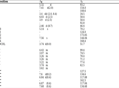
Table 1: 1H NMR (600 MHz) and 13C NMR (150MHz) Spectral data for Compound
1in (CD3OD; δ in ppm, J in HZ)
Position δH δC
1 5.32 d 93.2
3 7.41 d(2.0) 154.0
4 - 108.6
5 3.4 dd (2.0, 8.4) 34.5
6 4.03 d (2.5) 58.4
7 3.9 d (2.5) 58.4
8 - 92.8
9 2.46 d (8.7) 46.4
10 5.14 s 70.2
11 - 124.0
12 - 173.05
13 7.56 s 144.04
14 - 168,4
OCH3 3.74 d(8.0) 51.7
1' 4.43 m 99.4
2' 3.07 m 74.5
3' 3.26 m 78.4
4' 3.26 m 71.2
5' 3.22 m 77.4
6' 3.74 m 62.5
3.62 m
1" - 127.5
2" 7.6 d(8.2) 134.6
3" 6.84 d(8.4) 117.08
4" - 162.5
5" 6.87 (8.4) 117.04
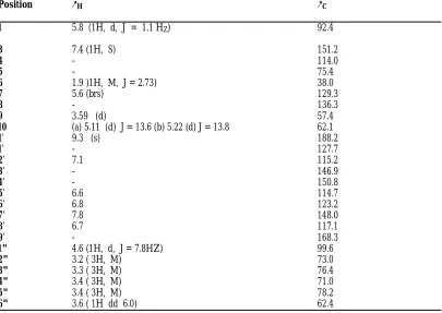
Table 2: 1H NMR (600MHZ) and 13C NMR (150M HZ) Spectral Data for Compound 2
(CD3OD; δ in ppm, J in HZ)
Position δH δC
1 5.8 (1H, d, J = 1.1 HZ) 92.4
3 7.4 (1H, S) 151.2
4 - 114.0
5 - 75.4
6 1.9 )1H, M, J = 2.73) 38.0
7 5.6 (brs) 129.3
8 - 136.3
9 3.59 (d) 57.4
10 (a) 5.11 (d) J = 13.6 (b) 5.22 (d) J = 13.8 62.1
1' 9.3 (s) 188.2
1' - 127.7
2' 7.1 115.2
3' - 146.9
4'
- 150.8
5' 6.6 114.7
6' 6.8 123.2
7' 7.8 148.0
8' 6.7 117.1
9' - 168.3
1" 4.6 (1H, d, J = 7.8HZ) 99.6
2" 3.2 ( 3H, M) 73.0
3" 3.3 ( 3H, M) 76.4
4" 3.4 ( 3H, M) 71.0
5" 3.4 ( 3H, M) 78.2
Table 3: 1H NMR (600MHZ) and 13C NMR (150 MHZ) Spectral Data for Compound
3 (CD3OD; δ in ppm, J in HZ).
POSITION δH COSY δC
1 4.74(1H, d J = 9.6) H - 9 94.2
3 6.42 (1H, dd, J = 6) H – 4, H - 5 142.3
4 5.11(1H, dd J=6, 4.6) H-3, H-5, H-6 103.6
5 2.61(1H, M) H-3, H-4, H-6 36.1
6 3.84 (1H, dd J =8.6) H-1”, H-5, H-7 84.2
7 3.62 (1H, brs) H - 6 58.7
8 - - 68.2
9 2.72(1H, dd J=9.7) H -1 42.6
10a 3.90 (1H, d, J=12.6) H -10b 63.02
10b 4.76 (1H, d, J=12.6) H – 10a
Glycosyl
1' 4.98 (1H, d, J=7.8) H - 2' 98.6
2' 5.23 (1H, dd, J=9.2) H - 1' 73.4
3' 4.94 (1H, t, J=9.5) H 4' 71.2
4' 5.23 (1H, t, J=9.2) H – 3', H - 5' 68.7
5' 3.63 (1H, M) H-6'a,b H-4 72.6
6'a 4.15 (1H, dd, j=12.3) H-5', H-6b 62.4
6'b 4.34 (1H, dd, J=12.4) H-5', H -6a
Rhamnosyl
1" 4.96 (1H, brs) H – 2" 103.4
2" 5.34 (1H, dd, J=3.5) H – 1", H.3" 70.9 3" 5.66 (1H, dd, J=9.6) H-2", H-4" 69.3 4" 5.41 (1H, t, J=9.8) H-3", H-5" 72.0 5" 4.09 (1H, dd, J=6.2) H-4", H-6" 62.5
6" 1.27 (3H, d, J=6.3) H-5" 17.9
Cinnamoyl
1"' - 136.2
2"' 7.45(d) 129.1
3"' 7.32 (d) 128.4
4"' - 150.4
5"' 6.5 (d) 129.2
6"' 6.4 (d) 129.6
7"' 7.8 118.6
8"' 6.7 (d) 146.8
C = 0 - 168.4
Acknowledgements
References
Bernstein, J. S. (1994), Nuclear Magnetis Resonance in pharmaceutical Technology. In Swarbruck J and JC. Boyland (Eds). Encyclopedia of pharmaceutical Technology, vol. 10 pp 335–360. New York Marcel–Dekker.
Biswanath, D., Debashis, R.C. and Biskas, C. M. (2009). Naturally occurring iridoids, secoiridoids and their Bioactivity. An up dated review, Part 3. Chemical pharm. Bull. 57. 8: 765-796.
Brown, G.D. (2003). 13C-2H Correlation NMR Spectroscopy studies of the In vivoTransformation of Natural products from Artemisia annua. Journal of Phytochemistry Research. 5: 45 – 59.
Cogne, A. L., Queiroz, E. F., Marston A., Wolfender, J. L., Mavi, S., Hostettmann, K., (2005). On-line identification of unstable iridoids from Jamesbrihenia fodina by HPLC–Ms and HPLC NMR–Phytochem. Anal. 16, 429–439.
Dalziel J.M. (1999). Useful plants of tropical West Africa. Crown Agents London, (United Kingdom) PP. 432–434.
Dominquez, M., Marin, J. C., Esquivel, B., Cespedes, C. L (2007). Pensteminoside, an unusual catalpol–type iridoid from Pensterman gentianoides HBK (Plantaginaceae) phytochemistry 38, 957–963.
Francis, A. C. (2003). Organic Chemistry. Mc graw Hill University of Virginia Fifth Edition. New York. U.S.A. P. 1011-1346.
Furniss H, Smith T. Vogels textbooks of practicals Organic Chemistry.5th Ed, Longman Singapore publishers Ltd, 1994, 131–152, 395–400.
Gousiadou, C., Karioti, A., Heilmann, J. Skaltsa, H. (2007). Iridoids from Scutellaria albida ssp albida, Phytochemistry 68, 1799–1804.
Hui Y., Bei J., Zhi, N.A, Yun pin G. and Han D.S (2000). Two New Iridoid glucoside from Clerodendrum serratum. Chinese Chemical letters , Vol.11, no.3 pp 231-234
Hosny, M., Rosazza, J. P. N., (1998). Glycosides A – L., twelve acylated iridoid glycosides from Gmelina arborea .J. Nat. Prod. 61. 734 – 742.
Ihsan, C., Hassan K. and Otto S. (2001). Iridoid Glycoside from Trichosantha. Journal of Natural product Research. 64:60-64.
Ik- Hwi, K., Satoru, T.,Yukio H., Tomoyo H. and Koichi T. (2004). New Quassnoids, Javanicolides C and D and Javanicosides B-F, from seed of Brucea javanica. Journal of Natural Product Research. 42: 863-868.
Ilyas M. Pateh U.U., Musa A. M and Mohammed M.(2014). Steroidal and triterpenoidal saponins from the stems bark extract of Stachytarpheta angustifolia Mill (vahl) Verbenaceae J. of Scientific and innovative research 3(2)1-10
Jian-Ming, J., XI-Chui, L. and Chon, R. (2003). Three New hexogen Glycosides from Fermented Leaves of Agaves americana. Journal of Asian Natural Product Research. 5:95 – 103.
Jinju, M. H. (1990). African Traditional Medicine. A case study of Hausa Medicinal Plants and Therapy. Gaskiya Corpn. Ltd. Zaria. Nigeria P. 43 – 50.
Jun-Mian, T., Hong – Ping, H.E., Ying T. D., Xian – Wen, Y., Zhu-Ling, G. and Xiao-Jiang, H.(2008).Three New Lignan Glycoside from Mananthes patentiflora. Journal of Asian Natural Product Research. 3: 230 -244.
Kemp W. (1991). Organic Spectroscopy. Macmillan Education Ltd. Sound Mills Basing Stole, Hampshire, United Kingdom. P. 186-197
Lan S., Chen G., Guang S., Wang W., Li Z., Chen H., Liu Y and Pei Y. (2009). Steroidal Saponins from Tribulus terrestris. ELSEVIER, 74: 399-403.
Masaki, K., Hide, K. and Daisuke, U. (2001). Isolation and Structure Elucidation of korolkoside, a bis-iridoid Glycoside from Lonicera korolkovii. Journal of Natural Product Research. 64:1090 – 1092.
Mohammed M, Musa A .M., Adeiza A.A., Musa S.H and Lande L.(2013). Bioactive caffeic glycoside ester and Antimicrobial activity of various extracts from the leaf of Stachytpheta angustifolia mill vahl verbenaceae. Journal of pharmacognosy and phytochemistry. 3, 77-85
Mohammed H, Tomofumi M, Veroni que L and Mavic-aleth ( 2003)Twonew biologically active Triterpeniod Saponin Acylated with Salicylic acid from Albizia adianthifolia. J Nat Prod 66:372-377.
Neerja T. Akhilesh Y.K., Pooja S., Krauna S., Ram K.V and Madan M.G (2008). Iridoid glycoside from Gmelina arborea. J. phytochem 69, 2387 -2390.
Shengmin, S., Xianfang, C., Nangun, Z., Jin-Woo, J. and Chi-tang, H.O. (2003). Iridiod Glycoside from the leaves of Morinda Citrifolia. Journal of Natural Product. 64: 799–800.
Shu – Hua, Q., Si Zhang, H., Zhi, H. Z., Jian H. and Qing – Xin, L. (2004). New Briaranes from South China Sea Gorgonian Junceella juncea. Journal of Natural Product. 8: 1907–1910.
Silverstein R. M. Bassler, G.C and Morril T.C (1991). Spectrometric identification of organic compounds, fifth edn. John willey and sons Inc. Singapore.
Song, Y., Li S. L., Wu M. H., Li, H. J., Li, P., (2006). Qualitative and quantitative analysis of Iridoid glycosides in the lower buds of Lonicera sp. by capillary H P L
Sophon, R., Kasan S., Nongmy, J., Narongsak, C. and Amoro, P. (2002). Crystal structure of Ipolamiide Monohydrate from Stachytarpheta indica. Japan society for Analytical chemistry. 5: 1063-1068.
Sticher, O. (1977). In: Wagner H., Wolff P. (Eds) New Natural products and plant Drugs with Pharmaceutical, Biological or Therapeutical activity. Springer, New york. Pp. 145-176. coupled with Ms detector Anal. Chem. Acta 564, 211-218,
Watt, J.M. and Breyer – Brandwijk, M.G. (1963). Medicinal and Poisonous plants of Southern and Eastern Africa. E. S Living Stone Edinburg Publishers. P. 1046 – 1088.
Wei L., Xian L., Jia Y., Da- Li M. and Ning L. (2008). Two New triterpeniod Saponins from the carpophores of Xanthoceras Sorbifolia Journal of Briange Research. 10:285-290.
Ya-Ching, S., Chung-Ling, L., Shih –Chao, C., Ashraf, T.K., Chinlien, K. and Chin-hsin, W. (2004). Vibsane Diterpeniods from the leaves and flowers of Viburnum odoratissismum. Journal of Natural Product. 67: 74–77.
Yong-Qin, Y. and Ling, Y. K. (2008): Ether soluble resin glycoside from the root of Ipomea batatas, Journal of Asian Natural Product Research. 10:245 - 252 ..
Yoshiyasu, F., Yuka, M., Yoshiko, K., Ih-shung, G., Hironobu T. and Tomoyuki E. (2004). Iridoid Glycosides and P- coumaroyl Iridoids from Viburnum luzonicum and their cytotoxicity. Journal of Natural Product. 67: 1833-1838.
Zhou X.Q., Bi Z.M., Li P., Tang D, Cai H.X. (2007). A new Iridoid glycoside from Gardenia jasminoides. Chin chem. Letter 18, 1221 - 1223