UDC 577.218
AnAlysis of AurorA kinAses genes
expression points on their distinct roles
in prostAte cAncer development
O. MaNkOVSka1, G. GeraShcheNkO1, e. rOzeNBerG1, e. StakhOVSky2, O. kONONeNkO2, yu. BONdareNkO3, V. kaShUBa1,4
1Institute of Molecular Biology and Genetics, National academy of Sciences of Ukraine, kyiv; 2National cancer Institute, Ministry of health of Ukraine, kyiv;
3Institute of Urology National academy of Medical Sciences of Ukraine, kyiv; 4department of Microbiology, tumor and cell Biology (Mtc),
karolinska Institutet, Stockholm, Sweden;
e-mail: [email protected]
received: 28 May 2019; Accepted: 18 October 2019
aurora kinases a and B play a crucial role in the regulation of mitosis, while aurora c controls meiotic division. these proteins showed controversial behavior upon the development of epithelial tumors. Our aim was to examine if there are any differences in expression of Aurora kinases genes in malignant and non-malignant tumors and non-tumor tissues; to compare their expression with clinical characteristics of pa-tients and expression of other prostate cancer-associated genes. Quantitative rt-Pcr was used to determine aurora a-c genes expression in 33 prostate adenocarcinomas (t), paired conventionally normal tissues (N), and 17 ade nomas (a). relative expression values (re) for genes studied were estimated using 2-ΔCt and 2-ΔΔCt method. the kruskal-Wallis with correction on multiple comparisons, according to the Benjamini-hochberg procedure with Fdr = 0.2, dunn-Bonferroni post hoc test and Spearman rank correlation analysis were used for statistical analysis. As turned out, RE values for AURKA were found to be significantly lower in samples of T group in comparison with A group. Moreover, significant up-regulation of AURKC expression levels were detected for t 3-4 stage compared to t 1-2 stage. re values of aUrkc in t group were positively correlated with the tumor stage, while aUrkB re demonstrated a negative correlation with the tumor stage. We also found significant correlations between AURK genes expression levels and prostate cancer-associated genes in t group. We suppose that all these data point to probable involvement of aurora kinases’ genes in prostate carcinogenesis.
k e y w o r d s: prostate cancer, aurora kinases, eMt genes, tMPrSS2-erG fusion transcript, PteN expression .
P
rostate cancer (PCa) is one of the most com-mon oncological diseases in men, the second after lung cancer. In the last decades, in-tensive research was attempted to investigate diag-nostic and progdiag-nostic PCa markers, novel therapy targets, mechanisms of the development of castra-tion-resistant PCa (CRPC), etc. This increased our knowledge and improved treatment approaches. Nevertheless, the reasons, lying under genetic and epigenetic events during prostate cancer initiation and progression remain not fully understood. Greatheterogeneity of PCa sets many challenges in a way of prediction of prostate cancer evolution and choice of appropriate therapy [1].
Somatic point mutations are less common in PCa than in most other solid tumors. The more general event for PCa is DNA copy-number varia-tions. While indolent and low Gleason tumors have relatively not many such alterations, more aggressive primary and metastatic tumors have extensive oc-currence of copy-number variations, genome-wide [2]. A huge number of chromosome rearrangements,
© 2019 Mankovska O. et al. This is an open-access article distributed under the terms of the Creative Commons Attribution License, which permits unrestricted use, distribution, and reproduction in any medium, provided the original author and source are credited.
leading to large insertions, duplications, transloca-tions and formation of aberrant gene fusions are usual events during prostate cancer progression [3]. One of the possible drivers of genomic rearrange-ments is an aberrant expression of key mitotic regu-lators, among which are kinases of an Aurora family [4, 5]. Kinases of the Aurora family are the highly conserved serine/threonine kinases, the major regu-lators of the cell cycle, particularly spindle formation and chromosome segregation. Both, loss of function and overexpression of their genes can lead to malig-nant transformation via deviations in mitotic divi-sion, which occur as direct or indirect consequences of inappropriate functioning of the Aurora kinases [6].
Aurora A is encoded by the aUrka gene, which is localized on chromosome 20q13.2. There are 14 transcript variants of aUrka, encoding proteins from 9 to 46 kDa. This kinase is involved in several events during cell division, namely, the centrosome separation, microtubule formation and stabilization at the mitotic spindle, ensuring the ac-curate chromosome segregation. Aurora A regulates progression from G2 to M phase through Bora-me-diated activation of Plk1 [7]. It was found, that this kinase, when overexpressed can transform NIH 3T3
cells and Rat1 fibroblasts in vitro [8].
Overexpres-sion and amplification of the Aurora A kinase gene
were described for a set of human cancers, including breast, urogenital, hematolymphoid, and also CNS lesions [9]. In gastric cancer, this is associated with aneuploidy, cell survival and taxane drug resistance [10].
The aUrkB gene is localized on chromosome 17p13.1, encoding the second Aurora kinase, Auro-ra B, consisting of 344 amino acids. This protein is a member of chromosomal passenger complex, func-tions at the beginning of mitosis and concentrates in the proximity of centromeres and kinetochores, where chromosome attachment to microtubules of the mitotic spindle is happening. In anaphase, Auro-ra B shifts to the spindle equator. AuroAuro-ra B regulates the interaction of kinetochores and chromosomes,
segregation of sister chromatids and cytokinesis [11]. Recently, it was shown that Aurora B is an upstream regulator of the end-on conversion process [12]. Ele-vated levels of Aurora B were reported in prostate cancer cell lines and prostate tissues from patients with PCa [13].
Aurora C is a less studied member of Aurora kinase family, although it was discovered in the late 90s [14]. The aUrkc gene is mapped on chromo-some 19q13.43, and 3 transcript variants exist, en-coding proteins of 275–309 residues. This protein
is rather specific for germ cells and works during
meiotic cell division. It is associated with Aurora B and Survivin in vivo [15]. Its function is very similar
to the Aurora B kinase, but, probably, it has different
regulation (due to the absence of N-terminal domain with regulatory motifs). Moreover, overlapping in functions is not absolute, and those two Aurora ki-nases are unique in this sense. For instance, it was demonstrated, that Aurora C plays a role in cell cycle progression, while the Aurora B role is restricted to maintaining of SAC (spindle assembly checkpoint) [16]. The functional role of AURKC in cancer is not well understood yet, but an increase of its levels in tumors is frequently associated with cell prolifera-tion, migration and bad prognosis. Overexpression of Aurora C in NIH-3T3 cell lines results in abnormal
cell division, centrosome amplification and appear -ance of multi-nuclear cells [17]. Those events are strongly associated with malignant transformation, therefore, the aUrkc gene was considered as onco-gene and Aurora C is a potential target for anticancer therapy [6].
Although increased levels and hyperactivity of kinases from the Aurora family are usually re-ported to be associated with tumor development and progression, and also with poor prognosis, there are
few reports, confirming this idea in a case of PCa.
For example, it was shown by immunostaining that
AURKA, AURKB and AURKC were significantly
reduced in PCa tissues, compared with cells of pros-tate nodular benign hyperplasia. However, no
sig-nificant differences in gene expression were found
at mRNA levels. This could be explained by muta-tions in genes, encoding Aurora kinases [18]. From the other hand, it was shown that Aurora A and B are expressed at higher levels in precancerous
le-sions and PCa of different stages, compared with
non-cancerous tissues [19].
Therefore, the aim of our research was to exa-mine the expression of Aurora kinase genes at the Abbreviations: Ar – Androgen receptor; cdnA –
mRNA level in PCa, surrounding non-cancerous prostate tissue and benign prostate tumors (adeno-mas). Also, we wanted to investigate whether there is any correlation between expression of aUrk genes with presence or absence of a TMPRSS2-ERG fu-sion transcript, PTEN status and also expresfu-sion of prostate cancer-associated genes (PCAG) [20-22]. We believe, our study will shed some light on the role of Aurora kinases in PCa development.
materials and methods
a collection of prostate tissues samples. Sam-ples of cancer tissue and paired conventional nor-mal tissue (CNT) (at an opposite side of tumor) were frozen in liquid nitrogen immediately after surgical resection, performed at the National Cancer Institute (Kyiv, Ukraine). Benign prostate tumors (prostate adenoma samples) were collected at the Institute of Urology (Kyiv, Ukraine) after radical prostatectomy; samples were frozen in liquid nitrogen, as described above. All the samples were collected in accordance with the Declaration of Helsinki and the guidelines, issued by an Ethics Committee of the Institute of Urology, the National Cancer Institute and the Ethics Committee of the Institute of Molecular Biology and Genetics (protocol number: 16, December 3, 2018). Altogether, 33 samples of prostate adenocarcinoma
of different Gleason score and stages, 33 paired CNT
samples and 17 samples of benign prostate tumors (adenomas) were collected and analyzed. Tumor samples were characterized, according to an
Inter-national System of Classification of Tumors and the
World Health Organization (WHO) criteria. Clini-cal and pathologiClini-cal characteris tics (CPCs) of PCa samples are presented in Table 1. The presence and/ or absence of a TMPRSS2/ERG fusion was detected earlier in our laboratory for the same cohort of pa-tients [20].
total rNa isolation and cdNa synthesis. 50– 70 mg of frozen tissues were mashed to a powder in the liquid nitrogen. Total RNA was extracted by TRI-reagent (SIGMA), according to the manufac-turer’s protocol. The total RNA concentration was analyzed by a spectrophotometer (NanoDrop Tech Experimental technologies Inc., USA). The quality of the total RNA was determined in a 1% agarose gel by band intensity of 28S and 18S rRNA (28S/18S
ratio). cDNA was synthesized from 1 μg of the to -tal RNA, following by treatment with RNase free
DNase I (Thermo Fisher Scientific, USA), using Re -vertAid H-Minus M-MuLV Reverse Transcriptase
(Thermo Fisher Scientific, USA), according to the
manufacturer’s protocol.
Quantitative polymerase chain reaction (qPcr). Gene expression levels of Aurora kinases genes (AURKA, AURKB and AURKC) were de-tected by qPCR, using HOT FIREPol EvaGreen qPCR Mix Plus (Solis BioDyne, Estonia) and CFX96 Real-Time PCR Detection System (Bio-Rad, USA) under the following conditions: 95 °C – 12 min, following 40 cycles of 95 °C – 15 s, 60 °C – 20 s, elongation 72 °C – 20 s. Sequences of primers for
aUrka, aUrkB and aUrkc genes were described in [23]. TBP has been used as reference gene for
relative quantification [21]. 2-ΔCT and 2-ΔΔCT methods,
described earlier [21], were used for the relative gene expression (RE) level calculation and analysis.
Statistical analysis. The Kolmogorov-Smirnov test was used to analyze the normality of distribution. The Kruskal-Wallis test and Dunn-Bonferroni post
hoc test were performed to determine RE differen ces
between pairs of sample groups under multiple com-parisons in 2-ΔCt. The Wilcoxon Matched Pairs test was performed to compare RE in prostate
adenocar-cinoma and paired CNT. RE fold differences in 2-ΔΔCt model were considered significant when expression
changes were more than 2-fold. The Fisher exact test
was performed to monitor differences between sam -ple groups. The Spearman's rank correlation test was
used to find the putative correlations between RE
and CPCs of prostate tumors and also correlations between RE of the studied genes. All calculations were performed, using a STATISTICA 10 software.
results and discussion
RE of genes, encoding Aurora kinases (aUrka ,
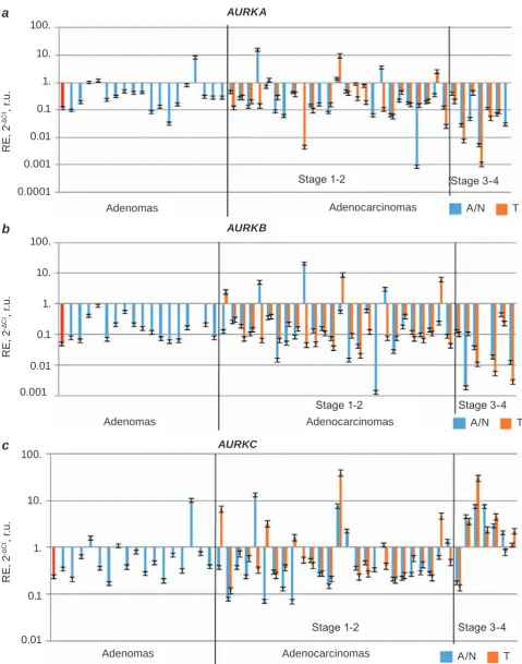
aUrkB, aUrkc) was detected in prostate ade-nocarcinomas (T), paired conventionally normal prostate tissues (N) and benign prostate tumors (ade-nomas) (A), see Fig. 1. Expression of aUrk genes in peripheral blood of healthy donors served as an additional positive control (red column, Fig. 1).
The statistical analysis of the obtained data did not support the Gaussian distribution of RE of the studied genes in all groups. Therefore, we used non-parametric criteria for further analysis.
Since Aurora kinases are considered as poten-tial oncogenes, we were aimed to investigate the
Wil-Fig. 1. relative expression (re) of aUrka (a), aUrkB (b) and aUrkc (c) in adenocarcinomas, conventio-nally normal prostate tissues and benign prostate tumors (adenomas). t/N pairs were distributed, according to the stage advancement. adenomas were placed randomly. In legend: a – adenomas: N – conventionally normal prostate tissue; T – adenocarcinoma. The first column (in red) – RE of AURK genes in peripheral blood (an additional control)
AURKA
R
E,
2
-ΔC
t, r.
u
.
0.0001 0.001 0.01 0.1 1. 10. 100.
a
Adenomas Adenocarcinomas A/N T
Stage 1-2 Stage 3-4
AURKC
R
E,
2
-ΔC
t, r.
u
.
0.01 0.1 1. 10. 100.
c
Adenomas Adenocarcinomas A/N T
Stage 1-2 Stage 3-4
AURKB
R
E,
2
-ΔC
t, r.
u
.
0.001 0.01
0.1 1. 10. 100.
b
Adenomas
Stage 1-2 Stage 3-4
coxon Matched Pairs Test applied to 33 T/N pairs.
However, no significant differences have been found
between T and N samples.
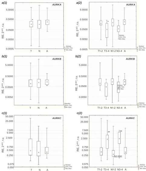
Next, we examined RE of aUrk genes, com-paring these values in malignant (tumors) and non-malignant (conventionally normal tissues and
ade-nomas) lesions. The significant differences in RE
were found for aUrka in tumors and adenomas (P = 0.047), with lower expression in tumors (Fig. 2,
a(1)). We proposed then that more pronounced dif-ferences may be expected by taking into account tu-mor stages as this is a tutu-mor characteristic known as the most clinically valuable one. For this purpose, the samples have been divided into 5 groups: tumors at stages 1/2 and 3/4 (T1/2 and T3/4 respectively), paired normal samples called N1/2 and N3/4 de-pending on the tumor samples and adenoma samples
called A (Fig. 2). The difference in RE of Aurora
kinases genes was analyzed based on the calcula-tions using Kruskal-Wallis test and Multiple com-parison test (Dunn-Bonferroni post hoc method). As one can see in Fig. 2, a(2), RE of aUrka gene is much lower in tumors at the stage 3-4, than that in adenoma tissues (P= 0.036). When comparing the expression of aUrkc gene, the N1/2 and N3/4
groups have different expressions with higher value
corresponding to N3/4 tissues (P = 0.021). Finally, no
differences between the groups under investigation
were observed for aUrkB expression.
Considering in addition another important prostate cancer characteristic called Glisson score
(GS), we found no differences between the high
and low GS samples in expression of Aurora kinase genes.
It is well known that the deviations in cell divi-sion processes can lead to genomic rearrangements and the formation of gene fusions. In particular, this concerns the expression of fusion transcripts, some of which were described for their carcinogenic ef-fect. Previously we have reported [17] about the presence of the most common prostate cancer fusion transcript, TMPRSS2-ERG, observed in the investi-gated samples [17]. Here the samples were grouped according to the presence or absence in them of TMPRSS2-ERG fusion transcript, T and N with fu-sion (F+); T and N without fufu-sion (F-), respectively. Their comparison with each other and with adeno-mas by using the Kruskal-Wallis and the multiple comparison (Dunn-Bonferroni post hoc method) tests characteris tics allows us to make a conclusion about lower aUrka expression in F- tumor samples
when compared to adenoma samples (P = 0.0325).
No other significant differences in expression were
observed for fusion transcript positive and negative samples.
PteN levels were assessed in these samples earlier as well [18]. So, T, N and A samples were divided into groups with the low and high PteN
expression. We found that aUrka expression in adeno carcinomas with high PteN levels was
sig-nificantly lower than in adenomas with low PteN
levels (P = 0.033).
To establish an unchanged expression level, we analyzed RE of aUrk genes in early stage conven-tional normal tissues (N1/2), advanced stages con-ventional normal tissues (N3/4) and adenomas. We consider that only N1/2 and adenoma samples have normal unchanged RE in aUrk genes. It was re-solved to admit those values as average unchanged expression level of studied genes in prostate tissue. We estimated changes in RE of aUrka, aUrkB
and aUrkc expression compared to taken average unchanged RE.
In T3-4 group, 63% had more than 2-fold lower expression of aUrka and aUrkB compared to ave rage unchanged RE, among which 50% of sam-ples even had more than 3-fold lower expression of those genes. In contrast, level of aUrkc expression in the same groups was higher than average un-changed expression of this gene. Compared to it, in 75% of samples from T3-4 group we detected 2-fold higher aUrkc RE. Moreover, 3-fold higher levels of aUrkc occurred in 63% of samples from this group. For N3-4 group the changes were even more pronounced: 87.5% of those samples have more than 3-fold higher expression of aUrkc in comparison with average unchanged expression level. Summa-rizing, the group of conventionally normal tissues from patients with high tumor stage (N3-4), but not the tumor tissues from those patients (T3-4) demon-strated the strongest alterations in expression of
aUrka and aUrkc genes.
ex-R
E
, 2
-ΔC
t, r.
u
.
T N A 0.0005
0.0050 0.0500 0.5000 5.0000
Median 25%-75% Min-Max
a(1)
Median 25%-75% Min-Max
Median 25%-75% Min-Max
R
E
, 2
-ΔC
t, r.
u
.
T1-2 T3-4 N1-2 N3-4 A 0.0005
0.0050 0.0500 0.5000 5.0000
a(2)
AURKA AURKA
R
E
, 2
-ΔC
t, r.
u
.
T N A 0.0050
0.0500 0.5000 5.0000
Median 25%-75% Min-Max
b(1)
Median 25%-75% Min-Max
Median 25%-75% Min-Max
R
E
, 2
-ΔC
t, r.
u
.
T1-2 T3-4 N1-2 N3-4 A 0.0050
0.0500 0.5000 5.0000
b(2)
AURKB AURKB
Fig. 2. Differences in RE of AURK A, AURKB and AURKC in adenocarcinomas, conventionally normal prostate tissues and benign prostate tumors (adenomas). a – aUrka expression; b – aUrkB expression; c – aUrkc expression; 1 – samples were grouped into 3 groups: cancer tissue (t), paired conventional normal tissues (N) and adenomas (a); 2 – samples were grouped into 5 groups, based on the stage of disease: the stage 1 and 2 (t1-2), the stage 3 and 4 (t3-4), paired conventional normal tissues from patients with the stage 1 and 2 tumors (N1-2), paired conventional normal tissues from patients with the stage 3 and 4 tumors (N3-4), and adenomas (A). *Significant RE differenced in T comparing to A; **significant RE differenced in T1-2 and N1-2 comparing to t3-4 and N 3-4 accordingly
0.050
Median 25%-75% Min-Max
R
E
, 2
-ΔC
t, r.
u
.
T1-2 T3-4 N1-2 N3-4 A 2.500
5.000 7.500 50.000
c(2)
AURKC
50.000 25.000
0.075 0.250 0.500 0.750
R
E
, 2
-ΔC
t, r.
u
.
T N A
Median 25%-75% Min-Max
c(1)
AURKC
0.050 2.500
5.000 7.500 50.000
25.000
pression (rs = -0.344, P < 0.05) in adenocarcinoma group. Expression of AURKC, however, correlated with stage positively (rs = 0.372, P < 0.05). Thus, here we observe increasing of AURKC expression with tumor progression, accompanied by decreasing of aUrkB RE level in high-grade tumors.
correlation of re of aUrk a, B, c and levels of prostate cancer-associated genes. Spearman rank correlation analysis of aUrk genes RE have
shown a significant correlation between aUrkB
and aUrka genes in T (rs = 0.365, P < 0.05). Ex-pression of PcaG genes, previously studied in our group [18, 19], also correlated with expression of the Aurora kinases genes in adenocarcinoma group (Ta-ble 2). Levels of AR 1 isof, caSP3 and OcLN corre-lated positively with expression of aUrka. aUrkB
expression positively correlated with Vdr levels. RE of MkI67 and NkX3.1 correlated with both, the aUrka and aUrkB, but not with the aUrkc
expression. Of note, aUrkc expression positively
CPC Genes
aUrka aUrkB aUrkc
GS -0.093 -0.219 0.119 Stage -0.201 -0.344* 0.372* PSA ng/ml -0.067 0.010 0.039 Age -0.026 0.176 0.204 Fusion status 0.278 0.106 -0.056
PteN re 0.142 -0.112 -0.020
t a b l e 1. correlation between cPc and aUrk gene expression (rs values)
Note: *P < 0.01
t a b l e 2. correlation between expression of eMt and aUrk genes (rs values)
Note: *P < 0.01, **P < 0.05
PcaG
genes
aUrk genes
aUrka aUrkB aUrkc
ar 1 isof 0.561* 0.331 -0.109
caSP3 0.361** 0.234 0.065
MkI67 0.562* 0.647* -0.025
NkX3.1 0.358** 0.349 -0.113
OcLN 0.355** 0.210 -0.221
Vdr 0.296 0.488* 0.025
INSr a 0.162 0.006 -0.397**
cyP17a1 0.104 0.159 0.370**
correlated with cyP17a1 (rs = 0.370, P < 0.05) and negatively with INSra levels (rs =-0.397, P < 0.05).
As was already mentioned above, PCa is the extremely heterogeneous disease at the level of or-gan, patient and population [24]. Despite the appre-ciable amount of investigations on prostate cancer nowadays, the data reported on the expression of Aurora kinases in PCa cell lines and clinical samples are incomplete and contradictory so far.
Here we stress first of all that the absence of differences between tumor and normal pairs from
the patients with prostate cancer can be explained by pathological processes in normal surrounding tis-sues in a case of aberrant signals from tumor cells and microenvironment. However, those tissues are closely connected with tumor and cannot be treated as absolutely normal. Based on our results, one can conclude about the presence of pathological altera-tions in those samples rather than the absence of
differences between malignant and non-malignant
tissues [25, 26].
Significantly higher levels of aUrka mRNA
in adenomas tissues, which often carry out inflam -matory process, is explained by the studied property
of Aurora A to participate actively in inflammation.
One of the possible ways for Aurora A to induce
car-cinogenesis is namely through promotion of IκBα-mediated activation of NFκB [27]. Therefore, high
levels of aUrka transcripts in adenomas can be
the part of intensive inflammation process. During
tumor development, cancer cells have already a
great amount of different phenotypes with various
genomic and epigenetic alterations, resulting in their uncontrolled proliferation and survival. We hypothe-size, that they acquire another intensive stimulus for proliferation and, consequently, in high stage tu-mors, the primary drivers of cell division lose their position and express at lower levels. Nevertheless, we should take into account that we observe these results only at the mRNA level, but we did not check the presence of the protein products in studied sam-ples. Stability of Aurora A and B is neatly regulated by ubiquitination and proteosome degradation [28]. Ubiquitination is often disrupted in cancer cells, leading to inappropriate degradation of key tumori-genesis players [29]. Thus, lower levels of mRNA do not always imply low levels of active proteins, that will be an issue of further investigations.
Con-sequently, it should result in overexpression of genes from AR axis [30], but was not evidently connected with overexpression of ar gene. Jones et al. [31]
demonstrated a direct influence of Aurora kinase A
on constitutively active variant of androgen recep-tor, ar-V7, expression and regulation of its splicing. However, these data do not explain the calculated correlation between expression of ar 1 isof and
aUrka. Nevertheless, it allows us to make sugges-tions about the possibility of the mutual regulation of these genes.
OcLN gene, the common marker of normal epithelial cells, encodes occludin, the protein, which involved in disruption of tight junctions and actin reorganization [32]. Aurora A kinase also acts in a similar manner, participating in structural changes
in actin filaments and lamellipodia formation [33].
Correlation between OcLN and aUrka genes’ expression can occur because of involvement of their products in the same cellular processes, e.g. enhancing cells motility and migration. Co-regu-lation or mutually adjustment of their expression should be further investigated. However, elevated levels of transcripts of those genes and/or their pro-tein products can potentially serve as marker of metas tatic and invasive phenotype of such tumor.
In our results, expression of Aurora kinases A and B genes positively correlated both with MkI67
and NkX3.1 expression. As MkI67 encodes marker of proliferation KI67 and commonly reported to be expressed together with other proliferation markers (which includes Aurora kinases’ genes) [34], we suppose that our results confirm previous data. On the other hand, NkX3.1 is a well-known tumor suppressor gene, which encodes NK3 homeobox 1 (NKX3.1) protein. Expression of NkX3.1 is common for normal epithelial cells. It is one of the AR target genes and is often loss in prostate cancer. Moreo-ver, NKX3.1 and AR proteins directly regulate each other in a feed-forward regulatory loop [35]. Some controversial features in the behavior of the present results can be attributed to upregulation of AR ex-pression by Aurora A kinase. Elevated amounts of androgen receptor cause increased expression of
NkX3.1 gene. It might be an ordinary cellular re-sponse to intensive proliferation, i.e. NKX3.1 is used as a “gate-keeper” to block cell proliferation [36]. In cells where expression of NkX3.1 is not loss via de-letions or methylation of its promoter, expression of this gene can be increased proportionally in answer to abnormal proliferation.
A mutual regulation for aUrka and aUrkB
gene expressions by c-Myc was reported previously by Hollander et al. [37]. As one can easily see, our results, demonstrating positive correlation between AURKA and AURKB transcript levels in prostate tissues, support previous observations and can be ac-cordingly explained by mutual regulation of aUrka
and aUrkB gene expressions.
Expression of aUrkB did not exhibit any
sig-nificant differences between the groups of samples. However, different mechanisms can be used to ex -plain negative correlation between expression of Aurora B gene with tumor stage and decreased level of its transcript (similarly to aUrka), compared to unchanged average expression level, in high stage tumors.
At first, cells, deficient of crucial cell division
regulator, may acquire a great amount of mutations, and decreased aUrkB expression can be not the consequence or feature of high grade tumor, but the driver and reason of tumor progression. Secondly, overlapping function of Aurora C and Aurora B can explain positive correlation of aUrkc and nega-tive of aUrkB with tumor stage. Unfortunately, the methods used in the present work do not give access to unambiguous conclusion concerning the mecha-nism of gene correlations.
Strong positive correlation between aUrkB
and Vdr (Vitamin D receptor) genes’ expression we explained as example of two independent co-existing processes in prostate cancer development. Both of them are connected with tumor stage. It was previously reported, that high level of VDR protein product is associated with lower tumor stage and more favorable prognosis for prostate cancer patients [38]. In our opinion, it could be two indirectly con-nected events in PCa.
We also found the positive correlation between
is also associated with еру develop ment of CRPC. It
encodes oncogenic isoform of insulin receptor and activates cellular pathways in response to binding with insulin-like growth factor (IGF) or insulin. Binding with both ligands promotes cell growth, proliferation and avoidance of apoptosis [41]. It is suggested that INSRA signaling is one of the pos-sible mechanism of androgen-independent PCa de-velopment [42]. On the other hand, expression of steroidogenic enzymes, such as CYP17A1, are also regarded to CRPC [40]. We propose that here we ob-serve two mutually exclusive directions of PCa pro-gression to CRPC.
Some decades ago, it was shown the existence
of the meiotic specific genes, which are activated
in mitotically dividing somatic cells and become the powerful drivers of carcinogenesis. Such de-viations contribute to illegitimacy in chromosome segregation and exchange of genetic material, ane-uploidy and other cell division failures, which can
produce different populations of tumor cells. Aurora
kinase C and CYP17A1 are two proteins that are ac-tive in germ cells, and they are putaac-tive oncogenes. Co-expression of them in PCa supports a soma-to-germline oncogenic model of carcinogenesis. This model arose as an alternative to embryologic theory of cancer and explained several aspects, which could not be explained by stem cell-like model, e.g. abnor-mal mitosis, rapid genomic and epigenetic evolution, altered DNA repair, chromosomal end protection, etc. [43].
We demonstrated that aUrka and aUrkc are
differentially expressed in PCa, compared with ade -noma, especially at advance stages of the disease. Beside an altered expression pattern of aUrk in prostate cancers, we also found a correlation of their
expression and different molecular characteristics of
tumors, and with prostate cancer-associated genes. Our data suggest that genes, encoding Aurora ki-nases, could be involved in prostate carcinogenesis.
Funding. This work was funded by the State Budget Program “Support for the Development
of Priority Areas of Scientific Research” (Code:
6541230) and National Academy of Sciences of
Ukraine in a frame of budget topic “Identification
of novel biomarkers for diagnostics of malignancies ad development of gene therapy approaches using model systems”
acknowledgements. We thank Dr. Elena Kashu-ba and Dr. Sergiy Mankovsky for their great support in preparing the text of the manuscript.
Conflict of interest. Authors have completed the
Unified Conflicts of Interest form at
http://ukrbio-chemjournal.org/wp-content/uploads/2018/12/ coi_disclosure.pdf and declare no conf lict of interest .
АнАліз експресії генів кінАз родини aurora вкАзує нА їхню різну роль в розвитку рАку передміхурової зАлози
О. С. Маньковська1, Г. В. Геращенко1, Є. Е. Розенберг1, Е. О. Стаховський2, О. А. Кононенко2, Ю. Н. Бондаренко3, В. І. Кашуба1,4
1Інститут молекулярної біології
і генетики НАН України, Київ;
2Націнальний інститут раку, Міністерство
охорони здоров’я України, Київ;
3ДУ «Інститут урології НАМН України», Київ;
4Відділ мікробіології, онкології та клітинної
біології, Каролінський інститут, Стокгольм, Швеція;
e-mail: [email protected]
Кінази Aurora A і B відіграють важливу роль у регуляції мітозу, тоді як Aurora C контро-лює мейотичний поділ. Ці протеїни проявляли суперечливу поведінку за розвитку епітеліаль
-них пухлин. Нашою метою було дослідити, чи існують відмінності в експресії генів кіназ ро
-дини Aurora в злоякісних та незлоякісних пух
-линах та непухлинних тканинах; порівняти їх експресію з клінічними характеристиками па
-цієнтів та експресією інших генів, пов’язаних із раком передміхурової залози. Кількісну RT-PCR було використано для визначення експресії генів Aurora A-C у 33 аденокарциномах передміхуро
-вої залози (Т), в парних умовно нормальних тка
-нинах (N) та 17 аденом (A). Значення відносної експресії (ВЕ) для досліджуваних генів оцінюва
-ли за допомогою 2-ΔCt та 2-ΔΔCt методу. Для статис -тичного аналізу було використано тест Круска
-вала негативну кореляцію зі стадією пухлини. Також виявлено значну кореляцію між рівнями експресії генів aUrk та генами, пов’язаними з раком передміхурової залози, в групі аденокар
-цином. Зроблено припущення, що одержані дані вказують на ймовірне залучення генів кіназ ро
-дини Aurora в канцерогенез передміхурової за
-лози.
К л ю ч о в і с л о в а: рак передміхурової залози, кінази родини Aurora, гени EMT, злитий транскрипт TMPRSS2-ERG, експресія PteN.
references
1. Tolkach Y, Kristiansen G. The Heterogeneity of Prostate Cancer: A Practical Approach.
Pathobiology. 2018; 85(1-2): 108-116.
2. Cancer Genome Atlas Research Network. The Molecular Taxonomy of Primary Prostate Cancer. cell. 2015; 163(4): 1011-1025.
3. Jaratlerdsiri W, Chan EKF, Petersen DC, Yang C, Croucher PI, Bornman MSR, Sheth P, Hayes VM. Next generation mapping reveals novel large genomic rearrangements in prostate cancer. Oncotarget. 2017; 8(14): 23588-23602. 4. Do TV, Hirst J, Hyter S, Roby KF, Godwin AK.
Aurora A kinase regulates non-homologous end-joining and poly(ADP-ribose) polymerase function in ovarian carcinoma cells. Oncotarget.
2017; 8(31): 50376-50392.
5. Kalsbeek D, Golsteyn RM. G2/M-phase checkpoint adaptation and micronuclei formation as mechanisms that contribute to genomic instability in human cells. Int J Mol Sci. 2017; 18(11). pii: E2344.
6. Tang A, Gao K, Chu L, Zhang R, Yang J, Zheng J. Aurora kinases: novel therapy targets in cancers.
Oncotarget. 2017; 8(14): 23937-23954.
7. Lindon C, Grant R, Min M. Ubiquitin-Mediated Degradation of Aurora Kinases. Front Oncol.
2016; 5: 307.
8. Fu J, Bian M, Jiang Q, Zhang C. Roles of Aurora kinases in mitosis and tumorigenesis. Mol cancer res. 2007; 5(1): 1-10.
9. Lehman NL, O'Donnell JP, Whiteley LJ, Stapp RT, Lehman TD, Roszka KM, Schultz LR, Williams CJ, Mikkelsen T, Brown SL, Ecsedy JA,
Poisson LM. Aurora A is differentially expressed
in gliomas, is associated with patient survival in glioblastoma and is a potential chemotherapeutic target in gliomas. cell cycle. 2012; 11(3): 489-502.
10. Kamada K, Yamada Y, Hirao T, Fujimoto H, Takahama Y, Ueno M, Takayama T, Naito A,
Hirao S, Nakajima Y.
Amplification/over-expression of Aurora-A in human gastric
carcinoma: potential role in differentiated type
gastric carcinogenesis. Oncol rep. 2004; 12(3): 593-599.
11. Krenn V, Musacchio A. The Aurora B Kinase in Chromosome Bi-Orientation and Spindle Checkpoint Signaling. Front Oncol. 2015; 5: 225.
12. Shrestha RL, Conti D, Tamura N, Braun D, Ramalingam RA, Cieslinski K, Ries J, Draviam VM. Aurora-B kinase pathway controls the lateral to end-on conversion of kinetochore-microtubule attachments in human cells. Nat commun. 2017; 8(1): 150.
13. Chieffi P, Cozzolino L, Kisslinger A, Libertini S,
Staibano S, Mansueto G, De Rosa G, Villacci A, Vitale M, Linardopoulos S, Portella G, Tramontano D. Aurora B expression directly correlates with prostate cancer malignancy and
influence prostate cell proliferation. Prostate.
2006; 66(3): 326-333.
14. Kimura M, Matsuda Y, Yoshioka T, Okano Y. Cell cycle-dependent expression and centrosome localization of a third human aurora/Ipl1-related protein kinase, AIK3. J Biol chem. 1999; 274(11): 7334-7340.
15. Yan X, Cao L, Li Q, Wu Y, Zhang H, Saiyin H, Liu X, Zhang X, Shi Q, Yu L. Aurora C is directly associated with Survivin and required for cytokinesis. Genes cells. 2005; 10(6): 617-626.
16. Quartuccio SM, Schindler K. Functions of Aurora kinase C in meiosis and cancer. Front cell dev Biol. 2015; 3: 50.
17. Khan J, Ezan F, Crémet JY, Fautrel A, Gilot D, Lambert M, Benaud C, Troadec MB, Prigent C. Overexpression of active Aurora-C kinase results in cell transformation and tumour formation.
PLoS One. 2011; 6(10): e26512.
18. Nna E, Madukwe J, Egbujo E, Obiorah C, Okolie C, Echejoh G, Yahaya A, Adisa J, Uzoma I. Gene expression of Aurora kinases in prostate cancer and nodular hyperplasia tissues.
Med Princ Pract. 2013; 22(2): 138-143.
20. Mevs LV, Gerashchenko GV, Rosenberg EE, Pikul MV, Marynychenko MV, Gryzodub OP, Vozianov SO, Stakhovsky EA, Kashuba VI.
Detection of prostate specific ETS fusion
transcripts in cancer samples. Biopolym cell.
2017; 33(4): 256-267.
21. Gerashchenko GV, Mankovska OS, Dmit-riev AA, Mevs LV, Rosenberg EE, Pikul MV, Marynychenko MV, Gryzodub OP, Stakhov-sky EO, Kashuba VI. Expression of epithelial-mesenchymal transition-related genes in prostate tumours. Biopolym cell. 2017; 33(5): 335-355. 22. Gerashchenko GV, Mevs LV, Chashchina LI,
Pikul MV, Gryzodub OP, Stakhovsky EO, Kashuba VI. Expression of steroid and peptide hormone receptors, metabolic enzymes and EMT-related genes in prostate tumors in relation to the presence of the TMPRSS2/ERG fusion.
exp Oncol. 2018; 40(2): 101-108.
23. Baldini E, Arlot-Bonnemains Y, Sorrenti S, Mian C, Pelizzo MR, De Antoni E, Palermo S, Morrone S, Barollo S, Nesca A, Moretti CG, D'Armiento M, Ulisse S. Aurora kinases are expressed in medullary thyroid carcinoma (MTC) and their inhibition suppresses in vitro growth and tumorigenicity of the MTC derived cell line TT. BMc cancer. 2011; 11: 411.
24. Shoag J, Barbieri CE. Clinical variability and molecular heterogeneity in prostate cancer.
asian J androl. 2016; 18(4): 543-548.
25. Egeblad M, Nakasone ES, Werb Z. Tumors as organs: complex tissues that interface with the entire organism. dev cell. 2010; 18(6): 884-901. 26. Aran D, Camarda R, Odegaard J, Paik H,
Oskotsky B, Krings G, Goga A, Sirota M, Butte AJ. Comprehensive analysis of normal adjacent to tumor transcriptomes. Nat commun.
2017; 8(1): 1077.
27. Briassouli P, Chan F, Savage K, Reis-Filho JS, Linardopoulos S. Aurora-A regulation of nuclear factor-kappaB signaling by phosphorylation of IkappaBalpha. cancer res. 2007; 67(4): 1689-1695.
28. Willems E, Dedobbeleer M, Digregorio M, Lombard A, Lumapat PN, Rogister B. The functional diversity of Aurora kinases: a comprehensive review. cell div. 2018; 13: 7. 29. Senft D, Qi J, Ronai ZA. Ubiquitin ligases in
oncogenic transformation and cancer therapy.
Nat rev cancer. 2018; 18(2): 69-88.
30. Shu SK, Liu Q, Coppola D, Cheng JQ. Phosphorylation and activation of androgen
receptor by Aurora-A. J Biol chem. 2010; 285(43): 33045-33053.
31. Jones D, Noble M, Wedge SR, Robson CN, Gaughan L. Aurora A regulates expression of AR-V7 in models of castrate resistant prostate cancer. Sci rep. 2017; 7: 40957.
32. Suárez-Causado A, Caballero-Díaz D, Bertrán E, Roncero C, Addante A, García-Álvaro M, Fernández M, Herrera B, Porras A, Fabregat I, Sánchez A. HGF/c-Met signaling promotes liver progenitor cell migration and invasion by an epithelial-mesenchymal transition-independent, phosphatidyl inositol-3 kinase-dependent pathway in an in vitro model. Biochim Biophys acta. 2015; 1853(10 Pt A): 2453-2463.
33. Wang LH, Xiang J, Yan M, Zhang Y, Zhao Y, Yue CF, Xu J, Zheng FM, Chen JN, Kang Z, Chen TS, Xing D, Liu Q. The mitotic kinase Aurora-A induces mammary cell migration and breast cancer metastasis by activating the
Cofilin-F-actin pathway. cancer res. 2010; 70(22): 9118-9128.
34. Ding L, Zhang Z, Xu Y, Zhang Y. Comparative study of Her-2, p53, Ki-67 expression and clinicopathological characteristics of breast cancer in a cohort of northern China female patients. Bioengineered. 2017; 8(4): 383-392. 35. Tan PY, Chang CW, Chng KR, Wansa KD,
Sung WK, Cheung E. Integration of regulatory networks by NKX3-1 promotes androgen-dependent prostate cancer survival. Mol cell Biol. 2012; 32(2): 399-414.
36. Decker J, Jain G, Kießling T, Sander P, Rid M, Barth TTF, Möller P, Cronauer MV, Marienfeld RB. Loss of the Tumor Suppressor NKX3.1 in Prostate Cancer Cells is Induced by Prostatitis Related Mitogens. J clin exp Oncol.
2016; 5: 3.
37. den Hollander J, Rimpi S, Doherty JR, Rudelius M, Buck A, Hoellein A, Kremer M, Graf N, Scheerer M, Hall MA, Goga A, von
Bubnoff N, Duyster J, Peschel C, Cleveland JL,
Nilsson JA, Keller U. Aurora kinases A and B are up-regulated by Myc and are essential for maintenance of the malignant state. Blood. 2010; 116(9): 1498-1505.
38. Hendrickson WK, Flavin R, Kasperzyk JL, Fiorentino M, Fang F, Lis R, Fiore C, Penney KL,
Ma J, Kantoff PW, Stampfer MJ, Loda M,
cancer progression. J clin Oncol. 2011; 29(17): 2378-2385.
39. Montgomery RB, Mostaghel EA, Vessella R, Hess DL, Kalhorn TF, Higano CS, True LD, Nelson PS. Maintenance of intratumoral androgens in metastatic prostate cancer: a mechanism for castration-resistant tumor growth. cancer res. 2008; 68(11): 4447-4454.
40. Kmeťová Sivoňová M, Jurečeková J, Tatarková Z,
Kaplán P, Lichardusová L, Hatok J. The role of CYP17A1 in prostate cancer development: structure, function, mechanism of action, genetic variations and its inhibition. Gen Physiol Biophys. 2017; 36(5): 487-499.
41. Heidegger I, Kern J, Ofer P, Klocker H, Massoner P. Oncogenic functions of IGF1R and INSR in prostate cancer include enhanced tumor growth, cell migration and angiogenesis.
Oncotarget. 2014; 5(9): 2723-2735.
42. Weinstein D, Sarfstein R, Laron Z, Werner H. Insulin receptor compensates for IGF1R inhibition and directly induces mitogenic activity in prostate cancer cells. endocr connect. 2014; 3(1): 24-35.
43. McFarlane RJ, Wakeman JA. Meiosis-like functions in oncogenesis: a new view of cancer.