Coronal sealing ability of different sealers on teeth obturation and
the effectiveness of two different obturation techniques
Received: 10/5/2011 Accepted: 1/12/2011
Media A. Saeed * Intesar S. Toma* Dara H. Saeed *
Background and objectives: The purpose of this study was to evaluate the effect of two sealers on coronal seal and the coronal sealing ability of two obturation techniques.
Methods: Forty extracted single–rooted human teeth were instrumented with the step-back technique and were irrigated with 5.25% sodium hypochlorite (NaOCl). The smear layer was removed by washing with 10 ml of 17% ethylene diamine tetra-acetic acid (EDTA) and 10 ml of 5.25% (NaOCl). All instrumented root canals were randomly divided into two experimental groups; Group A: 20 roots epoxy resin sealer (AH26) was used, Group B: 20 roots Zinc oxide euginol (ZOE) sealer was used. Each group of main groups were subdivided randomly into two obturation groups; 1. 10 roots obturated by thermo plasticized technique (obtura II), 2. 10 roots obturated by cold lateral compaction tech-nique. The root surfaces were then coated with nail polish except for the coronal, then dye penetration study was conducted and samples were examined under the stereomicro-scope. Data had been collected from three independent examiners and statistically ana-lyzed using student t-test.
Results: There were significant differences between AH26 with ZOE in group obturated by obtura II and when obtura II technique compared with the cold lateral compaction tech-nique in group of AH26 sealer.
Conclusion: Epoxy resin sealer (AH26) and obtura II gave the best results in coronal seal-ing when compared with Zinc oxide euginol sealer and cold lateral compaction technique. Keywords: Coronal sealing, obturation, sealers.
Abstract
*Department of Conservative Dentistry, College of Dentistry, Hawler Medical University, Erbil, Iraq.
Introduction
An important consideration in endodontics is the ultimate seal of root canalsboth at coronal and apical ends to prevent micro-leakage that may cause root filling to fail. 1,2 Several in vitro experiments have demon-strated that some microorganisms can penetrate the coronal portion of root canal fillings and eventually reach the apical re-gion in some cases. 3,4 Hence, preventing coronal leakage is essential to the success of root canal treatment 5. Microleakage in root canals is complicated as many vari-ables may contribute, such as anatomy and
instrumented size of the root canal,
irrigating solutions, root filling techniques, physical and chemical properties of the sealer, and the infectious state of the ca-nal. 1 The use of sealers during root canal
obturation is essential for success and re-duces the amount of microleakage ob-tained, regardless the technique of obtura-tion used. 6,7 Studies showed that leakage
may occur at the interfaces between the sealer and dentin, sealer and guttapercha and in spaces within the sealer, thus the quality of the filling depends largely on the sealing ability of the sealers. 8 Kopper et
al. 9 showed that the resin- based root
leakage than zinc oxide – eugenole based sealers. Many different obturation tech-niques have been introduced ranging from solid core filling of gutta percha to its sof-tening techniques with either solvents or heat in the attempt to secure the best pos-sible obturation of the endodontic space and to increase the quality of sealing. Lat-eral cold condensation has been approved to be a very popular and clinically effective filling technique. It has the advantage of simple execution, conservative canal preparation and controlled filling. 2,10,11
While many thermoplasticized procedures and devices have been evaluated to im-prove the three-dimensional sealing of the root canal space and produce a more ho-mogeneous canal seal 11,12. Budd et al,
1991, reported that obtura II technique is superior to lateral condensation in adapting the filling material to the canal walls and effective in the obturation of accessory ca-nals. 13
The purpose of this study is to compare the coronal sealing ability of two endodon-tic sealers in concert with the quantity of microleakage associated with thermoplasti-cized injectable obturation technique with the lateral cold condensation.
1. Sample selection and preparation The spots were obtained from freshly ex-tracted teeth. The external tissue debris, calculus, soft tissue and the clotted blood were removed with scaler and tooth brush under running tab water and collected in a special container containing distilled water. The collected teeth was subjected to de-coronation process. After that, each root with a single canal would be opted and checked by a stainless steel k-file (size # 15) (Densply, Maillefer, Switzerland) to
ver-ify the canal patency. The stainless steel k-file (size # 15) must reach the apical
terminus and appear from the root apex slightly and tightly (just seen). Any root which not fulfilled this criterion had been discarded. After that the working length was calculated using stainless steel k-file
(size # 15) and the working length deter-mined by kept it 1 mm short of the apex. 2. Instrumentation techniques
The instrumentation was started with hand k-file (#15) (Densply, Maillefer, Switzer-land), which was introduced to the full working length and used in watch-winding action and was repeated for #20, #25, and #30 and the K-file #30 was considered the master apical file. Flaring of the canal to (#45) and recapitulation was executed with (# 30). The coronal third of each root was flared up to a 2-3 Gates Glidden bur (Antaeos, Germany) with a low-speed handpiece. After using each file, the canal was irrigated with 10 ml of 5.25% prepared NaOCl (Fas, Iraq) and in completion each canal was irrigated with 10 ml of 17% EDTA ( META, Korea) and 10 ml of 5.25% NaOCl to remove the smear layer ( or-ganic and inoror-ganic remnants). Finally, the root canals were dried with paper points. 5
3. Sample grouping
The selected forty roots were divided by simple random method into 2 groups; Group A: in 20 roots AH26 sealer was used, Group B: in 20 roots ZOE sealer was used. Then each group was subdivided randomly into 2 subgroups; 1. 10 roots ob-turated by obtura II, 2. 10 roots obob-turated by cold lateral compaction technique. 4. Sealer mixing and application
In group A (20 canals), the powder and liquid of AH26 ( Densply, Germany) were mixed on a cold dry clean glass slap in ac-cordance with the manufacturing instruc-tions (two to three volume units of powder with one volume unit of resin) to a homoge-nous consistency. While in group B (20 canals), the powder and liquid of ZOE (Technical and General Ltd, UK) were mixed on slap according to the manufactur-ing instructions (three drops of the liquid dropped on the glass slap and the powder added until saturation of drops and till a middle concentration paste achieved). In both groups a finger spreader (size #30) (St. Dent, Austria) was used to apply sealer to the canals.
5. Obturation techniques
First sub group of each main groups obtu-rated by injecting thermo-plasticized gutta-percha mass using the obtura II device via its needle which was inserted into the ca-nals in three steps. As gutta-percha was injected, the tip felt raised, at this point, the hand was removed from the gun to stop gutta-percha ejection, the tip was removed from the canal and as soon as possible, cold finger pluggers (sizes # 30, # 35, and # 40) (JS Dental, China) were introduced inside the canals in such away they reached (to the coronal two thirds of the canal, to the coronal one third and to cor-onal 2-3 mm from the orifice respectively) for vertical compaction. While second subgroup of each main group obturated with the cold lateral compaction technique (LCT). The master gutta-percha cone (size # 30) was inserted into the canal
by a tweezer. As soon as possible, finger spreader (size # 30) was introduced inside the canals in such away it reached 2mm short of the full working length with com-plete freedom and was used to adapt the master gutta-percha cone laterally to the proximal walls of the canals and to provide a space for the accessory gutta-percha cones. Standardized gutta-percha cones (size # 15) were used as accessory cones .The technique was considered com-pleted when the spreader had no more space to penetrate the canal orifice. Finally, excess gutta-percha was removed using a heated Ash no. 49 aided with spirit lamp and was compacted later on vertically us-ing heated endodontic fus-inger plugger. 6. Dye penetration study
The root surfaces in all groups were cov-ered with 2 layers of nail varnish, except for the coronal 2 mm. After the filling process all samples were stored in saline solution at 37°C (body temperature) for 72 hours. All specimens were centrifuged at 30g for 5 minutes in 2% methylene blue dye solution to allow the dye to enter into unfilled space of the canal. The specimens were washed out under running tap water for 5 minutes,
then each root was marked at the middle of mesial and distal sides with a longitudinal line by a permanent pen marker, then the roots sliced into two halves buccal and lin-gual using a diamond cutting disc (Komet, Germany) via slow-speed conventional hand-piece with water coolant. 5 Each half
was affixed on microscopic slide by sticky wax (Vevy, Switzerland) to allow its exami-nation under stereomicroscope
7. Stereomicroscope evaluation
The slides with samples were examined under the stereomicroscope (2x) by three pertinent endodontic experts (blinded to the
groups). They measured the linear dye penetration along the canal filling interface, from the coronal to the apical part of the root canal. For each sample three reading were taken so each group becomes thirty. The criteria for the stereomicroscopic evaluation were as following:14
• Score 0: No dye penetration. • Score 1: 0-0.9 mm dye penetration. • Score 2: 1-1.9 mm dye penetration.
Score 3: 2-2.9 mm dye penetration.
Stereomicroscopic evaluation
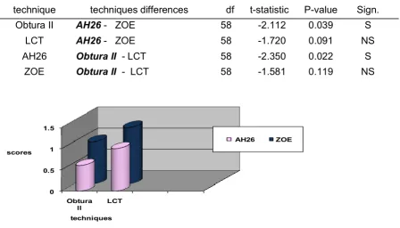
By using paired t- test, there were signifi-cant differences between AH26 with ZOE in group obturated by obtura II. Also there were significant differences when obtura II technique compared with the b
Results lateral compaction technique in group of
AH26 scores at p< 0.05. While there was non-significant difference between the other group scores at p> 0.05, (table 1, 2 and fig.1). Generally groups of AH26 gave best results than groups of ZOE, especially when AH26 group obturated by obtura II.
Obturation technique
Sealers
No. of samples
Mean of scores
SD Std. Error Mean
Obtura II AH26 30 0.6 ± 0.621 0.113
LCT AH26 30 1.00 ± 0.694 0.126
Obtura II ZOE 30 1.00 ± 0.830 0.151
LCT ZOE 30 1.33 ± 0.802 0.146
Table 1: The descriptive statistic of stereomicroscope reading
Table 2: t-test for difference between the groups (stereomicroscope)
Figure1: Bar chart showing the techniques of stereomicroscope scores
technique techniques differences df t-statistic P-value Sign.
Obtura II AH26 - ZOE 58 -2.112 0.039 S
LCT AH26 - ZOE 58 -1.720 0.091 NS
AH26 Obtura II - LCT 58 -2.350 0.022 S
ZOE Obtura II - LCT 58 -1.581 0.119 NS
0 0.5 1 1.5
Obtura ll LCT scores
techniques
Achieving an adequate coronal seal is one of the most important goals in endodontics, but there is wide variation in the sealing ability of different endodontic materials, 5
since root canal micro leakage is a com-plex subject because many variables may influence infiltration, such as root filling technique, physical and chemical proper-ties of sealers and presence or absence of smear layer. 15 Many in vitro methods have been used to evaluate the sealing qualities of endodontic filling materials, but most studies have used methylene blue dye be-cause it is inexpensive, easy to manipulate, has a high degree of staining, very small molecular weight even lower than that of bacterial toxins, the same leakage as bu-tyric acid and shows a deeper penetration than other dyes therefore was used in this study. 5,8,16 Air entrapped within the root canal filling material or inside the root canal system may inhibit penetration of dye into the pores and gaps. Oliver and Abbott stated that after centrifugation at 3,000 rpm for 5 minutes, dye penetration was 91.7%; dye penetration by passive immersion was 20.7%. For this reason, active dye penetra-tion tests have been employed whereby entrapped air is sucked out under a vac-uum or the dye penetration test may be performed under high pressure has occa-sionally been recommended; centrifugation was used in the current study. 5 Removal of the smear layer can be considered an es-sential step in successful root canal treat-ment. 17 For this reason, the smear layer was removed in this study before evalua-tion of the penetraevalua-tion and adaptaevalua-tion of root canal filling materials. The quality of the filling depends largely on the sealing ability of the sealers. The failure of various sealers may be due to their chemical com-position and physical properties (such as adhesiveness, dimensional stability, flow, solubility). 8
Generally, groups of AH26 sealer showed significantly less coronal leakage in com-parison to groups of ZOE sealer especially
Discussion when canals obturated by obtura II
al-though the result was non significant in group obturated by cold lateral compaction but gives better sealing. This result some what in agreement with some author's studies 18-20 as they showed that the epoxy
resin sealer had better sealing ability than ZOE sealer and the most efficient adhesion between sealer and root canal walls achieved when epoxy resin sealer was used. Studies showed that these findings may possibly be attributed to the fact that resin based sealers has low contraction and solubility in comparison with ZOE-based in addition to it has resin compo-nents in their formulations, which improve their adherence to the intra canal dentin walls and is an important factor for leakage prevention. 21,22 About obturation tech-niques, groups of obtura II showed signifi-cantly less coronal leakage in comparison to groups of cold lateral compaction espe-cially when AH26 used as a sealer. this result some what in agreement with some authors studies 23,24 as they showed that
the cold lateral compaction show higher leakage than thermoplasticized technique. While this result disagreed with some stud-ies, 25 it is concluded that the thermo
plasti-cized technique present the higher level of infiltration than cold lateral compaction. The factors that may be considered in ob-taining these conflicting results are: tech-nique of instrumentation, type of sealer used, sealer thickness, type and concen-tration of chelating agents used, and the technique used to remove the smear layer. The concept of injectable thermo-plasticized gutta-percha arose following the demonstration that gutta-percha in a heated state, when mechanically forced under pressure, would three dimensionally fill the root canal system more effectively and quicker than lateral or vertical conden-sation. 26 Injected thermo-plasticized
gutta-percha can adapt more effectively to ir-regularities in the canal. 27 On the other
canal irregularities and there is an inade-quate dispersion of the sealer leading to the formation of voids in and around the gutta-percha points. Therefore this draw-back, may have contributed to the high leakage score seen in this group. 24
AH26 was produced better coronal seal specially when canals obturated by obtura II. Obtura II as obturation technique was produced better coronal seal than lateral compaction technique although there was non significant difference in groups of ZOE when used as a sealer.
1. Kimura Y, Yonaga K, Yokoyama K, Matsuoka E, Sakai K, Matsumato K. Apical leakage of obtu-rated canals prepared by Er. YAG laser. J Endod 2001; 27: 567-70.
2. Mittal N, Dewan N, Gupta P and, Sharma GMK. In -vivo radiographic evaluation of sealing ability of root canals with various obturation techniques. Endodontology 2002; 14: 46-51.
3. Siqueira J, Rocas I, Lopes H, De Uzeda M. Cor-onal leakage of two root canal sealers containing calcium hydroxide after exposure to human sa-liva. J Endod 1999; 25(1):14–6.
4. Wolanek G, Loushine R, Weller R, Kimbrough W, Volkmann K. In vitro bacterial penetration of en-dodontically treated teeth coronally sealed with a dentin bonding agent. J Endod 2001; 27(5):354– 7.
5. Bodrumlu E, Tunga U. Coronal Sealing Ability of a New Root Canal Filling Material. JCDA 2007; 73 (7):623-623c.
6. Cohen S., Burns R. Pathways of the pulp. 8th ed. Mosby. 2002.
7. Gavrea I, Gray G, Hooper S, Jagger D. Compari-son of the quality of root filling achieved using five different obturation techniques. Endodontology 2007; 19(2): 5-13.
8. Verissimo D, do Vale M. Methodologies for as-sessment of apical and coronal leakage of endo-dontic filling materials: a critical review. J Oral Sci 2006; 48:93-8.
9. Kopper P, Vanni J, Della Bona Á, Figueiredo J, Portos. In vivo evaluation of the sealing ability of two endodontic sealers in root canals exposed to the oral environment for 45 and 90 days. J Appl Oral Sci. 2006; 14 (1):43-8.
10. Katić D, Karlović I, Karlović Z, Janković B. Leak-age of Different Canal Obturation Techniques. Acta Stomatol Croat 2006; 40(1): 28-34.
11. Ravanshad S, Khayat A. An In-vitro Evaluation of Apical Seal Ability of Thermafil Obturation Ver-sus Lateral Condensation. J Dent TUMS 2004; 1 (4): 48-55.
12. Goldberg F, Artaza L, De Silvio A. Effectiveness of different obturation techniques in the filling of simulated lateral canals. J Endod 2001; 27: 362-4.
13. Ibarrola J, Reynolds R, Hermsen M, Howard J, Knowles K. Evaluation of three methods of obtu-ration using the obtura II system. Braz J Oral Sci. 2002; 1(3):126-128.
14. Ponce B, Izquierdo C, Sandoval V, Reyes B. Comparative study of apical microleakage be-tween lateral compaction and vertical compaction with system B. Rev Odont Mex Journal. 2005; 9 (2): 65-72.
15. Cobankara F, Adanir N, Belli S, Pashley D. A quantitative evaluation of apical leakage of four root – canal sealers. . Int Endod J 2002; 35, 979-84. .
16. Roggendorf M, Ebert J, Petschelt A, Frankenber-ger R. Influence of Moisture on the Apical Seal of Root Canal Fillings With Five Different Types of Sealer. JOE 2007; 33: 31-33
17. Guerisoli D, Marchesan M, Walmsley A, Lumley P, Pecora J. Evaluation of smear layer removal by EDTAC and sodium hypochlorite with ultra-sonic agitation. . Int Endod J 2002; 35, 418–421. 18. De Almeida W, Leonardo M, Tanomaru Filho M,
Silva L. Evaluation of apical sealing of three en-dodontic sealers. Int Endod J 2000; 33: 25-7. 19. Suprabha B, Sudha P, Vidya M. A comparative
evaluation of sealing ability of root canal sealers. Indian J Dent Res. 2002; 13(1): 31-6. Abs. 20. Mitic A, Mitic N, Tosic G. Apical leakage of root
canal system obturation materials. Serbian Den-tal J 2005; 52: 90-6.
21. Dultra F, Barroso J, Carrasco L, Capelli A, Guerisoli D, Pecora J. Evaluation of apical micro-leakage of teeth sealed with four different root canal sealers. J Appl Oral Sci. 2006: 14(5): 341-5.
22. Aminsobhani M, Ghorbanzadeh A, Bolhari B, Shokouhinejad N, Ghabraei N, Assadian H, Alig-holi M.. Coronal microleakage in root canals ob-turated with lateral compaction, warm vertical compaction and GuttaFlow system . IEJ 2010; 5 (2):83-7.
23. Brosco V, Bernardineli N, Moraes I. “In vitro” evaluation of the apical sealing of root canals obturated with different techniques. J Appl Oral Sci 2003; 11(3): 181-5.
24. Rajeswari P, Gopikrishna V, Parameswaran A, Tina G, Kandaswamy D. In-vitro evaluation of apical micro leakage of thermafil and obtura-heated gutta percha in comparison with cold lat-eral condensation using fluid filtration system. Endodontology 2005; 17:24-31.
Conclusion
25. Bezerra A., Caldeira C., Prokopowitsch I., Andrade W. A comparative study of the apical sealing achieved by different obturation tech-niques in canals prepared with niti automatized instrumentation. ECLER Endo. 2000; 2(3). 26. Shetty K, Chandra S. An In-vitro Evaluation of
Volumetric Dye Penetration Observed with Quick Fil, Lateralcondensation, Modifiedtrifecta, Single Cone, and No Obturation Preceded by Sodium Hypochlorite and E.D.T.A. Endodontology 2006:5 -10.