2(3): 69-78, 2012 SCIENCEDOMAINinternational
www.sciencedomain.org
Effect of Gamma Irradiation on Antibacterial
Properties of Sea Crab Shell Chitosan
F.C.K. Ocloo
1*, A. Adu-Gyamfi
1, E. A. Quarcoo
1, Y. Serfor-Armah
2,
D. K. Asare
1and C. Owulah
11
Biotechnology and Nuclear Agriculture Research Institute, Ghana Atomic Energy Commission, P.O. Box LG 80, Legon, Ghana.
2
National Nuclear Research Institute, Ghana Atomic Energy Commission, P.O. Box LG 80, Legon, Ghana.
Authors’ contributions This work was carried out in collaboration between all authors. FCKO designed the study, performed the statistical analysis, wrote part of the protocol, and wrote part of the first draft of the manuscript. AAG also wrote part of the protocol and part of the first draft. EAQ helped in the irradiation of the chitosan solution. YSA, DKA and CO managed the analyses of the study. All authors read and approved the final manuscript.
Received 12thJune 2012 Accepted 14thSeptember 2012 Published 18thOctober 2012
ABSTRACT
Effect of gamma irradiation on antibacterial activities of chitosan is described. Chitosan was prepared from crab shells via demineralization, deproteinization, decoloration and deacetylation. Chitosan solutions (2%) were prepared in 1% acetic acid and irradiated at 0, 5, 15 and 25 kGy. The degree of deacetylation and viscosity-average molecular weight of the chitosan were determined. Susceptibility tests of E. coli and S. parathyphiagainst the chitosan were determined. E. coli was more susceptible to lower concentrations of chitosan solutions. Irradiated chitosan in solutions exerted a slightly faster inhibition on both E. coli and S. parathyphi than the unirradiated chitosan solution, but there was no difference observed between irradiated and unirradiated chitosan in solutions after 48 hours of incubation. The degree of susceptibility of both E. coli and S. parathyphi to irradiated chitosan in solutions was not significantly affected by the irradiation dose.
Keywords: Antibacterial activity; susceptibility; inhibition; molecular weight; chitosan; irradiation.
1. INTRODUCTION
Chitosan, an amino polysaccharide, has received much attention as a functional biopolymer for many diverse applications in food, pharmaceutical and cosmetics (Kumar, 2000; Shahidi et al., 1999). In many of these applications, specific molecular weights (Mw) of polysaccharides are required. Chitosan with an average Mw in the range of 5–10 kDa possesses strong bactericidal and superior biological activities (Kittur et al., 2003). Chitosan of 20 kDa prevents progression of diabetes mellitus and exhibits higher affinity for lipopolysaccharides compared to 140 kDa chitosan (Kondo et al., 2000). Chitooligomers have special antimicrobial activity (Begona and Ruth, 1997; Zheng and Zhu, 2003) and antitumour activity (Qin et al., 2002).
Recently, the antimicrobial and antioxidant activities of chitosan and its derivatives have attracted attention. It is of great interest to degrade chitosan into low molecular weight fragments under appropriate conditions, as these low molecular weight chitosans possess useful biological activities. The antibacterial effects of chitosan and chitosan oligomers are reported to be dependent on the molecular weight, degree of deacetylation (DD), and the type of bacterium (Uchida et al., 1989; Jeon et al., 2001; No et al., 2002; Tsai et al., 2002). The mechanism of antimicrobial activity of chitosan and derivatives is still yet to be elucidated. However, chitosan molecules are reported to be stacked over the microbial cell surface, blocking the nutrients (Shon, 2001) or bind to DNA as such inhibiting transcription or permeability of the microbial cell wall (Tharanathan and Kittur, 2003). Kumar et al. (2007) reported of higher bactericidal activity against Bacillus cereus and Escherichia coli for homogeneous low molecular weight chitosans (LMWC) of molecular weight 9.5–8.5 kDa, obtained by pronase catalyzed non-specific depolymerization (at pH 3.5, 37ºC) than native chitosan.
Low-molecular weight chitosan can be prepared by chemical, radiation, or enzymatic degradation of the high-molecular weight polymer. Radiation can provide a useful tool for degradation of different polymers. In the reaction, no other chemical reagents are introduced and there is not a need to control the temperature, environment or additives (Feng et al., 2008). Specifically, radiation can induce reactions such as chain scissions of the 1-4 glycosidic bonds which cause a reduction in molecular weight of the polymer and negligible cross-linking (Lim and Tung, 1997). Recent work by Feng et al. (2008) proved that lowering the molecular weight of chitosan increased antioxidant activity. However, research is necessary to determine the antimicrobial activity of chitosan as the molecular weight is reduced. The objective of this study was to assess the effect of gamma irradiation on the antibacterial activity of chitosan.
2. MATERIALS AND METHODS
2.1 Materials
2.1.1 Sample collection and preparation
blender, sieved to a particle size of 90 μm and then packaged in polyethylene bag for storage at ambient temperature until used.
2.1.2 Reagents and media
All reagents used in the study were from Sigma-Aldrich Chemie GmbH (Taufkirchen, Germany). These reagents were used without any further purification. Microbiological media were from Oxoid, United Kingdom.
2.2 Isolation/Production of Chitosan
Chitosan was produced from crab shells using the methodology of No et al. (1989) with some modification.
2.2.1 Demineralization
Crab shells were demineralized with 1N HCl for 30 minutes at ambient temperature with a solid to solvent ratio of 1: 15 (w/v) (No et al., 1989) with constant stirring and then filtered under vacuum. The retentate was washed for 30 minutes with tap water and oven-dried.
2.2.2. Deproteinization
Demineralized shells were deproteinized with 3.5 % (w/w) NaOH solution for 2 hours at 650C with constant stirring at a solid to solvent ratio of 1: 10 (w/v) (No et al., 1989). The sample was filtered under vacuum, and the retentate washed with tap water for 30 minutes and oven-dried.
2.2.3 Decoloration
Demineralized and deproteinized Crab shells (crab chitin) were decolorized with acetone (1:10) for 10 minutes and dried for 2 hours at ambient temperature, followed by bleaching with 0.315 % (v/v) sodium hypochloride (NaOCl) solution (containing 5.25% available Chloride) for 5 minutes at ambient temperature with a solid to solvent ratio of 1: 10 (w/v), based on dry shell (No et al., 1989). Samples were then washed with tap water and dried under vacuum for 3 hours until the powder was crispy.
2.2.4 Deacetylation
Crab chitin was refluxed for 6 hours at 100ºC using 50% concentrated sodium hydroxide solution (NaOH) with a solid to solvent ratio of 1: 15 (w/v). The resulting chitosan was washed to neutrality with tap water, rinsed with hot distilled water (90ºC), filtered, and dried at 60ºC for 24 hours in the oven.
2.3 Radiation
2.4 Analysis
2.4.1 Degree of deacetylation
Degree of deacetylation was determined for unirradiated chitosan. Film prepared from the sample was used to study the degree of deacetylation (DD). The film was prepared by casting 1.0% w/v chitosan in 1% acetic acid solution, followed by drying in a vacuum air for 12 hr. The film was then deprotonated by washing 3 times with methanol and kept in a desiccator for 12hr, then placed in sealed plate before scanning. The spectra of the chitosan was obtained using a FTIR (Fourier Transform Infrared Spectroscopy) (FTIR-8400S CE, Shimadzu Corporation, Japan) with a frequency range of 4000 – 400 cm-1. The degree of deacetylation of the chitosan was calculated using the baseline developed by Sabnis and Block (1997):
DD = 97.67 – [26.486 x (A1655/ A3450)]
where A1655and A3450are the absorbance at 1655 cm -1
of the amide-I band (a measure of the N-acetyl group content) and 3450 cm-1of the hydroxyl band as an internal standard to correct for film thickness.
2.4.2 Viscosity-average molecular weight (Mv) determination
Five chitosan concentrations of 0.20, 0.10, 0.05, 0.025 and 0.0125% (w/v) were prepared in 1% acetic acid for each of the irradiated chitosan samples (0, 5, 15 and 25 kGy). The relative viscosity measurement was performed by using an Ubbelohde capillary viscometer (size 1, Poulten, Selfe & Lee Ltd, England) at 25±1ºC. The intrinsic viscosity is defined as:
[η ] = (ηred)c→0
Which is the value of the reduced viscosity (η red) at zero concentration obtained from the linear plot of intrinsic viscosity against concentration. The viscosity molecular weight was calculated based on Mark Houwink equation (Chen and Hwa, 1996):
[η] = KMaor log [η ] = log K + a log M
where, [η ] is intrinsic viscosity, M is viscosity average molecular weight (in Dalton), K and a are empirical volumetric constants given by 8.93x10⁻⁴cm/g and a = 0.71 respectively.
2.4.3 Antibacterial property of chitosan samples
2.4.3.1 Bacterial culture
Stock culture of Escherichia coli (E. coli) and Salmonella parathyphi (S. parathyphi) were reactivated on Eosin Methylene Blue Agar and Xylose Lysine Deoxycholate Agar, respectively, to obtain 24 hr cultures. An inoculum concentration between 107 - 109 cfu/ml was made by inoculating colonies into sterile trypticase soya broth (TSB, Oxoid) and used for susceptibility test.
Susceptibility test was done by the tube dilution method described by Sugumar et al. (2010) with some modification. Stock solutions were 2% chitosan in solutions irradiated at 0, 5, 15 and 25 kGy. Varying concentrations of 0.02, 0.04, 0.06 and 0.2% were prepared for each dose treatment and the final pH adjusted to 6.8. One millilitre (1 ml) of the inocula was added to 9 ml of each chitosan solution prepared and incubated at 37ºC. One millilitre (1 ml) of the incubated mixture was taken into 9 ml of TSB at intervals of 0, 6, 12, 24 and 48 hrs for pour plating using Eosin Methylene Blue Agar (Oxoid, UK) for E. Coli and Xylose Lysine Deoxycholate Agar (Oxoid, UK) forS. parathyphi. Plates were incubated at 37ºC and counts were made between 18 – 24 hrs. For each chitosan solution, triplicate plating was undertaken.
2.5 Statistical Analysis
ANOVA was performed on the average-molecular weight data using MINITAB 14 (Minitab Inc., USA). The level of significance used was p<0.05 at 95% Confidence Intervals. Microsoft excel 2000 was used for graphical representation.
3. RESULTS AND DISCUSSION
3.1 Degree of Deacetylation
The degree of deacetylation as determined by FTIR was 80% (result not shown), which is comparable to values of 56 – 96% reported by No and Meyers (1995). Values ranging from 70 – 95% have also been reported (Canella and Garcia, 2001; Fernadez-Kim, 2004; Emi-Reynolds et al., 2007). Ocloo et al. (2011) have equally reported values of degree of deacetylation to be 76% for shrimp chitosan and 82% for commercial crab shell chitosan.
3.2 Viscosity-Average Molecular Weight (Mv) Determination
The molecular weight (Mv) of chitosan in solutions decreased significantly (p<0.05) with irradiation dose (Fig. 1) as a result of degradation. A sharp decrease was observed when irradiation dose was increased from zero (0) to 5 kGy. Similar findings were reported by Pasaphan et al. (2010).
3.3 Antibacterial Property of Chitosan Samples
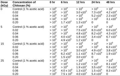
Table 1. Susceptibility ofE. colito irradiated chitosan solution at different concentrations Dose (kGy) Concentration of Chitosan (%)
0 hr 6 hrs 12 hrs 24 hrs 48 hrs
0 Control (1 % acetic acid) 0.02
0.04 0.06 0.20
> 106 > 106 > 106 > 106 > 106
> 106 > 106 > 106 > 106 1.7 x10⁴
> 106 3.0 x10⁵ > 106 > 106 1.3 x10⁴
> 106 7.3 x10⁴ 5.2 x10⁶ > 106 0
> 106 4.2 x10⁴ 2.3 x10⁵ 3.1 x10⁴ 0
5 Control (1 % acetic acid) 0.02
0.04 0.06 0.20
> 106 > 106 > 106 > 106 > 106
> 106 > 106 > 106 4.0 x10⁵ 4.9 x10⁵
> 106 7.5 x10⁵ 4.8 x10⁶ 1.8 x10⁴ 1.2 x10⁴
> 106 8.3 x10⁴ 8.2 x10⁵ 3.4 x10⁴ 0
> 106 6.0 x10⁴ 4.3 x10³ 6.7 x10³ 0 15 Control (1 % acetic acid)
0.02 0.04 0.06 0.20
> 106 > 106 > 106 > 106 > 106
> 106 > 106 > 106 6.2 x10⁴ 1.5 x10⁵
> 106 > 106 3.0 x10⁵ 1.1 x10⁴ 3.0 x101
> 106 > 106 4.9 x10⁵ 8.7 x10⁴ 0
> 106 6.3 x10⁵ 3.8 x10⁴ 1.9 x10³ 0 25 Control (1 % acetic acid)
0.02 0.04 0.06 0.20
> 106 > 106 > 106 > 106 > 106
> 106 > 106 > 106 4.0 x 10⁶ 7.5 x 10⁶
> 106 1.2 x10⁶ 1.9 x10⁴ 1.3 x10⁵ 4.0 x10⁴
> 106 > 106 3.5 x10³ 1.5 x10⁴ 5.4 x10²
> 106 8.1 x10⁴ 7.6 x10³ 8.3 x10³ 0 142.542 12.79 3.648 1.346 0 20 40 60 80 100 120 140 160
0 5 15 25
M
v
(k
D
a)
Fig 1: Effect of irradiation on viscosity-average molecular
weight (Mv) of chitosan solutions
Each value is the average of 3 counts
On the other hand, unirradiated chitosan solutions of 0.02 and 0.04% concentrations had no effect on the population ofSalmonella parathyphi after 48 hours incubation (Table 2). While the 0.06% concentration of unirradiated chitosan solutions gradually reduced population of S. parathyphi by 3 log units from 12 to 48 hours, the 0.2% concentration of unirradiated chitosan solution also reduced the population by 5 to 6 log units. Solutions of 0.02% concentration from irradiated chitosan (5, 15 and 25 kGy) had no inhibitory effect on the populations of S. parathyphi after 48 hours of incubation; however 0.04% concentration of chitosan solution irradiated samples (15 and 25 kGy) slightly reduced populations of S. parathyphi 1 to 2 log units after 24 to 48 hour. Solutions of 0.06% concentration from irradiated chitosan (5, 15 and 25 kGy) reduced populations of S. parathyphi by 1 to 3 log units after 24 to 48 hours. In the case of 0.2% concentration from irradiated chitosan in solution at 25 kGy, the population of S. parathyphi was reduced greatly by 1 to 6 log units from 6 to 48 hours.
Generally, all the irradiated chitosan solutions exerted a slightly faster inhibition (within 12 hours) on both E. coli and S. parathyphi than the unirradiated chitosan solution. However, there was no observable difference between the irradiated and unirradiated chitosan solutions after 48 hours of incubation.
The results of this study have shown that different concentrations of unirradiated and irradiated chitosan in solutions had varying degrees of inhibition against E. coli and S. parathyphi. However, 1% Acetic acid solution did not exert any noticeable effect on the population of the test isolates. These observations have demonstrated the antimicrobial activity of chitosan againstE. coli and S. parathyphi., confirming results reported by Chen et al. (1998), Rhoades and Roller (2000), Roller and Covill (2000) and Tsai et al. (2000).
The study has also shown that the degree of microbial inhibition of chitosan was dependent on its concentrations in solutions. This observation supports the findings of Seo et al. (2008). The study further showed that the degree of inhibition of the various chitosan solutions is dependent on the duration of incubation.
Despite the fact that there are several intrinsic and extrinsic factors that affect the antimicrobial activity of chitosan, the viscosity-average molecular weight has been identified as vital factor. Various studies have confirmed that irradiation of chitosan reduces the viscosity-average molecular weight (Yoksan et al., 2004; Feng et al., 2008) and that chitosan with lower molecular weight (of less than 10kDa) have greater antimicrobial activity than native chitosans (Uchida, 1989). In this study also, an irradiation dose of 5 kGy significantly decreased molecular weight of chitosan but this did not proportionately enhance the antimicrobial activity. Although both E. coli and S. parathyphiwere found be susceptible to irradiated and unirradiated chitosan solutions, the rate of inhibition of these test isolates was marginally increased by irradiation.
Table 2. Susceptibility ofSalmonella paratyphito irradiated chitosan solution at different concentrations
Dose (kGy)
Concentration of Chitosan (%)
0 hr 6 hrs 12 hrs 24 hrs 48 hrs
0 Control (1% acetic acid) 0.02
0.04 0.06 0.20
> 106 > 106 > 106 > 106 > 106
> 106 > 106 > 106 > 106 > 106
> 106 > 106 > 106 4.4 x 10⁴ 6.3 x 10⁵
> 106 > 106 > 106 1.9 x10⁴ 2.0 x101
> 106 > 106 > 106 6.4 x10³ 0 5 Control (1 % acetic acid)
0.02 0.04 0.06 0.20
> 106 > 106 > 106 > 106 > 106
> 106 > 106 > 106 4.0 x10⁶ 6.4 x10⁵
> 106 4.6 x 10⁶ > 106 8.6 x 10⁵ 2.5 x 10²
> 106 > 106 > 106 1.3 x10⁵ 1.0 x101
> 106 > 106 > 106 3.6 x10³ 0
15 Control (1 % acetic acid) 0.02
0.04 0.06 0.20
> 106 > 106 > 106 > 106 > 106
> 106 > 106 > 106 > 106 2.3 x10⁵
> 106 > 106 > 106 4.1 x10⁶ 4.5 x10²
> 106 > 106 4.7 x10⁵ 6.2 x10³ 4.0 x101
> 106 > 106 7.3 x10⁴ 1.8 x10³ 0
25 Control (1 % acetic acid) 0.02
0.04 0.06 0.20
> 106 > 106 > 106 > 106 > 106
> 106 > 106 > 106 > 106 3.1 x10⁵
> 106 > 106 7.6 x10⁶ 2.7 x10⁴ 3.1 x10⁴
> 106 > 106 4.1 x10⁶ 5.0 x10³ 4.2 x10²
> 106 > 106 8.6 x10⁵ 2.2 x10³ 0 Each value is the average of 3 counts
4. CONCLUSION
Microorganisms such as E. coli and S. parathyphi are susceptible to chitosan solution and this could be explored to improve food safety and stability. E. coliwas more susceptible to lower concentrations of chitosan solutions and the degree of inhibition of both E. coliand S parathyphiwas marginally increased by irradiation. Irradiation decreased molecular weight of chitosan but the degree of susceptibility of E. coli and S parathyphi to irradiated chitosan solutions was not significantly affected by the irradiation dose (0, 5, 15 and 25 kGy).
ACKNOWLEDGEMENT
COMPETING INTERESTS
Authors have declared that no competing interests exist.
REFERENCES
Begona, C.G., Ruth, D. (1997). Evaluation of the biological properties soluble chitosan and chitosan microspheres. International Journal of Pharmaceutics, 148, 231–240.
Canella, K.M.N., Garcia, R.B. (2001). Characterization of chitosan by gel permeation chromatography-influence of preparation method and solvent.Quim. Nova, 24(1), 13-17.
Chen, C.S., Liau, W.Y., Tsai, G.J. (1998). Antibacterial effects of sulfonated and N-sulfobenzoyl chitosan and applications to oyster preservation. J. Food Prot. 61, 1124– 1128.
Chen, R.H., Hwa, H.D. (1996). Effect of molecular weight of chitosan with the same degree of deacetylation on the thermal, mechanical and permeability properties of the prepared membrane. Carbohydr. Polym., 29, 353-358.
Emi-Reynolds, G., Zaki, S., Banini, G.K., Dogbe, S.A., Ofori-Appiah, M.A. (2007). Radiation processing and characterization of chitin and chitosan extracted from crab shells. Journal of the Ghana Science Association, 9(2), 18 – 24.
Feng, T., Li, J.D.Y., Hu, Y.,Kennedy, F.K. (2008). Enhancement of antioxidant activity of chitosan by irradiation.Carbohydrate Polymers, 73(1), 126-132.
Fernadez-Kim, S.O. (2004). Physico-chemical and functional properties of crawfish chitosan as affected by different processing protocols. M.Sc. Thesis, Louisiana State University, USA, 76.
Jeon, Y.J., Park, P.J., Kim, S.K. (2001). Antimicrobial effect of chitooligosaccharides produced by bioreactor. Carbohydr Polym, 44, 71–6.
Kittur, F.S., Vishu Kumar, A.B.,Tharanathan, R.N. (2003). Low molecular weight chitosans – preparation by depolymerization with Aspergillus niger pectinase and characterization. Carbohydrate Research, 338, 1283–1290.
Kondo, Y., Nakatani, A., Hayashi, K., Ito, M. (2000). Low molecular weight chitosan prevents the progression of low dose streptozotocininduced slowly progressive diabetes mellitus in mice. Biological & Pharmaceutical Bulletin, 23, 1458–1464.
Kumar, M.N.V.R. (2000). A review of chitin and chitosan applications. Reactive & Functional Polymers, 46, 1–27.
Kumar, A.B.V., Varadaraj, M.C., Gowda, L.R.,Tharanathan, R.N. (2007). Low molecular weight chitosans—Preparation with the aid of pronase, characterization and their bactericidal activity towards Bacillus cereus and Escherichia coli. Biochimica et Biophysica Acta, 1770, 495–505.
Lim, L.T., Tung, M.A. (1997). Vapor pressure of allyl isothiocyanate and its transport in PVDC/PVC copolymer packaging film.Journal of Food Science, 62, 1061–1066. No, H.K., Park, N.Y., Lee, S.H., Meyers, S.P. (2002). Antibacterial activity of chitosans and
chitosan oligomers with different molecular weights. Int J Food Microbiol, 74, 65–72. No, H.K., Meyers, S.P. (1995). Preparation and Characterization of Chitin and Chitosan-A
Review. Journal of Aquatic Food Product Technology, 4(2), 27-52.
Ocloo, F.C.K., Quayson, E.T., Adu-Gyamfi, A., Quarcoo, E.A., Asare, D., Serfor-Armah, Y., Woode, B.K. (2011). Physicochemical and functional characteristics of radiation-processed shrimp chitosan. Radiation Physics and Chemistry, 80, 837–841.
Pasanphan, W., Rimdusit, P., Choofong, S., Piroonpan, T., Nilsuwankosit, S. (2010). Systematic fabrication of chitosan nanoparticle by gamma irradiation. Radiation Physics and Chemistry, 79, 1095–1102.
Qin, C.Q., Du, Y.M., Xiao, L., Li, Z.,Gao, X.H. (2002). Enzymic preparation of water-soluble chitosan and their antitumor activity. International Journal of Biological Macromolecules, 31(3), 111–117.
Rhoades, J., Roller, S. (2000). Antimicrobial actions of degraded and native chitosan against spoilage organisms in laboratory media and foods. Appl. Environ. Microbiol.,66, 80– 86.
Roller, S., Covill, N. (2000). The antimicrobial properties of chitosan in mayonnaise and mayonnaise-based shrimp salads. J. Food Prot., 63, 202–209.
Sabnis, S., Block, L.H. (1997). Improved infrared spectroscopic method for the analysis of degree of N-deacetylation of chitosan. Polym Bull., 39, 67-71.
Seo, S., King, J.M., Prinyawiwatkul, W., Janes, M. (2008). Antibacterial activity of ozone-depolymerized crawfish chitosan.J Food Sci., 73(8), M 400-4.
Shahidi, F., Arachchi, J.K.V., Jeon, Y.J. (1999). Food applications of chitin and chitosans. Trends in Food Science & Technology, 10, 37–51.
Shon, D. H. (2001). Chitosan oligosaccharides for functional foods and microbial enrichment of chitosan oligosaccharides in soy-paste, Proceedings of the International workshop on Bioactive Natural Products, Tokyo, Japan, 56–66.
Sugumar, G., Ramesh, U., Selvan, A. (2010). Susceptibility of Crab Chitosan against Staphylococcus aureus. Bioresearch Bulletin, 1, 7-9.
Tharanathan, R.N., Kittur, F.S. (2003). Chitin—The undisputed biomolecule of great potential, Crit. Rev. Food Sci. Nutr. 43, 61–87.
Tsai, G.J., Wu, Z.Y., Su, W.H. (2000). Antibacterial activity of a chitooligosaccharide mixture prepared by cellulase digestion of shrimp chitosan and its application to milk preservation. J. Food Prot., 63, 747–752.
Tsai, G.J., Su, W.H., Chen, H.C., Pan, C.L. (2002). Antimicrobial activity of shrimp chitin and chitosan from different treatments and applications of fish preservation. Fish Sci, 68, 170–7.
Uchida, Y., Izume, M., Ohtakara, A. (1989). ‘Preparation of chitosan oligomers with purified chitosanase and its application’. In: Skjåk-Bræk G et al., editors. Chitin and chitosan: sources, chemistry, biochemistry, physical properties and applications. London, UK: Elsevier Applied Science, 373–82.
Yoksan, R., Akashi, M., Miyata, M., Chirachanchai, S. (2004). Optimal γ-Ray dose and irradiation conditions for producing low-molecular-weight chitosan that retains its chemical structure.Radiation Research, 161(4), 471-480.
Zheng, L.Y.,Zhu, J.F. (2003). Study on antimicrobial activity of chitosan with different molecular weights. Carbohydrate Polymers, 54, 527–530.