On the Nature of the Resting Frog Skin
Potential*
PRISCILLA M. WINN, NANCY S. LaPRADE, WILLIAM R. TOLBERT AND ERNST G. HUF
Department of Physiology, Medical College of Virginia, Richmond, and
Department of Physics, University of Richmond
I. SHORT CONTEMPORARY HISTORY
Investigations on the electrical properties of frog muscle, nerve, and skin belong to the oldest in the history of bioelectricity. Interest in the nature of the resting P.D. of frog skin was heightened by the discovery that there occurs in the epidermis of this tissue "active ion transport" (Huf, 1935; 1936; Us-sing, 1949), suggesting a possible relationship between the electrical and the chemical events. There is, as yet, no completely satisfactory explanation of the frog skin po-tential. Many investigators assume that there are at least two electro-genic layers within the multilayer epidermis, and numerous specula-tions on the nature of the skin P.D. have been offered on the basis of the two (or more) layer concept (Steinbach, 1933; Greven, 1941; Fukuda, 1942; 1944; Meyer and Bernfeld, 1946; Koefoed-Johnsen and Ussing, 1956; 1958; and others. For earlier investigators, see Steinbach, I.e., and Greven, I.e.). A rather penetrating analysis of the electrochemical behavior of frog skin has been presented by Linderholm (1952; 1954). He came to the conclusion that a single-layer concept was adequate to ex-plain the electrical and diffusion properties of skin. Fukuda's work is of particular significance. He
*
Supported by Public Health Serv-ice grants GM 03545 andGM-K6-16,687.
116
showed that the electrogenic outer layert requires the presence of Na+, but not of K+, whereas the inner layer depends on the presence of K+ in the adjacent bath. Fukuda sug-gested that the nature of the skin P.D. is intimately related to the preferential Na+ permeability of the outer layer, and the preferential K+ permeability of the inner layer.
Greven (1941) and Linderholm (1952; 1953) have proposed phys-ico-chemical models of skin which explain quite well the experimen-tally found relationship between change in Na Cl concentration in the outside bath and skin P.D. Both investigators calculated and found a P.D. change approximately 35 mv for a tenfold concentration change, excluding measurements at relatively high ionic strength ( µ,
=
0.1). Greven and Linderholm have not studied the electrical response of the inside to changes in ionic concentrations. The model of the skin proposed by Koefoed-Johnsen and Ussing (1956; 1958) gives emphasis to the already mentioned preferential permeability of the outer and the inner layer for Na+ and K+, respectively. When anion penetration was experimentally cir-cumvented (by replacingc1-
by 1h t In this paper the expressions "outer layer" and "inner layer" are used rather loosely. Nothing can be said with certainty about their loca-tion. The assumption of their presence is made because certain observations make it likely that such layers (or barriers) exist in the epidermis.so
:-
)'
these investigators found that the skin P.D. changed by nearly 59 mv when the outside Na+ concentration, or the inside K+ con-centration, was changed by a fac-tor of 10. Therefore, Koefoed-Johnsen and Ussing regarded the total skin P.D. as the sum of two Nernst diffusion potentials which are generated at the Na+ permeable, and the K+ permeable outer and inner layer, respectively. In other words, in their experiments, the outer layer behaved like a nearly perfect reversible Na+ electrode, and the inner layer like a nearly perfect K+ electrode. It is interest-ing to note that prior to this it was claimed that the inner layer behaved like a reversible H+ electrode (Meyer and Bernfeld, 1946). Flem-ing (1957) has tried to confirm this without success. Subsequent work has only in part confirmed the observations of Koefoed-John-sen and Ussing on sulfatet skins. Disagreement exists especially about the response of the outer layer to changes in Na+ concentration. Lind-ley and Hoshiko (1964) and Cereijido and Curran (1965) have reported a P.D. change of approxi-mately 35 mv for a tenfold concen-tration change. This agrees with our measurements given below.P. M. WINN, N. S. LaPRADE, W. R. TOLBERT AND E. G. HUF
made above. Ottoson et al. (1953) were the first to apply this method to frog skin. They were followed by Engbaeck and Hoshiko (1957). The latest report is by Chowdhury and Snell (1964) who may be con-sulted for additional references on this topic. So far, the results have not been in complete agreement with each other. When a slow, in-ward penetration of the epidermis is made with the microelectrode, one to four P.D. steps have been observed, but their exact location in the epidermis is not certain. The electrode becomes increasingly posi-tive with advancement of the tip, relative to the outside bath if both sides of the skin are exposed to salt solutions. Chowdhury and Snell ( 1964) are the only investigators who have obtained a nearly con-tinuous and smooth potential pro-file. They are inclined to interpret discrete P.D. steps as the result of some distortion of the cellular and tissue structure by the advancing microelectrode. A recent statement by a group of competent and ex-perienced investigators (Leh et al., 1965) strikes a note of warning to use great caution in the interpreta-tion of data: " . . . the application of microelectrode techniques to frog skin is beset with formidable technical difficulties from the stand-point of adequate control." In this paper, therefore, more confidence is placed in results which were ob-tained with the classical technique of P.D. measurement on intact skin using agar bridges and calomel half cells.
II. STATEMENT OF PROBLEMS. EXPERIMENTS
Upon closer inspection of each of the papers cited in section I, it becomes clear that the interpreta-tion of the data rests upon a great number of explicit and implicit as-sumptions. This, of course, is in the accepted tradition of scientific writing, but it also explains why the nature of the skin P.D. still is in a state of considerable
contro-versy. The review of the pertinent literature has led us to carry out the following experiments, some of which deal with the controversial quantitative aspects of the electrical responses of the skin to changes in Na+ and K+ concentration, and the effect that substitution of Na+ by Mg2+ has on these responses. Meas-urements of Qo., and of Na+, K+, and c1- content in skin were made to evaluate the extent of damage, if any, to the skins exposed for several hours to sulfate solutions of rather unphysiological composition. Stud-ies were also undertaken on the electrical response of osmotically and metabolically damaged skins to find out whether the Na+ re-sponse can be diminished or abol-ished, if only transiently, without affecting the K+ response, or vice versa.
Methods
The experiments were performed during all seasons, except winter, on belly skin of large frogs (R. pipiens) . The skin was mounted in a two chamber (each 18 ml) cell made of lucite. The skin area was 4.9 cm2 • Continuous mixing of the fluid (25 C) was achieved by using circulating pumps (20 ml per min). Skin P.D.'s were measured in the conventional way with calomel half cells, millivolt recorders (Varian Associates, Model G-10; Sargent, Model SR) and occasionally Keith-ley Electrometer, Model 600A. Careful attention was given to asymmetry and junction P.D.'s in the system. They were either ab-sent or played only a minor role, and when used for corrections did not significantly alter the observa-tions and conclusions drawn from the data. Measurements on skins were started about 1 hour after mounting of the skins while ex-posed to sulfate solutions, pH 8, containing Na
=
100; K=
10; Tris (hydroxymethyl) amino meth-ane = 10, ,ueq per ml. Keeping constant the composition of the solution at one side of the skin, theNa+ and K+ concentration of the solution at the other side was al-tered, lowering [Na+] and elevating [K+], but keeping [Na+]
+ [K+
] con-stant. Total osmolarity: 135 mil-liosmols per liter by the method of freezing point depression. [Na+]=
110 (no K) will be designated as Na1; lower [Na+] will be designated as Na,, and Na2/Na1 will be desig-nated as r. [Na+]o and [Na+]1 stand for sodium concentration in the solutions at the outside and at the inside of the skin, respectively. So-lutions were changed at about 10 min intervals when fairly stable new P.D. levels were usually seen. The data on skins which gave less than 90% recovery in Na= 100, K
=
10 were discarded. Usually the re-sponse of the outside was tested before testing the inside, but no dif-ferences in results were found due to the order of testing. P.D. will designate the potential difference across the whole skin (inside +). .6:V=
(P.D.). - (P.D.),, i.e., the difference in P.D.'s at Na2 and Na1.Oxygen uptake measurements
(20 C) on fresh skin samples
(120 mg) were carried out with the Warburg method. The belly skin was cut into several pieces which were randomly placed into Warburg flasks containing solutions of various compositions. Estima-tions of Na+ and K+ in skin were done as described earlier (Huf et al., 1955). For c1- estimations, the method of van Slyke and Send-roy ( 1923) was employed. A drop of picric acid was added to the standard solutions to simulate the yellow color of skin digests.
Electrical Response of the Outside
(Outer Layer
of the
Epidermis;
June
1963 through
June 1964)
Studies on 19 skins ( 63 meas-urements) gave results (table 1, col. 3) which fitted the computer calculated regression equation:
P.D.
NATURE OF RESTING FROG SKIN POTENTIAL
TABLE 1
Dependence of frog skin P.D. on varying composition of salt solution at the epithelial side. Belly skin of Rana pipiens. Na1
=
110; K=
0. Na2=
lower Na• concentration, as given in column 1. Composition at the dermal side of the skin was kept constant: Na 110; K=
10. THAM 10; pH 8, 25 C. Com-mon anionso
,,_
.
1 2 3
1
_
41 5
Solution pH 8
P.O.
Na+ K+ THAM* r = Na,/Na, (inside+) 6.V
a
t
= PK/PNaµEq/ml mv mv
110 0 IO 1.000 92 0.410
75 35 10 0.682 84 - 8 0.365
35 75 10 0.318 73 - 19 0.280
10 100 10 0.091 53 -39 0.167
2 108 10 0.018 30 -62
o.011i
* Tris (hydroxmethyl) amino methane.
t Calculated from a = (r0.59 - r)/(1 r); see section Illa.
t Comparable to the value given by Lindley and Hoshiko (1964); see in-troduction.
TABLE 2
Electrolyte composition of fresh skin and experimental skin (after use). Ex-perimental skins were soaked for one hour in sulfate solutions containing, in µEq/ml, Na: 110; K 10. During the experiments the skins were exposed, in sequence, to sulfate solutions pH7 of decreasing Na concentration (110 ~ 0 µEq/ml), and increasing K concentration (10 ~ 60 µEq/ml). Once or twice in each experiment Mg SO, (50 µEq/ml) replaced Na or K. All solutions were isosmotic. Time of study, March and April 1963.
µEq/gm dry wt. at end of Testing Duration of experiment
Experiment inside (i) experiment - -- - - - %
No. outside (o) (hrs) Na K Cl H20
- - - - -Fresh Skins
(Huf et al.,
1955) 254 164 215 74
- - · -Experimental
Skins 2 i 3 321 7.2 79.8
3 i 4 195 12.0 80.6
4 i 22 471* 19.0* 83. l *
5 i 6 196 21.8 82.2
6 o, i 6 181 6.0 79.8
7 i, 0 4 324 80.3
14 o, i 5 229 210 78.6
15 o, i 6 366 210 81.0
I
- - -
-Average 310 198 11. 7 80.5
* Not
included in the average value.118
with confidence limits of about
± 4 mv. In 3 of the 19 cases, the salt solutions contained 2 mM per liter CaSO,. The results were not different from those seen when Ca++-free solutions were used. An-other series on six skins (24 meas-urements) was performed with Mg++ containing solutions of the fol-lowing composition (µ.osmols per ml): Na•:K•:Mg++ = 75: 10:25; 35:10:65; 10:10:90; 2:10:98. The regression line was:
P.D.
=
23.3 log [Na+]o+
17.4(
2)
Substituting Mg++ for Na• increased the ionic strength of the solutions by factors varying from 1.4 to 2.5. No significant difference in freezing point depression was found, how-ever, between solutions with and without Mg++. A Beckman sodium electrode 39278 in conjunction with
a Beckman Model 76 Expanded
Scale pH meter was used to check the Na• activities in the solutions. We consistently found that for a tenfold change in [Na•], the P.D. of the Na• electrode changed by 53 to 54 mv.
Electrical Response of the Inside
(Inner
Layer of the
Epidermis;
June
through August 1963)
From experiments on nine skins ( 31 measurements) it was found that a tenfold change in [K •] 1, changed the skin P.D. on the aver-age by 57 mv. In five additional
experiments (20 measurements)
P. M. WINN, N. S. LaPRADE, W. R. TOLBERT AND E. G. HUF
TABLE 3
Oxygen consumption of frog skin in solutions of varying composition. All solutions were buffered with 5 mM/1 THAM. Each solution was tested on six to eight skins of different frogs. Qo2 data are given as mean values ± one standard deviation of the mean.
Composition of Solutions
Time of Milliosmoles /1
Series Exp. pH Na K Mg S04 [Fe(CN)G] Sucrose from t.C0
Milliosmoles /liter
H August 6 100
H 1963 7 100
H
s
1001 April
s
1002 June
s
503 1963
s
04
s
06 May
s
507 1963
s
509
s
0s
April 8 505 1963
s
0Metabolic, Electrolyte
Measurements on Skin
(March and April
1963)
10 0 10 0 10 0
10 0 60 0 60 50 60 0
10 50 10 0 110 0
10 0 60 0
Seven skins which had been used in 3 to 6 hours experiments were analyzed for Na+, K+ and c1- (table 2). The average results were: Na+,
310; K+, 198; c1-, 11.7 µ,eq per gm dry wt, and H20, 80.5%. Except for the per cent water, which was about 9% above control values,
(Huf et al., 1955) there was no
significant alteration in the Na+ and
K+ content of whole skin. It should be noted that about 5 % of the c1-in fresh skin remained in skin which was kept for several hours in chlo-ride-free sulfate solutions. Skins in
Na+
+ K
+ sulfate solutions showednormal respiration rate 0.53 ± 0.04 ml 02 per mg per hr (table 3). No
significant decrease in Qo2 was seen
during a period of five hours. The
lowest Qo,, (0.28 ± 0.02) was seen in Na+ free K"SO. solution. This is
in agreement with Zerahn's work
(1956). Skins in solutions
contain-ing 50 Na, 10 K, and either 50 milliosmols per liter Mg++ or su-crose had the same Qo.,: 0.36 ±
55 0 0 135
55 0 0 135
55 0 0 135
55 0 0 135
55 0 0 136
so
0 0 13730 0 55 135
so
0 0 13S30 0 55 136
55 0 0
0 15 75 13S
0 15 75 136
0.05 and 0.41 ± 0.02 for skins in Mg++ and sucrose solutions, respec-tively. In the foregoing, all errors are expressed as one standard error of the mean of eight meas-urements for each case. These ob-servations suggest that skins in sulfate solutions do not suffer se-vere metabolic alterations which, in other media, are readily detect-able by the method of whole skin analysis (Huf et al., 1955; 1957; Huf and Doss, 1959).
Electrical
Response of
Osmotically or Chemically
Damaged Skin
(March through May 1964)
Results obtained on 5 of 11 ex-periments with skins which were osmotically damaged by soaking
for several hours in bicarbonated
water (Winn et al., 1964) are
shown in figure 1. The
experimen-tal conditions, other than those mentioned under
Met hods
,
are given in the legend. At the end of the experiments the epidermis could easily be separated from the corium.In some of these experiments the
Ionic Strength
µ Qo,
ml X mg dry wt.-1 X hr-1
0.165 0.54 ± 0.033
0.165 0.53 ± 0.043
0.165 0.52 ± 0.040
0.165 0.55 ± 0.033
0.165 0.51 ± 0.039
0.290 0. 32 ± 0.02S
0.090 0.34 ± 0.026
0.290 0.36 ± 0.047
0.090 0.41 ± 0.019
0.165 0.2S ± 0.022
0.150 0.52 ± 0.037
0.150 0.45 ± 0.036
outside (normally negative relative to the inside) became slightly posi-tive. This was the case when the K2SO. concentration at the inside was high, giving rise, probably, to a K+ diffusion potential. Similar re-sults were obtained on 30 skins which were trea~ed before and dur-ing the testdur-ing with 0.02 M diethyl malonate, or 0.02 M NaF, or 0.001 M quinone (Eubank et al., 1962). In all experiments both sides of the skin failed
concurrentl
y
to respond in the manner typical for fresh skin.Ill. DISCUSSION AND INTERPRETATIONS
Response of the Outer Layer
It has been found by Koefoed-Johnsen and Ussing (1956; 1958)
that the epidermis of the brown frog (R.
temporaria)
behaves likean almost ideal Na+ electrode over
NATURE OF RESTING FROG SKIN POTENTIAL
[No]0
)Jeq/ml
[K]; .,ueq/ml
110
75
35
110
75
35
Fig. 1-Results obtained on five skins which were osmotically damaged by soaking for several hours in bicarbo-nated water. Semi-log plot of change in P.D. (abscissa) with changing out-side [Na•]o (keeping inside solution constant at 110 Na 10 K), and, fol-lowing this, with changing inside [K•J.: (keeping outside solution constant at 110 Na lOK). Sum of [Na•]
+
[K•] in the test solutions was kept constant 110. Soaking periods: A and D, 2 hours; B and C, 1 Y2 hours; E, 4 hours.for the skin of the leopard frog
(R. pipiens) and the bullfrog (R.
catesbiana). When placed in
sul-fate Ringer's solution, these skins gave a P.D. change of only about 36 mv (23 mv, if Mg2• was pres-ent in the solutions) for a tenfold change in Na• concentration, in-stead of the expected P.D. change of 59 mv. The results of the ex-periments presented above suggest that this is typical for the normal fresh skin and is not related to a poor physiological condition of the skin membrane. Skins which devi-ated greatly from the ideal Na•
electrode behavior (see above) per-formed quite well when the dermal side was tested for response to po-tassium, i.e., a P.D. change of al-most 59 mv for a tenfold change in K• concentration was obtained. In experimentally damaged skin, both sides failed concurrently to respond in the manner typical for fresh skin. This, it would seem,
rules out the possibility that an in-creased "sulfate-shunt" (Ussing and Windhager, 1964) was the cause for the non-Nernstian behavior of the outer layer of fresh skin. One would expect that skins in poor physiological condition leading to increased anion permeability would fail in their Na• and K• responses,
like the experimentally damaged skins. It should be mentioned here that, in the experiments with meta-bolic inhibitors, every stage and de-gree of damage was applied, rea-soning that in mildly poisoned skins a transient isolation of the Na• from the K• response might occur. This, however, was never achieved. The skins exposed for many hours to solutions of rather unphysiologi-cal composition had a respiration rate and a Na• and K• content comparable to control skins. It must be granted that the method of whole skin analysis is not a very sensitive method to detect altera-tions in skin electrolytes. On the other hand, the same method per-mits a demonstration of gain in Na•
and loss in K• in metabolically poisoned skins. It cannot completely
be ruled out that the skin of R.
temporaria behaves differently from
the skin of the other species men-tioned above. For instance, the skin of R. temporaria is thinner than the skin of R. pipiens. The same is true, however, for the skin of R. pipiens as compared to the skin of R. catesbiana, and yet, there
is no difference in the Na• re-sponse in the skins of the last men-tioned species. An entirely satis-factory explanation for the electrical behavior of the outer layer of frog skin when the outside Na• concen-tration is changed is not available at the present time. Any discussion of this topic should include the fol-lowing thoughts: a) Significance of K• leakage from the epidermis; b) Greven's skin model; c) Linder-holm's skin model; d) Koefoed-Johnsen and Ussing's skin model. The role of K• leakage is discussed first because this relates more im-mediately to the experiments de-scribed above.
a) K• leakage from the
epider-mis. It is well known that the
epi-dermis shows leakiness to potassium (Steinbach, 1937; Huf et al., 1952;
Huf and Wills, 1953; Bricker et al.,
1963; Klahr and Bricker, 1964) . A skin with K• leakage would be analogous-to a glass Na• electrode with a "potassium error." It would explain qualitatively why a P.D. change of less than 59 mv per ten-fold change in Na• concentration is to be expected. The following quantitative considerations will show that K• leakage seems to play some role, but it does not fully ac-count for difference between the ideal 59 mv and the actual 36 mv P.D. change.
If one chooses the Goldman-Hodgkin-Katz equation for calcu-lations of ratios of the permeability coefficients, a
=
PK/ PN., one ob-tains from equation (3) below (Lindley and Hoshiko, 1964):RT [Na2
J
A V
=
-
ln - (1 - a)+
aF Na1
P. M. WINN, N. S. LaPRADE, W. R. TOLBERT AND E. G. HUF
Equating (3) with (1), applied to Na1 and Na.,, one obtains
ro.59 _ r a =
-1 - r
The limiting value of a for r can be obtained as:
0.59 lim r. - r
,_, 1 - r
d( 0.59 - )
lim r r
,_, d(l - r)
1 - 0.59
=
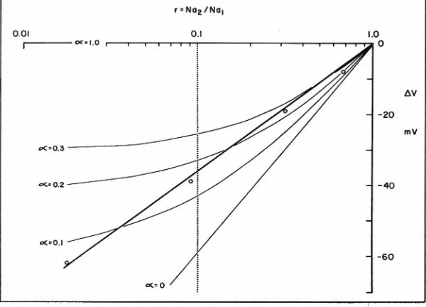
0.41The thin curves in figure 2 show the predicted relationships between
..6. V and r, calculated from equa-tion ( 3) by assuming several va l-ues for a. The heavy line is the regression line fitted to the experi-mental data (table 1, col. 4). The graph suggests that the linear semi-log relationship between Na./Na1
=
r, and ..6. V may be the result of a decrease of a with decrease of r.Whereas this possibility cannot be excluded, it is, intuitively, a some-what remote possibility. A better argument against this possibility comes from the following facts. From the studies of Cereijido et al.
(1964) and those of Winn et al. (1964), a PNa for the outer layer of the order of 8
x
10-• cm per sec may be calculated, valid for [Na+]o=
100. PNa increases rapidly with decreasing [Na]0• From the data of Huf and Wills (1953) and those of Klahr and Bricker ( 1964),a rough estimation of PK (cor-rected for skin P.D. where needed)
shows that PK is probably below 1 X 10 .... cm per sec. From these data, values for a may be calculated over a wide range of [Na]0 , all of which are far below the a values shown in table 1, col. 5. It is doubt-ful, therefore, that the high values
for a shown in table 1 have any real meaning.
b) Greven's skin model (1941).
Greven made the assumption that the outer layer of the frog skin con-tains "Festions," A, so that the
"membrane" condition may be rep-resented as m+A+n-, where m+ and
n- are the diffusible ions, e.g., Na+ and
c1
-
in the skin. When a NaCl0.01
!),.V
mV
oC•O
Fig. 2--Semi-long plot (heavy line) of the change of difference in skin P.D. (LlV) with changing r
=
Na2/Na1 in the outside solution. Na1 is the original Na+ con-centration = 110, K = O; Na. is the Na+ concentration of the subsequently used test solutions. The sum of [Na+]+
[K+] was kept constant at 110. Inside solution 110 Na, 10 K. a= PK/PN •.gradient is imposed upon the skin in the direction: outside ~ inside (c1
>
c.), a P.D. (or E) is gen-erated which can be expressed as the sum of a diffusion potential and two Donnan potentials (one on eachside of the membrane). This P.D. can be calculated by applying the theory of Teorell-Meyer-Sievers. One obtains:
E = RT
[u
In X2+
AuF x,
+
Au+
.!
In (x,+
A)(x2 -A)]
2 (x, - A)(x2
+
A)in which
u =
x,
UK - UA UK+ UA
V4C, 2
+
A2;V4C2 2 + A2
To test the validity of this model,
values for UK and UA were taken from physicochemical tables.
c.
was kept constant, 120 mM per liter N aCI; Ci was varied from 120 to 0.47 mM per liter NaCl. Valuesfor A were assumed, ranging from 60 to 0.001 meq per liter. All measured P.D. values were cor-rected for the asymmetry potential which existed when C1
=
c2=
120mM NaCl per liter. It was found (see fig. 9 of Greven's paper) that the behavior of the model was in remarkably good agreement with the behavior of the skin. The model also predicted one maximum in the P.D./log C1 curve. This maximum was also seen in experiments, if NaCl was used. It occurred in solu-tions of NaCl ;;::::; 30 mM per liter. Below this concentration, a tenfold change in C1 gave a P.D. change of about 40 mv. It is known that maxima are not seen if sulfate Ringer's of comparable ionic
strength solutions are used. An ex-planation for this has been given by Linderholm (1952; 1954; see below).
c) Linderholm's skin model
(1952; 1954). Greven's assumption of fixed charges in frog skin has been criticized by Linderholm, who gives reasons which make it
NATURE OF RESTING FROG SKIN POTENTIAL
likely that fixed charges are of
sig-nificance (see also Linderholm,
1960). Considering the well-known specificity of the response of the
outer layer to Na+, Linderholm
sug-gested that the form of the P.O./
log c1 curve may have something
to do with the active transport of
Na• ion involving a specific carrier. His skin model is described as
fol-lows: "The frog skin membrane is
supposed to be inhomogeneous in so far as there are some parts of
the membrane, where active
trans-port does not take place but where
both Na and other ions diffuse
through the skin as passive ions,
maybe through fine pores . . . . The other part of the membrane con-tains a sodium carrier, and here the active transport takes place. It
may be thought of as a liquid
membrane, essentially impermeable to other ions than those transported by the carrier." Applying principles of electrochemistry to this model, Linderholm could derive the fol-lowing equation for the P.O. (or cp) of a "hypothetical frog skin" sepa-rating two NaCl solutions:
G Na - Gel RT
a _ In a2
GaNa +Gel F ai
The meaning of the symbols is as follows: 'fa N•
=
effective active transport potential; G's=
partial ion conductances; a1 and a2=
ac-tivities of the NaCl solutions at the outside and the inside of the skin. The model expresses the P.O. as the algebraic sum of a fraction of'Pa N", and a diffusion potential. rp. "" is itself C1 dependent; it decreases with increasing Ci, although not quite linearly with respect to log c,.
Linderholm has shown that the be-havior of model and skin are in good agreement. The model also predicts a maximum in the rp/log c1 curve (see previous section). Lind-erholm found that in skins with high total conductance the maxi-mum was often at low
c,
,
and viceversa. The model has not yet been tested for the case GCI
<
<
GaN
.,
or G80 •«
G."'. Applying simple alge-bra, however, it can be seen that cp remains the algebraic sum of (ci-dependent) rp/ ' and a diffusion po-tential. This feature of the model
makes it useful for a quantitative
analysis of the electrical response
of the outside of the skin to changes
in the outside electrolyte
concen-tration.
d) The Koefoed-Johnsen and
Ussing skin model (1956; 1958)
The attractiveness of this two-layer concept lies in the fact that it attempts to explain the skin P.O.
in terms of two intra-epithelial Na•
and K• diffusion potentials (see also
Andersen and Zerahn, 1963; Han-sen and Zerahn, 1964). The claim
that skin in sulfate Ringer's
solu-tion gives a nearly 59 mv change
for a tenfold change in Na•
concen-tration of the outside bath has never
been confirmed (see sections I and
II). In other words, it still has to be shown that the rp/log c1 relation-ship is quantitatively predictable
from the model for skin in sulfate,
or in chloride Ringer's.* All
in-vestigators seem to agree that the rp/log C1 relationship for skin in sulfate Ringer's does not have a
maximum in a solution
approach-ing an ionic strength of µ,
=
0.1.The reason for this may be found
in the much higher total
conduct-ance of sulfated skins, as compared to skins in chloride Ringer's (Cereijido and Curran, 1965). This would shift a possible maximum to
a higher c1 (see Linderholm, 1952;
1954).
Re
s
ponse of
the Inner Layer
When Linderholm (1952; 1954)
*
We have found that when the sodium concentration on the outside was lowered from N a1 to Na,, skins in sulfate-Ringer's almost followed the law ~V=
RT/F Inv Na,/Nai. and ~V=
RT/F In -«Na,/Na1 in the presence of Mg2• (equations ( 1)and (2) respectively), rather than the Nernst law ~V
=
RTIF ln(Na,/Na1).proposed his version of the
one-layer concept of the skin P.O., he
did not consider the interesting work of Fukuda (1942). He showed
that, upon removal of K+ from
chloride-Ringer solution at the
in-side of the skin, the total skin P.O. rose and, upon stepwise increase
of the K• concentration, the P.O.
stepwise decreased. This is perhaps
better explained if one assumes the
involvement of a second
electro-genic layer in the generation of the
total skin P.O. Koefoed-Johnsen
and Ussing have extended Fukuda's
work, using sulfate-Ringer's instead of chloride-Ringer's. They noticed
that the inner layer behaved very
nearly like a reversible K•
elec-trode. A tenfold change in K•
con-centration at the inside of the skin gave a skin P.O. change of about
59 mv. This result was confirmed
by Cereijido and Curran (1965);
our own measurements reported in section II are also in agreement with those of Koefoed-Johnsen and
Ussing. Any hypothesis on the
na-ture of the electrical response of
the inner layer must, of course,
take into consideration the
mecha-nism of active Na• transport, which
may be located in this region.
Stud-ies on electrolyte distribution and
active ion transport in frog skin under varying metabolic conditions (Huf et al., 1957) have suggested that a metabolically forced 1 : 1 Na•µ K+ exchange may be an es-sential step in the mechanism of
active sodium transport according
to the following sequence of reac-tions:
outside bath-7Na+ + RKm+1Nan-i -7RKmNan+K+f-inside bath
leakage f- K+ + RKmNan
R is a "carrier," which may have
a definite but as yet unknown
ad-P. M. WINN, N. S. LaPRADE, W. R. TOLBERT AND E. G. HUF
jacent areas. The reasons for the
assumption of attachment of sev-eral atoms of Na• and K• to R are
given in the quoted paper. Opera-tion of a carrier system involving the structure RKmNa. implies that no active transport takes place if either the K+, or the Na• concen-tration, or both, are too low in the transport compartment. This is in agreement with the experimental data of Huf and Wills ( 19 51) , Ussing ( 1954), and Curran and Cereijido ( 1965). Figure 3A is a
simplified version of the model
shown in figure 4 of the paper of Huf et al. (1957). On the basis of these experimental results and assumptions, several speculative models describing the electrical re-sponse of the inner layer to changes in the K• concentration on the
in-side of the skin may be constructed.
a) Figure 38 shows the well-known model of Koefoed-Johnsen and U ssing ( 19 5 8) . It is assumed that the inner layer is the seat of the Na• pump, generating the force EN. on Na• crossing this border. Because of the K• electrode be-havior of the inner border and also because of the equivalence rule (short circuit current = net Na• flux, Ussing and Zerahn, 1951), Koefoed-Johnsen and Ussing have assumed that EN. is kept electro-neutral by means of a 1: 1 Na• PK• exchange across the inner border (cell membrane). For a sulfated skin in steady state, therefore, this model visualizes the existence of an electroneutral, K•-coupled Na• pump and a Nernst-type K• diffu-sion potential across the inner border.
b) An alternative model, equally lacking unequivocal experimental support but preferred by us, is the following one.
t
Implicit in thet First presented on May 7, 1965 at the 43rd Meeting of the Virginia Academy of Sciences, Richmond, (Va.
!. Sci. 16: 391, 1965). It is interesting
to note that Cross et al. have pre-sented similar arguments and support-ing data on frog muscle (!. Physiol.
181: 865-880, 1965).
c
0K+ LEAKAGE
Cl
- - - c 1
B
Fig. 3A-Active Na• transport model by Huf et al. (1957). R is a hypothetical polyvalent metabolically supported carrier forming a complex RKmNa,. which can exchange one ion for the other when energy transfer occurs. 3A depicts an
electrogenic Na• pump, since K• is assumed to recycle only within the transport
compartment, and does not cross the "inner layer." See also the similar Klahr
and Bricker model (1964). 3B) Frog skin model by Koefoed-Johnsen and
Us-sing (1958) depicting an electroneutral Na• pump. 0.c.m. =outer cell membrane.
I.c.m. = inner cell membrane. 3C) Hypothetical electrical equivalent circuit rep-resenting the open frog skin. The scheme is essentially a combination of the model proposed by Linderholm (1952; 1954), and the active Na transport model
suggested by Huf (3A). The two-layer concept ("o" and "i"), rather than
Linder-holm's one-layer concept has been adopted. (Section I and III of this paper). Without the K• parameters, the scheme is identical with Linderholm's model (19 54), with the main resistances (or conductances, G) located in both layers.
The subscripts A and p indicate "active" and "passive" respectively. ENa
(Linder-holm's </>AN•) is the true transport potential of the sodium pump. In accordance with the data of Huf et al. (1957), K• is treated as if it were present in two
compartments: pump potassium, and bulk potassium. The K battery (EK) is
124
NATURE OF RESTING FROG SKIN POTENTIAL
model discussed above seems to be the assumption that all cell potas-sium is functionally in one com
-partment. It has been shown, how-ever, (Huf et al., 1957; 1959) that transcellular active Na• transport and cellular Na•-K+ balance are separable (e.g., with fluoroacetate, or changing temperature), but not entirely separate mechanisms, sug-gesting the presence of K+ in the cell in at least two functionally dif-ferent compartments: pump
potas-sium which need not cross the in-ner border, and bulk potassium
which, if it crosses this border freely, may account for the K+ elec-trode behavior of the inner border of the epidermis. For the normal skin in steady state, bulk K+ may be kept in electrochemical balance by an electrogenic Na• pump in accordance with the Ussing-Teorell equation
c/>;n/cf>out(K+ flux ratio)
= exp. (E - EK)F/RT.
When the membrane potential
(bulk K+ concentration across the inner border), the K+ fluxes are equal. The P.D. drop seen at the inner border when [K•], is in-creased is a transient, not a steady state phenomenon, as the important experiments of Klahr and Bricker (1964) have shown. In their studies, using sulfate solutions, skins re-gained 40 to 120% of the original steady state P.D. within about 1 hour.+ Huf et al. (1955) also have observed recovery and mainten-ance of the P.D. of skins in chlo-ride Ringer's at elevated [K•]. This
was associated with elevation in K+
accumulation in the non-chloride space. These observations are con-sistent with the interpretation of
t
The short circuit current also fellsharply and transiently when [K•],
was raised.
the electrical response of the inner layer to K+ as follows: Steady state P.D.:ENa ± O; P.D.: shortly after tenfold increase of [K•],: EN.-59, transiently, leading slowly to the original steady state P.D. :EN. ± 0 (ideal recovery). For steady state conditions, this model, therefore, visualizes the existence of an elec-trogenic, K•-coupled Na• pump
which operates with a fraction of the total cell K•. In doing so, this mechanism effects transcellular ac-tive Na• transport and maintenance of cellular K balance without the appearance of a K+ diffusion po-tential. Under non-steady state con-ditions a Nernst-type K+ diffusion potential does appear, which, how-ever, is transient in nature.
c) There is no proof that there exists any coupling, tight, ionic (a) or loose, electrical (b), between active transcellular Na• transport and cellular K+ balance. Both proc-esses may occur independently of each other. This view is supported by several facts, among them the observation (Huf et al., 1957; Cur-ran and Cereijido, 1965) that cer-tain drugs when applied in low concentration inhibit only Na•
transport. When used in higher con-centration, however, the skins loose K+ and gain Na•. This suggests ac-tive uptake of K+ into the cells, independent of active transcellular Na• transport, to balance K+ loss via a diffusion pathway. Steinbach (1937) over 30 years ago had al-ready published data in favor of "potassium secretion" in the in-side ~ outside direction. A model of the skin such as this would not be incompatible with the K+ elec-trode behavior of the inner layer.
IV. SUMMARY
1. In this paper some of the high-lights of research on the nature of the resting frog skin potential have been presented. Reviewing a period of about 30 years, it was the inten
P. M. WINN, N. S. LaPRADE, W. R. TOLBERT AND E. G. HUF
through their experimental work,
have tried to find unequivocal s olu-tions to such problems as follows: a) The number and location of electrogenic layers (barriers) within
the rather complex epidermis. b) The characterization of these bar-riers in terms of specific permea-bility properties. c) The electrical response of the two sides of the
skin to changes in ionic
concentra-tions in the solutions at the skin
surfaces. d) The role of intra-epi-dermal active ion transport in the generation of the skin P.D. e) The correlation between active ion transport, skin P.D. and intra-epi-thelial (intracellular) electrolyte distribution.
Highly refined methods of study are now widely in use. This, of course, is unavoidable and
neces-sary, to find conclusive answers to the problems mentioned. On the other hand, one must be on guard
about possible pitfalls when apply-ing such refined techniques. The
study of the P.D. profile within the epidermis, using microelectrodes is beset with difficulties (see section I). Another example is the study of permeabilities of diffusion bar-riers within the epidermis by the method of applying radioisotopes to opposite surfaces of the skin. The analysis of data requires the consideration of such knotty prob-lems as coupled flows and isotope interactions (Kedem and Essig,
1965). Although progress is made in these areas, it must be admitted that at this time no completely sat-isfactory explanation of the resting frog skin P.D. can be given.
A summary of viewpoints pre-sented in this article is shown in Figure 3C. This tentative skin model is essentially a modification of the scheme suggested by Linderholm (1954). If the skin is in steady state, K+ movement may not con-tribute to the skin P.D., except that the small outward K+ leakage may be a factor. As has been pointed out in section III, the Linderholm skin model gives a reasonably good quantitative explanation for the
electrical response of the outside of the skin to changes in the Na•
concentration. The model shown in 3C raises the problem of the na-ture of the coupling between EN.
and EK at the inside (inner layer) of the skin; it may vary from an electrical (in open skin) to an ionic (shorted skin) coupling if cellular K+ is to be maintained in either case. The model describes the P.D. as a function of ion concentrations in the solutions at the two sides of the skin membrane. The model does not explain the skin P.D. in terms of intra-epidermal electrolyte gradients and the P.D. profile. The solution of this problem still lies in the future.
2. A reinvestigation was made on the electrical response of the outside and the inside of the skin to Na• and K• sulfate in the pres-ence and absence of Mg++. Fresh skins, metabolically poisoned skins, and osmotically damaged skins were used. Skin electrolytes and skin respiration were measured to evaluate possible tissue damage in skins kept for hours in sulfate solu-tions of rather unphysiological composition. The results are briefly summarized in section IL
REFERENCES
ANDERSEN, B. AND K. ZERAHN. Method
for non-destructive determination of
the sodium transport pool in frog
skin with radiosodium. Acta Physiol.
Scand. 59: 319-329, 1963. BRICKER, N.
s.,
T. BIBER AND H. H.UssING. Exposure of the isolated frog skin to high potassium
con-centrations at the internal surface.
I. Bioelectric phenomena and
so-dium transport. l. Clin. Invest. 42:
88-99, 1963.
CEREIJIDO, M., F.
c
.
HERRERA,w.
J.FLANIGAN AND P. F. CURRAN. The
influence of Na concentration on
Na transport across frog skin. l.
Gen. Physiol. 47: 879-893, 1964.
CEREIJIDO, M. AND P. F. CURRAN.
In-tracellular electrical potentials in
frog skin. l. Gen. Physiol. 48:
543-557, 1965.
CHOWDHURY, T. K. AND F. M. SNELL.
A microelectrode study of electrical
potentials in frog skin and toad
bladder. Biochim. et Biophys. Acta
94: 461-471, 1965.
CURRAN, P. F. AND M. CEREIJIDO. K
fluxes in frog skin. l. Gen. Physiol.
48: 1011-1033, 1965.
ENGBAECK, L. AND T. HosHIKo.
Elec-trical potential gradients through frog skin. Acta Physiol. Scand. 39:
348-355, 1957.
EUBANK, L. L., E. G. HUF, A. D.
CAMPBELL AND B. B. TAYLOR.
Ef-fects of redox systems on active ion transport in frog skin. l. Cellular
Comp. Physiol. 59: 129-144, 1962.
FLEMING,
w.
R. On the role ofhydro-gen ion and potassium ion in the
active transport of sodium across
the isolated frog skin. l. Cellular
Comp. Physiol. 49: 129-152, 1957.
FUKUDA, T. R. Ueber die Bedingungen
fiir das Zustandekommen des Asym-metriepotentials der Froschhaut.
lap. l. Med. Sci. Part 3. Biophysics.
8: 123-134, 1942.
FUKUDA, T. R. Sonderstellung des
N atriums bei der Potentialbildung
an der Froschhaut. lap. l. Med. Sci.
Part III. Biophysics. 10: 77-86, 1944.
GREVEN, K. Ein Beitrag zum Problem des Ruhestroms der Froschhaut.
Arch. ges. Physiol. (Pfluger) 244: 365-405, 1941.
HANSEN, H. H. AND K. ZERAHN.
Con-centration of lithium, sodium and
potassium in epithelial cells of the
isolated frog skin during active
transport of lithium. Acta Physiol.
Scand. 60: 189-196, 1964.
HuF, E.G. Versuche uber den Zusam-menhang zwischen Stoffwechsel, Po
-tentialbildung und Funktion der Froschhaut. Arch. ges. Physiol.
(Pfluger) 235: 655-673, 1935.
HuF, E. G. Dber aktiven Wasser-und Salztransport durch die Froschaut.
Arch. ges. Physiol. (Pfluger) 237:
143-166, 1936.
HuF, E. G. AND J. WILLS. Influence
of some inorganic cations on active
salt and water uptake by isolated
frog skin. Am. l. Physiol. 167:
255-260, 1951.
HUF, E. G., J. WILLS AND M. J.
CooLEY. The significance of the
anion in active salt uptake by
iso-lated frog skin. Arch. ges. Physiol.
(Pfluger) 255: 16-26, 1952.
HuF, E. G. AND J. WILLS. The
rela-tionship of sodium uptake, potas
sium rejection and skin potential in
isolated frog skin. J. Gen. Physiol.
36: 473--487, 1953.
HUF, E. G., J. P. WILLS AND M. F.
ARRIGHI. Electrolyte distribution
and active salt uptake in frog skin.
J. Gen. Physiol. 38: 867-888, 1955.
HuF, E. G., N. S. Doss AND J. P.
WILLS. Effects of metabolic
inhibi-tors and drugs on ion transport and
oxygen consumption in isolated frog
skin. J. Gen. Physiol. 41: 397-417,
1957.
HUF, E. G. AND N. S. Doss. Effect of
temperature on electrolyte metabol
-ism of isolated frog skin. J. Gen.
Physiol. 42: 525-531, 1959.
KEDEM, 0. AND A. ESSIG. Isotope
flows and flux ratios in biological membranes. J. Gen. Physiol. 48: 1047-1070, 1965.
KLAHR,
s.
AND N.s.
BRICKER. On theelectrogenic nature of active sodium
transport across the isolated frog
skin. J. C/in. Invest. 43: 922-930,
1964.
KoEFOED-JOHNSEN, V. AND H. H.
Us-SING. Nature of the frog skin
po-tential. XXth International
Physiol-ogy Congress, p. 511, 1956 (Abstr).
KoEFOED-JoHNSEN, V. AND H. H.
Us-SING. The nature of the frog skin
potential. Acta Physiol. Scand. 42:
298-308, 1958.
LEB, D. E., C. EDWARDS, B. D.
LIND-LEY AND T. HosHIKO. Interaction
between the effects of inside and
outside Na and K on bullfrog skin
potential. J. Gen. Physiol. 49: 309-320, 1965.
LINDERHOLM, H. Active transport of ions through frog skin with special
reference to the action of certain diuretics. Acta Physiol. Scand. 27
(Suppl. 97): 1-144, 1952.
LINDERHOLM, H. On the behavior of
the "sodium pump" in frog skin at
various concentrations of Na ions
in the solution on the epithelial
side. Acta Physiol. Scand. 31: 36 -61, 1954.
LINDERHOLM, H. The frog skin as a
model of the kidney tubules for studies of active transport of ions and the action of diuretics. Nieren
-symposion (K. Kramer and K. J.
Ullrich, eds.) Gottingen 1959.
George Thieme Verlag, Stuttgart 1960. p. 1-19.
LINDLEY, B. D. AND T. HosHIKO. The
effects of alkali metal cations and
126
NATURE OF RESTING FROG SKIN POTENTIAL
common anions on the frog skin
potential. J. Gen. Physiol. 47:
749-771, 1964.
MEYER, K. H. AND P. BERNFELD. The
potentiometric analysis of mem-brane structure and its application to living animal membranes. J.
Gen. Physiol. 29: 353-378, 1946.
OTTOSON, D., F. SJOSTRAND,
s.
STEN-STROM AND G. SVAETICHIN. Micro
-electrode studies on the E.M.F. of
frog skin related to electron
micro-scopy of the dermo-epidermal
junc-tion. Acta Physiol. Scand. 29 (Suppl.
106): 611-624, 1953.
STEINBACH, H. B. The electrical
po-tential difference across living frog
skin. J. Cellular Comp. Physiol. 3: 1-27, 1933.
STEINBACH, H. B. Potassium in frog
skin. J. Cellular Comp. Physiol. 10:
51-60, 1937.
UssING, H. H. The active ion tran
s-port through the isolated frog skin
in the light of tracer studies. Acta
Physiol. Scand. 17: 1-37, 1949.
UssING, H. H. Ion transport across biological membranes. In Ion Transport Across Membranes. H. T.
Clarke (ed.) New York: Academic
Press, Inc., 1954, p. 3.
USSING, H. H. AND K. ZERAHN. Ac
-tive transport of sodium as the
source of electric current in the short-circuited isolated frog skin.
Acta. Physiol. Scand. 23: 110-127, 1951.
UssING, H. H. AND E. E. WINDHAGER.
Nature of shunt path and active
sodium transport path through frog
skin epithelium. Acta. Physiol.
Scand. 61: 484-504, 1964.
VAN SLYKE, D. D. AND J. SENDROY,
JR. The determination of chlorides
in blood and tissues. J. Biol. Chem. 58: 523, 1923; quoted from: Peters, J. P. and D. D. Van Slyke.
Quanti-tative Clinical Chemistry. Methods.
Baltimore: Williams and Wilkins
Company, Vol. 2, 1932, p. 835.
WINN, P. M., T. E. SMITH, A. D.
CAMPBELL AND E.G. HuF. Sodium
diffusion in epidermis and cerium
of frog skin and in Ringer-agar gel.
J. Cellular Comp. Physiol. 64: 371
-388, 1964.
ZERAHN, K. Oxygen consumption and
active sodium transport in the iso
-lated and short circuited frog skin.
Acta Physiol. Scand. 36: 300-318,