www.fm.viamedica.pl
Address for correspondence: Ashraf Y. Nasr, MD, Anatomy Department, King Abdulaziz University, PO Box 80205, Jeddah 21589, Kingdom of Saudi Arabia, tel: +966 (2) 6401000 ext. 20477, fax: +966 (2) 4601000 ext. 20121, e-mail: [email protected] *This study was done in the Anatomy Department, Faculty of Medicine, King Abdulaziz University, Jeddah, Kingdom of Saudi Arabia.
The radial artery and its variations:
anatomical study and clinical implications*
A.Y. Nasr
1, 21Anatomy Department, Faculty of Medicine, Zagazig University, Zagazig, Egypt 2King Abdulaziz University, Jeddah, Kingdom of Saudi Arabia
[Received 24 September 2012; Accepted 10 October 2012]
Background: Background: Background: Background:
Background: To describe the radial artery and its variants in origin, branching pattern, mode of termination, and measurements of its length and external diameter.
Material and methods: Material and methods: Material and methods: Material and methods:
Material and methods: One hundred upper limbs of 30 men and 20 women adult cadavers were used in this study. The cadavers were obtained from the Anatomy Department, Faculty of Medicine, King Abdulaziz University, Jeddah, Saudi Arabia. The axillary region, arm, forearm, and hand of each limb were dissected to clarify the course and branches of the radial artery. This anatomi-cal descriptive study was conducted between September 2010 and August 2012 after approval of the Ethical Committee.
Results: Results: Results: Results:
Results: The mean distance of the normal origin of the radial artery as one of two terminal branches of the brachial artery was 38.7 ± 9.5 mm in men and 36.5 ± 8.5 mm in the upper limbs of women below the intercondylar line, and variant origin of the radial artery was found in eight limbs. The mean of radial artery length was 226.2 ± 21.7 mm in men and 209.9 ± 13.9 mm in women and that of its external diameter was 3.3 ± 0.7 mm in men and 3.2 ± 0.66 mm in women at 1 cm distal to its origin; 3.1 ± 0.73 mm in men and 3.0 ± 0.66 in women at 2 cm proximal to the styloid process of the radius. The radial artery showed different branching patterns and three modes of termination. Conclusions:
Conclusions: Conclusions: Conclusions:
Conclusions: Knowledge of radial artery description and its variants has great importance in different clinical fields and basic medical studies. (Folia Morphol 2012; 71, 4: 252–262)
Key words: radial artery, branches, variations, cadaver
INTRODUCTION
The radial artery (RA) is the smaller of the two ter-minal branches of the brachial artery (BA) in the cubit-al fossa, medicubit-al to the biceps tendon. It ascends from the BA in the cubital fossa approximately 1.0 cm be-low the bend of the elbow opposite the neck of the radius and is a more direct continuation of the BA. After its origin it traverses through the lateral aspect of the forearm approaching its lower end where it enters the palm to anastomose with the deep branch
of the ulnar artery to complete the formation of the deep palmar arch. The proximal RA courses underneath the muscle belly of the brachioradialis muscle, and its middle part lies near the superficial branch of the ra-dial nerve. The distal third of the RA becomes superfi-cial and is positioned anterior to the radius and pronator quadratus muscle between the tendons of the bra-chioradialis and flexor carpi radialis [26].
access due to its superficial course that make it eas-ily accessible and effectively compressed for haemo-stasis induction, early patient ambulation, and in-creased post-operative patient comfort. Also it has a relative lack of local vascular complications asso-ciated with the femoral approach [6]. Moreover, RA has been used in coronary artery bypass grafting, in cosmetic surgeries as forearm flaps, and in renal dialysis by making an autogenous fistula [21].
The anatomical variations of the upper limb ar-terial pattern are common and have been previous-ly reported by several investigators [17, 19]. Diver-sions of the RA from its normal anatomical pattern as regards its origin or its course constitute the larg-est group of vascular variations of the upper limbs [28]. Such variations may interfere with diagnostic, therapeutic, and surgical interventions [1].
New interest in RA anatomy is being generated due to the increased use in different coronary inter-ventions. The ease of access, high success rate, ease of care for nursing staff, given rich collateral circulation of the human hand, and low risk of thrombosis are the reasons for the popularity of the RA. These ana-tomic features of the RA are the main determinants for the feasibility of using it as a route for coronary intervention [33]. Moreover, in this condition, the RA had close proximity to the cephalic vein that might produce dangerous complications during the intrave-nous injection of medications [22]. The variant high origin of the RA, defined as RA arising either from the brachial or axillary artery (AA) proximal to the antecu-bital fossa, has been found in 2.4% to 14.3% of upper extremities [33, 34]. Opposite origin of the radial and ulnar arteries to the usual arrangement, defined as the origin of the RA from the medial and of the ulnar artery from the lateral side of the brachial artery, has been rarely reported [29]. Absent RA, with an estimat-ed incidence of 0.03%, is rare [5]. The RA is smaller compared to the brachial and femoral arteries. Accord-ing to Yoo et al. [34], the mean radial inner diameters of patients dictate that about 40.5% of female and 68.3% of male patients can physically accept a 6 Fr arterial sheath (mean radial diameter, 2.69 ± 0.4 mm in males and 2.43 ± 0.38 mm in females).
The study of the anatomic distribution and varia-tions of the RA has great importance to achieve the best results and to avoid possible complications after diagnostic, therapeutic and operative interven-tions. Thus, the present study aimed to describe the anatomical topography of the RA and determine the variant incidence of its origin, course, relation, branching pattern, and mode of termination in
hu-man cadavers with discussion of its morphological and clinical significance.
MATERIAL AND METHODS
One hundred upper limbs of 50 adult human ca-davers (30 males and 20 females) were used in this study. The specimens were obtained from the Anato-my Department, Faculty of Medicine, King Abdulaziz University, Jeddah, Saudi Arabia. The cadavers were placed in a supine position and their upper limbs were abducted to 90° to straighten their arteries and ex-tend their elbow and wrist joints. After thoracotomy a red-coloured latex was injected into the left subcla-vian and brachiocephalic branches of the aortic arch to clear the course and distribution of RA as well as to give accurate data about its external diameters. The right and left upper limbs of each cadaver were dis-sected from the axillary region down to the hand in-cluding the arm, cubital fossa, forearm, and the ana-tomical snuffbox. The skin and fasciae of the dissect-ed regions were incisdissect-ed and reflectdissect-ed to expose the deep structures. Both pectoralis major and minor muscles were dissected from their origins and reflect-ed on the lateral side to expose the axillary vessels and branches of the brachial plexus. The biceps muscle was retracted laterally to follow the course and branching pattern of the axillary and brachial arteries and their surrounding nerves. The brachioradialis muscle was displaced laterally to facilitate the handling, mobilisa-tion, and dissection of RA within the forearm. The an-atomical snuffbox was dissected to expose the RA down to the first dorsal interosseous space. The course and the branches of the RA in the forearm and hand were carefully dissected; their morphology and varia-tions were recorded. The flowing parameters of RA were measured in both right and left limbs of each cadaver. (1) Its original level in relation to the interepi-condylar line of the humerus, (2) its length in correla-tion with the forearm length, and (3) its external di-ameter at 1 cm distal to its origin and at 2 cm proxi-mal to the styloid process of the radius. In addition, any variant of its course and distribution and its branch-ing patterns within the forearm and hand and modes of termination were measured. The measurements were taken using a Vernier calliper (0.01 mm accura-cy) and measuring strap (Fig. 1). All data were tabu-lated regarding sex and side of the limb.
Statistical analysis
mea-surements were expressed as mean ± SD. Continu-ous variables were compared using Student’s t-test. A p value of < 0.05 was considered statistically sig-nificant. The approval of the medical Ethics tee was agreed by the Institutional Ethics Commit-tee of the Faculty of Medicine, King Abdulaziz Uni-versity. The study was conducted between Septem-ber 2010 and August 2012.
RESULTS
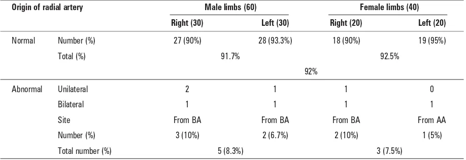
The incidence of normal and variant origin of the RA was summarised (Table 1). The classic origin of the RA, as a one of the two terminal branches of the BA within the cubital fossa below the level of the in-tercondylar line of the humerus, was seen in 92 out of 100 (92%) upper limbs. This pattern was repre-sented in 55 out of the 60 (91.7%) male upper limbs and 37 out of 40 (92.5%) female upper limbs. How-ever, the abnormal high-origin RA was observed in 8 (8%) out of 100 upper limbs, 5 male and 3 female. This abnormal origin was predominant on the right upper limbs where it was noticed in 5 right (3 male
and 2 female) upper limbs and 3 left (2 male and 1 female) upper limbs. In 7 (7%) out of 100 upper limbs the RA originated from the AA by two differ-ent forms while in a left female upper limb (1%) the RA arose from the AA.
The first variant of RA origin was seen in 4 limbs where theRA arose from the medial aspect of the upper third of the BA. Thereafter, it passed in close contact to the medial side of the BA within the arm down to the level of the intercondylar line where it crossed both the median nerve and BA superficially to the opposite side. Within the cubit-al fossa, this abnormcubit-al high-origin RA crossed the bicipital tendon superficially from medial to later-al to reach the medilater-al border of the brachioradilater-alis muscle. This artery showed normal course and dis-tribution within the forearm and hand. The course of this limb continued as the ulnar artery (Fig. 2).
In the second variant of RA origin, which was noticed in 3 limbs, the RA originated from the lateral aspect of the upper part of the BA. This abnormal RA crossed the median nerve superficially and descend-ed on its lateral side down to the cubital fossa where it passed alongside the medial aspect of the biceps tendon. Thereafter, it followed the normal course and distribution of the RA within the forearm and the hand. Meanwhile, the BA of this limb had normal course and relations within the arm down to the cu-bital fossa where it passed deep to the deep head of the pronator teres muscle and continued as the ul-nar artery within the forearm (Fig. 3).
A unique variant of RA origin was seen in a left female upper limb. The AA of this limb gave an abnormal arterial trunk from the anterior aspect of its second part at a distance 62 mm from the Figure 1. Light photograph of a left female upper limb showing
the methods of measurement of the radial artery parameters.
Table 1. The incidence of normal and variant origin of radial artery (%)
Origin of radial artery Male limbs (60) Female limbs (40)
Right (30) Left (30) Right (20) Left (20)
Normal Number (%) 27 (90%) 28 (93.3%) 18 (90%) 19 (95%)
Total (%) 91.7% 92.5%
92%
Abnormal Unilateral 2 1 1 0
Bilateral 1 1 1 1
Site From BA From BA From BA From AA
Number (%) 3 (10%) 2 (6.7%) 2 (10%) 1 (5%)
Total number (%) 5 (8.3%) 3 (7.5%)
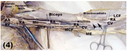
mid-clavicular point. This trunk gave the lateral tho-racic and thoraco-acromial branches and continued on the medial side of the median nerve as an abnor-mal RA down to the cubital fossa. Through its course within the axilla and arm, this abnormal RA passed superficially between the axillary and brachial arter-ies on its lateral side and the medial cutaneous nerve of the forearm and the axillary vein on its medial aspect. At the cubital fossa, the abnormal RA crossed both the median nerve and the BA superficially deep to the bicipital aponeurosis and brachialis muscle to become on their lateral side. Thereafter, it passed within the forearm and hand with normal course and distribution (Fig. 4).
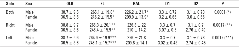
Different measurements of the RA were listed (Table 2). From these, we concluded marked differ-ences between male and female measurements, where the mean distance between the intercondy-lar line of the humerus and the RA origin was 38.7 ± ± 9.5 mm in males and 36.5 ± 8.5 mm in females. In addition, the mean length of the RA was 226.2 ± ± 21.7 mm in males and 209.9 ± 13.9 mm in females. Moreover, the external diameter mean was 3.3 ± ± 0.72 mm in males and 3.2 ± 0.66 in females at a distance of 1 cm distal to the origin of the RA while it was 3.1 ± 0.73 mm in males and 3.0 ± 0.66 mm in females at a distance of 2 cm proximal to the styloid process.
In the upper limbs of male cadavers, the mea-surements of right RA were slightly higher than those of the left ones. The mean distance of RA origin from the intercondylar line of the humerus measured 38.8 ± 9.7 mm in right and 38.7 ± 9.6 mm in left upper
limbs. The mean length of the forearm from the cen-tre of the lateral epicondyle to the distal end of the styloid process of the radius was 265.3 ± 20.1 mm in right upper limbs and 264.6 ± 19.9 mm in left upper limbs. The length of the right RA was 226.3 ± 22.0 mm, and it was 226 ± 21.8 mm in left upper limbs. The mean of the external diameter of the RA on both right and left upper limbs was 3.3 ± ± 0.7 mm at a distance of 1 cm distal to its origin while its value was 3.1 ± 0.7 mm at a distance of 2 cm proximal to the styloid process (Table 2).
However, in the upper limbs of female cadavers, the distance between the RA origin and the inter-condylar line of the humerus was equal in both right and left upper limbs, and its mean was 36.5 ± ± 8.6 mm. The mean length of the forearm was 246.4 ± 15.9 mm in right upper limbs and 246.1 ± ± 15.7 mm in left upper limbs. Moreover, the mean Figure 3. Light photograph of a right male upper limb showing the radial artery (RA) originating from the lateral aspect of the upper part of the brachial artery (BA). The median nerve (Mn) passes deep to the RA and crosses the BA; AA — axillary artery; R1, R2, R3 — roots of median nerve; MCN — musculocutaneous nerve; CBM — coracobrachialis muscle.
Figure 4. Light photograph of a left female upper limb showing the radial artery (RA) originating from the front of second part of the axillary (AA). The RA crosses the median nerve (Mn) and bra-chial artery (BA) superficially at the cubital fossa deep to the bi-cipital tendon and brachialis muscle; AV — axillary vein; MCF — medial cutaneous nerve of the forearm; CV — cephalic vein; LCF — lateral cutaneous nerve of the forearm; ME — medial epicondyle. Figure 2. Light photograph of a right female upper limb showing
length of the RA was 210 ± 14.2 mm in right upper limbs and 209.8 ± 14.1 mm in left upper limbs. The mean of the original external diameter of right RA was 3.07 ± 0.49 mm and those of the left upper limbs was 3.02 ± 0.48 mm. Its value at a distance of 2 cm proximal to the styloid process was 2.76 ± 0.49 mm in the right upper limbs and 2.74 ± 0.45 mm in the left upper limbs (Table 2).
A statistically significant difference was found between the forearm length and RA length of all the upper limbs of the male cadavers with those of the upper limbs of the female cadavers (p < 0.001). Moreover, a significant difference was noticed be-tween the length of the forearm of right male up-per limbs and those of female upup-per limbs (p = = 0.0017), and a significant difference was report-ed between the forearm length of the left male up-per limbs and those of left female upup-per limbs (p = = 0.0012). However, no significant difference was observed between all the measurements of right male and that of left male upper limbs or between the measurements of right female and those of left female upper limbs (Table 2).
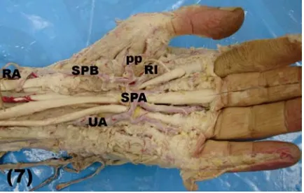
The branching patterns of the RA within the fore-arm and hand were fully described (Table 3). The ra-dial recurrent branch was the first branch originating from the RA (Fig. 5). This branch was present in 98 out of 100 (98%) upper limbs, and it was absent in 2 out of 100 (2%) upper limbs. This branch had 2 sources of origin; the most common 1 was the RA. This original pattern was found in 83 out of 100 upper limbs: 50 out of 60 (83.3%) male and 33 out 40 (82.5%) female upper limbs. Whereas, the variant original source was the BA that gave the origin of recurrent artery in 15 out 100 upper limbs: 9 male and 6 female upper limbs (Fig. 6). The second branch of the RA was the superficial palmar artery, which originated from the RA at the area of the wrist joint (Fig. 7). This branch Table 2. The radial artery measurements (mean ± SD)
Side Sex OLR FL RAL D1 D2 P
Both Male 38.7 ± 9.5 265.1 ± 19.8* 226.2 ± 21.7* 3.3 ± 0.72 3.1 ± 0.73 0.0001 (*) Female 36.5 ± 8.5 246.2 ± 15.5* 209.9 ± 13.9* 3.2 ± 0.66 3.0 ± 0.66
Right Male 38.8 ± 9.7 265.3 ± 20.1** 226.3 ± 22 3.3 ± 0.7 3.1 ± 0.7 0.0017 (**) Female 36.5 ± 8.6 246.4 ± 15.9** 210 ± 14.2 3.07 ± 0.5 2.76 ± 0.49
Left Male 38.7 ± 9.6 264.9 ± 19.9*** 226 ± 21.8 3.3 ± 0.7 3.1 ± 0.73 0.0012 (***) Female 36.5 ± 8.6 246.1 ± 15.7*** 209.8 ± 14.1 3.02 ± 0.48 2.74 ± 0.45
*significant difference between male and female (p < 0.05); **significant different at p < 0.05 between right male and female limbs; ***significant different at p < 0.05 between left male and female limbs; OLR — original level of radial artery; FL — forearm length; RAL — radial artery length; D1 — external diameter at 1 cm distal to the origin; D2 — external diameter at 2 cm proximal to the styloid process
was noticed in 95 out of 100 (95%) upper limbs and was absent in 5 out of 100 (5%) upper limbs. In 4 male and 2 female upper limbs, this branch showed a high origin from the RA. The third branch of the RA was the palmar carpal artery, which was observed in 77 out of 100 upper limbs (Fig. 5): 46 out of 60 male (76.7%) and 31 out of 40 female (77.5%) upper limbs. However, it was absent in 23 out of 100 upper limbs (Fig. 7): 14 out of 60 (23.3%) male and 9 out of 40 female (22.5%) upper limbs.
origi-nated from the RA in 51 out of 60 (85%) upper limbs, and it was absent in 4 limbs, while the superficial palmar arch gave the origin of this branch in 5 male upper limbs (6.7%). In female upper limbs, the radia-lis indicis branch was absent in 3 out of 40 (7.5%) upper limbs while it originated from the RA in 34 out of 40 upper limbs and in 3 upper limbs (7.5%) it arose from the superficial palmar arch. In addition to the previous branches, the RA gave from one to three muscular branches to the forearm muscles.
Finally, the RA showed 3 patterns of termination. The first pattern was in the form of a single branch
that anastomosed with the deep ulnar branch to form the deep palmar arch. While, in the second pattern, the RA divided into 2 terminal branches. However, in the third pattern of termination, the RA divided into 3 branches. The first (classic) pattern was seen in 52% of the specimens, the second pattern appeared in 45% of the upper limbs, and the third pattern was seen in 3% only (Table 3).
DISCUSSION
The variation of the original level of the RA may interfere with therapeutic, diagnostic, and surgical Table 3. The incidence of normal and variant origin of radial artery (RA) branches (%)
Branch All male limbs (%) All female limbs (%)
Present Absent RA Abnormal Present Absent RA Abnormal
RRA 98.3 1.7 83.3 15 97.5 2.5 82.5 15
SPA 95 5 89 6 95 5 90 5
PCA 77 23 77 0.0 77.5 22.5 77.5 0.0
FDM 88.3 11.7 88.3 0.0 87.5 12.5 87.5 0.0
PP 98.3 1.7 90 8.3 100 0.0 92.5 7.5
RI 93.3 6.7 85 8.3 92.5 7.5 85 7.5
T1 51.5 0.0 0.0 0.0 52.5 0.0 0.0 0.0
T2 45 0.0 0.0 0.0 45 0.0 0.0 0.0
T3 3.3 0.0 0.0 0.0 2.5 0.0 0.0 0.0
Branch Origin Male RA (60) Female RA (40)
Right (30) Left (30) Right (20) Left (20)
N % N % N % N %
RRA From RA 25 83.3 25 83.3 17 85 16 80
Absent 0.0 0.0 1 3.3 0.0 0.0 1 5
From BA 5 16.7 4 13.3 3 15 3 15
SPA From RA 26 86.7 27 90 18 90 18 90
Absent 2 6.7 1 3.3 1 5 1 5
High origin 2 6.7 2 6.7 1 5 1 5
PCA From RA 24 80 22 73.3 16 80 15 75
Absent 6 20 8 26.7 4 20 5 25
RI From RA 25 83.3 26 86.7 17 85 17 85
From SA 3 10 2 6.7 1 5 2 10
Absent 2 6.7 2 6.7 2 10 1 5
PP From RA 27 90 27 90 18 90 19 95
From SA 2 6.7 3 10 2 10 1 5
Absent 1 3.3 0.0 0.0 0.0 0.0 0.0 0.0
FDM Present 26 86.7 27 90 17 85 18 90
Absent 4 13.3 3 10 3 15 2 10
Termination Normal 16 53.3 15 50 9 55 10 50
pattern (2) Branch 3 43.3 14 46.7 0.0 45 9 45
(3) Branch 1 3.3 1 3.3 0.0 0.0 1 5
to 15.6% in cadavers and embryos [22] while it ranged from 8% to 24.4% in angiographic studies [29, 30].
In the present study, the incidence of normal origin of the RA was seen in 92 out of 100 upper limbs with left predominance while its abnormal high origin was observed in 8 out 100 upper limbs with male and right side predominance. The BA was the source of 7 out the 8 abnormal high-origin RAs while, the RA originated from the AA in only 1 left female upper limb.
In accordance with the results of the present study, an abnormal origin of the RA from the BA was found in 7% to 7.7% of the specimens [29]. However, in the literature, lower incidence of the abnormal origin of the RA from the BA was observed in 2.3% [32], in 2.4% [34], in 2.66% [8], in 3% [1], and in 4.17% [17]. However, the abnormal high or-igin of RA was observed in 12.5% [18], in 14% [22], and in 23% [21].
Similar to the results of the present study, a va-riant RA originating from the AA was found in 1% of the specimens [2, 8]. However, a higher incidence of the variant RA was seen in 3.25% [21] and in 2.66% [8].
The pattern and rate of the abnormal RA were different from one race to another; the axillary ori-gin of the RA was seen in 5% of African people while it was seen in 2.7% of the Caucasian population [6]. The incidence range of the high origin RA from the BA varied from 5.9% to 12.1% among Caucasians but it was observed in 2.3% of 304 Korean cadavers [32]. However, its prevalence was 0.33% among Sin-gaporean Chinese Cadavers [4]. The racial difference of the existence of abnormal origin of the radial had no clear explanation. Such abnormal origins of the RA might be the cause of failure of the transradial approach during surgical or radiological interven-tions. Thus, this variation must be kept in mind dur-ing any vascular, reconstructive, cardiac, ortho-paedic, or radiological manipulations.
The abnormal course of the RA is of interest to clinicians, particularly surgeons and radiologists. In the present study, the abnormal high-origin RA had a variant relation within the arm especially to the median nerve and the BA and to a lesser extent to the biceps and brachialis muscles. In agreement with the results of the present study, Natsis et al. [17] found 2 cases of high-origin RA with different cours-es within the arm, as well as 2 cascours-es in which the RA originated from the medial side of the BA above the intercondylar line. These arteries descended in front Figure 5. Light photograph of a right upper limb showing
branch-es of the radial artery (RA) within the forearm including the radial recurrent artery (RRA), muscular (open arrow heads) and palmar carpal branch (arrow); BA — brachial artery; Mn — median nerve; U — ulnar; AP — abductor pollicis longus tendon; EPB — extensor pollicis brevis tendon; PL — extensor pollicis longus; FCR — flexor carpi radialis.
procedures. High origin of the RA was the most com-mon arterial variation of the upper limb from either brachial or AA, where its incidence varied from 4.17% Figure 6. Light photograph of left male upper limb showing the origin of the radial recurrent artery (RRA) from the brachial artery (BA) and trifurcation of the BA; RA — radial artery; UA — ulnar artery; Mn — median nerve.
of the median nerve at the cubital fossa and crossed it anteriorly below the intercondylar line to pass within the front of the forearm in its normal ana-tomical course [8].
However, an unusual course of the RA was ob-served by Pelin et al. [19], who stated that the RA originated from the medial aspect of the upper part of the BA crossing the median nerve twice: once at its original level and secondly in the cubital fossa. Thereafter, the RA descended in its normal anato-mical course within the front of the forearm. Simi-larly, a case of high origin of RA with double crossing the median nerve within the arm was obseved [24].
The abnormal course of the RA was not only ob-served in the arm but also within the anatomical snuffbox as well. Superficial passage of the RA to the tendon of the extensor pollicis longus muscle within the anatomical snuffbox has been previously reported [8, 16]. Moreover, Patnaik et al. [18] found the RA at the base of the 2nd metacarpal where it
turned distally to pass through the 2nd
intermetac-arpal space between the 2 heads of the 2nd dorsal
interosseous muscle. Such anomalies were not ob-served in the present study. In agreement with the case of the abnormal RA of axillary origin of the present study, RA coursing behind the biceps brachii tendon was observed [9]. In the present study, the RA crossed the median nerve and BA superficially deep to the brachialis and biceps muscles at the front of the elbow joint.
The presence of such a superficial RA showed close proximity to the cephalic vein, which might produce dangerous complications during the intra-venous injection of medications and might interfere with the palpation of the normal radial pulse at the wrist with production of cannulation failure [16]. Moreover, in this condition, the RA had close proxi-mity to the cephalic vein that might produce dan-gerous complications during the intravenous injec-tion of medicainjec-tions [22].
Embryologically, the upper limb arteries deve-lop mainly from the lateral branch of the 7th
cervi-cal intersegmental artery. The arterial trunk grows outwards along the ventral axis line to terminate in the deep palmar plexus in the hand. The proxi-mal part of this trunk gives the axillary and brachi-al arteries, and its distbrachi-al part persists as the in-terosseous artery and the deep palmar arch. The RA sprouts from 2 arterial buds arising from the lateral side of the BA. These buds coalesce with each other while the ulnar artery develops from an
arte-rial bud just above the point at which the median artery arises. Later, the RA establishes a new con-nection with the main trunk at or near the level of the ulnar artery origin. The upper part of the devel-oping RA stem usually disappears to a large extent [15, 23]. The anomalies of the RA might be due to the persistence of its proximal segment with pro-duction of high origin [27]. Although knowledge of the exact factors responsible for each arterial variation is impossible, many changes may occur due to changes in the haemodynamic forces, foe-tal position within the uterus, genetic predisposi-tion, chemical factors, and developmental arrest at any stage [17, 21].
In the present study, the RA normally originat-ed as one of two terminal branches of the BA at a mean distance of 38.75 ± 9.65 mm (range 21.3– –57.7 mm) in male upper limbs and 36.5 ± 8.6 mm (range 22.6–51.5 mm) in female upper limbs be-low the intercondylar line of the humerus. How-ever, the BA terminated at a mean distance of 2.99 cm (ranging from 1.0 to 4.5 cm) distal to the inter-condylar line [18]. Meanwhile, the RA began about 1 cm below the bend of the elbow at the level of the neck of the radius [26]. However, abnormal branching pattern was found in 9% [29] and in 3.2% [22] of the specimens. The discrepancy of this distance might be due to racial difference or the number of specimens.
The results of the present study revealed that the mean length of the RA was 226.3 ± 22 mm in right and 226 ± 21.8 mm in left male upper limbs while in female cadavers it was 210 ± 14.2 in right and 209.8 ± 14.1 mm in left upper limbs. However, the mean length of radial was 22.99 ± 3.03 cm in males and 1.74 ± 0.53 cm in females [1]. Moreover, the range of RA lengths was 20–24 cm in males and 2 cm less in females [7].
The angiographic measurement of the luminal diameter of the RA at 2 cm proximal to the styloid process was 2.6 ± 0.5 mm and its range was 1.6 to 3.8 mm: 2.69 ± 0.4 mm in men and 2.43 ± ± 0.34 mm in women [33]. However, the mean dia-meter of right and left RA was 2.3 ± 0.4 mm (1.4–3.6 mm) and 2.2 ± 0.4 mm (1.2–3.1 mm), respectively, in males 2.3 ± 0.39 and in females 2.11 ± 0.29 [28]. However, the luminal inner RA diameter at 1–2 cm proximal to the styloid pro-cess using two-dimensional ultrasound was 2.6 ± 0.41 mm [34], which was more than that re-ported by Shima et al. [25], who, in a study using cadavers, reported that the average diameters of RA in proximal and distal portions were 2.3 and 2.2 mm, respectively. The reason for this discre-pancy might be due to using patients for one hand and cadavers for the other one. However, in the Japanese population it was 3.1 ± 0.06 in male and 2.8 ± 0.6 mm in female patients [11]. Know-ledge of RA diameter helps cardiologists and radio-logists to cannulate various sizes of sheaths dur-ing transradial coronary interventions and to im-prove microsurgical techniques.
Moreover, the mean diameter of the RA at the wrist was measured by electronic digital callipers in presence of complete and incomplete palmar arches. It was 3.1 ± 0.2 mm in the presence of complete arch and 2.6 ± 0.3 mm on the right and 2.7 ± 0.2 mm on the left side of the incomplete palmar arch [5].
Regarding the limb side, the baseline diameter of right RA was 2.58 ± 0.38 mm while whose of the left RA was 2.71 ± 0.32 mm. The authors added that the right RA, representing the dominant side, was significantly smaller than that of the non-domi-nant side [13]. However, no significant difference was reported in diameter between the left and right radial arteries [20].
Knowledge of the differentbranching patterns of upper limb arteries has clinicaland surgical sig-nificance. The branching pattern of the RA and its variations has been rarely studied [10]. In the present study, the radial recurrent artery originated from the RA in 83.3% of male and 82.5% of female speci-mens and from the BA in 15% the specispeci-mens. Mean-while, it was absent in 2% of the specimens. In dis-agreement with the results of the present study, the radial recurrent artery was a branch of the BA in 12% of the specimens [8]. Moreover, the palmar carpal artery was absent in 26.7% [8] of cases, while it was absent in 22.5% of the specimens of the
present study. Conversely, the absence of the first dorsal metacarpal was (12%) more than that seen by Gupta et al. [8], who reported that the first dor-sal metacarpal artery was absent in 9.3% of speci-mens. The differences between the results of these 2 studies might be related to the number of speci-mens used in each.
In the present study, the superficial palmar ar-tery was absent in 5% of cases, and originated from the RA at a higher level in 6% of the specimens. Simi-lar observations have been reported by others [8]. However, the high-origin superficial palmar artery from the RA was found in only 1 case [3]. Another variation was found where the RA gave a 2nd dorsal
metacarpal artery at the proximal end of the 2nd
in-termetacarpal space [18]. Such an observation was not seen in the present study and was not previous-ly recorded [8].
In the present study, the RA showed three dif-ferent terminal patterns: the classic termination as a single branch to form a deep palmar arch in 52% of cases, into 2 terminal branches in 45% of cases, and into three terminal branches in 3% of the specimens. However, the second pattern was seen in 52% of cases [9] while the third pattern showed 2 extremes: in 84.6% [2] and 2.7% of cas-es [8]. Moreover, division of the RA into 4 termi-nal branches was found in 15.3% of the specimens [2]. The previous pattern of RA termination was not seen in the present study. The difference of the termination pattern of the RA might be relat-ed to the number or the race of the cadavers usrelat-ed in the study.
With the progress of diagnostic and surgical techniques, the RA has great clinical significance where it is used in different surgical and radiologi-cal procedures such as the radial forearm flap in the reconstructive surgeries of the arm, as a graft for coronary bypass, and in the transradial ap-proach during coronary interventions [19]. The ac-ceptance of the transradial approach became more popular than the transfemoral or transbrachial approaches for coronary procedures as the RA had a superficially safe course for better haemostasis, not surrounded by major veins or nerves, and with good collaterals [30]. Moreover, the RA is used with increasing frequency to replace the great saphenous vein as a coronary bypass graft, with the belief that it provides improved long-term patency [31].
as the inability to measure blood pressure, misterpretation or difficulties in the angiograph in-tervention, puncture to the superficially located artery with production of finger gangrene and muscular contraction, iatrogenic damage to the artery during the surgical manipulation in ortho-paedic, plastic, or vascular surgeries, or the artery might be mistaken for a vein with production of false intra-arterial injection with severe bleeding or drug poisoning. Thus, the importance of such anatomical variations has grown with the exces-sive use of RA as a conduit in coronary bypass sur-gery [10]. However, the compression of the medi-an nerve by varimedi-ant RA could be confused with other causes of radiculopathy or neuropathies. Thus, knowledge of the exact arterial course and its relation to the nearest peripheral nerve have crucial importance when vascularised nerve ho-mografts are used [14].
The variation of the branching pattern of the RA has great significance in cardiac catheterisa-tion for angioplasty, pedicle flaps, or arterial graft-ing, where any abnormal positions or divisions must be identified before surgery. Thus, physicians should be aware of this abnormality before initi-ating the procedure [12]. Further study is advis-able to correlate the clinical significant of RA pa-rameters in comparison with those of the femoral artery for cardiac catheterisation, angiography, and coronary bypass operations.
CONCLUSIONS
Knowledge of normal and variant RA distribution and parameters provide surgeons and radiologists with the ability to make proper decisions that achieve better preoperative evaluation, surgical and radiolo-gical interventions, and good postoperative results.
ACKNOWLEDGMENTS
The author extends deep thanks to Professor Mohamed H. Badawoud, Chairman of the Anatomy Department, Faculty of Medicine, King Abdulaziz University for his great support and guidance. He also appreciates the efforts of all technical staff members.
REFERENCES
1. Bidarkotimath S, Ramakrishna A, Arunachalam K (2012) An anatomical study of primary pattern of arteries of upper limb with relevance to their variations. NUJHS, 2: 2249–7110.
2. de Rezende MR, Mattar Júnior R, Cho AB, Hasegawa OH, Ribak S (2004) Anatomic study of the dorsal arterial sys-tem of hand. Rev Hosp Clin Fac Med Sao Paulo, 59: 71–76.
3. Dhar P, Lall K (2008) An atypical anatomical variation of palmar vascular pattern. Singapore Med J, 49: 245–249. 4. Dong Z, Yi Z, Jun S, Eng-Ang L, Yip GW (2010) High origin of radial arteries: a report of two rare cases. Scientific World J,10: 1999–2002.
5. Fazan VP, Borges CT, Da Silva JH, Caetano AG, Filho OA (2004) Superficial palmar arch: an arterial diameter study. J Anat, 204: 307–311.
6. Franchi E, Marino P, Biondi-Zoccai GG, De Luca G, Vassanelli C, Agostoni P (2009) Transradial versus trans-femoral approach for percutaneous coronary proce-dures. Curr Cardiol Rep, 11: 391–397.
7. Guo WH (2006) Arterial grafting for coronary artery bypass surgery. 2nd Ed. Springer, Heidelberg, Berlin. 8. Gupta C, Ray B, Dsouza AS, Nair N, Pai SR, Manju M
(2012) A morphological study of variations in the branching pattern and termination of the radial artery. Singapore Med J, 53: 208.
9. Jelev L, Surchev L (2008) Radial artery coursing behind the biceps brachii tendon: significance for the transra-dial catheterization and a clinically oriented classifica-tion of radial artery variaclassifica-tions. Cardiovasc Intervent Radiol, 31: 1008–1012.
10. Joseph J, Ranjit D, Jatin D (2005) Superficial ulnar ar-tery. Eur J Cardiothorac Surg, 28: 495–496.
11. Loh YJ, Nakao M, Tan WD, Lim CH, Tan YS, Chua YL (2007) Factors influencing radial artery size. Asian Car-diovasc Thorac Ann, 15: 324–326.
12. Madhyastha S, Murlimanju BV, Jiji P, Saralaya VV, Rai A, Vadgaonkar R (2011) Morphological variants of the human superficial palmar arch and their clinical impli-cations. Morphol. Sci, 28: 261–264.
13. Madssen E, Petter H, Rune W (2006) Radial artery dia-meter and vasodilatory properties after transradial cor-onary angiography. Ann Thorac Surg, 82: 1698–1703. 14. Malcic-Gurbuz j, Gurunluoglu R, Ozdogmus O, Yalin A (2002) Unique case of trifurcation of the grachial ar-tery: its clinical significance. Clin Anat, 15: 224–227. 15. Moore KL, Dalley AF (2006) Upper limb. Clinically
oriented anatomy. 4th Ed. Lippincott Williams & Wilkins, Philadelphia.
16. Morris G, Rowe M, Delacure D (2005) Superficial dor-sal artery of the forearm: case report and review of the literature. Ann Plast Surg, 55: 538–541.
17. Natsis K, Noussios G, Paraskevas G, Lazaridis N (2009) Study of two cases of high-origin radial artery in hu-mans. Eur J Anat, 13: 97–103.
18. Patnaik VVG, Kalsey G, Singla RK (2002) Branching pat-tern of brachial artery: a morphological Study. J Ana-tom Soc India, 51: 176–186.
19. Pelin C, Zagyapan R, Mas N, Karabay G (2006) An un-usual course of the radial artery. Folia Morphol, 65: 410–413.
20. Riekkinen HV, Karkola KO, Kankainen A (2003) The ra-dial artery is larger than the ulnar. Ann Thorac Surg, 75: 882–884.
22. Rodriguez-Niedenführ M, Vazquez T, Parkin IG, Sanudo JR (2003) Arterial patterns of the human upper limb: up-date of anatomical variations and embryological de-velopment. Eur J Anat, 7: 21–28.
23. Satyanarayanana N, Sunitha P, Shaik MM, Satya-vathidevi P (2010) Brachial artery with high up division with its embryological basis and clinical significance. IJAV, 3: 56–58.
24. Shetty D S, Raghu J, Cliwyn S, Braganza S, Nayak B, Somayaji SN (2010) Presence of a median arterial arch associated with high origin of radial artery. IJAV, 3: 158–159.
25. Shima H, Ohno K, Michi K, Egawa K, Takiguchi R (1996) An anatomical study of the forearm vascular system. J Craniomaxillofac Surg, 24: 293–299.
26. Standring S (2008) Gray’s anatomy: the anatomical basis of clinical practice. 40th Ed. Churchill Livingstone, Elsevier, Edinburgh, London.
27. Swaroop N, Dakshayani KR (2011) The high origin of radial artery and its clinical significance. Anatomica Karnataka, 5: 32–35.
28. Tariq Ashraf, Ziauddin P, Sultana H, Muhammad AM, Fahad S, Javed A (2010) Size of radial and ulnar artery in local population. J Pak Med Assoc, 60: 817–819.
29. Uglietta JP, Kadir S (1989) Arteriographic study of vari-ant arterial anatomy of the upper extremities. Cardio-vasc Intervent Radiol,12: 145–148.
30. Valsecchi O, Vassileva A, Musumeci G, Rossini R, Tespili M, Guagliumi G, Mihalcsik L, Gavazzi A, Ferrazzi P (2006) Failure of transradial approach during coronary inter-ventions: anatomic considerations. Catheter Cardiovasc Interv, 67: 870–878.
31. Vollala VR, Nagahhooshana S, Bhat SM (2008) Trifur-cation of brachial artery with variant course of radial artery: rare observation. Anat Sci Int, 83: 307–309. 32. Yang HJ, Gil YC, Jung WS, Lee HY (2008) Variations of
the superficial Brachial artery in Korean Cadavers. J Korean Med Sci, 23: 884–887.
33. Yokoyama N, Takeshita S, Ochiai M, Koyama Y, Hoshino S, Isshiki T, Sato T (2000) Anatomic variations of the radial artery in patients undergoing trans-radial coronary intervention. Catheter Cardiovasc Interv, 49: 357–62.