www.fm.viamedica.pl
Address for correspondence: Dr. Marios Loukas, MD, PhD, Department of Anatomy, American University of the Caribbean, Jordan Road,
Anatomical variations of the superficial
and deep palmar arches
Marios Loukas
1, 2, Danny Holdman
1, Shelly Holdman
11Department of Anatomy, American University of the Caribbean, Sint Maarten, Netherlands Antilles 2Harvard Medical School, Boston, MA, USA
[Received 6 April 2004; Revised 21 February 2005; Accepted 22 February 2005]
The use of radial arteries as an arterial bypass conduit is an invasive procedure which is becoming popular among various medical centres. The greatest risk associated with harvesting the radial artery is ischaemia of the soft tissues of the hand. In this study we dissected 200 hands derived from 100 formalin-fixed cadavers in order to identify arterial patterns that will allow safe removal of the radial artery for use in bypass procedures. A complete superficial palmar arch (SPA) was found in 90% of the cases and divided into 5 types, while the remain-ing 10% possessed an incomplete palmar arch. Types of SPA are designated by the letter S. In type S-I (40%), the SPA is formed by anastomosis of the superfi-cial volar branch of the radial artery to the ulnar artery. Type S-II (35%) is formed entirely of the ulnar artery. Type S-III (15%) is formed by anastomosis of the ulnar and median arteries. Type S-IV (6%) is formed by anastomosis of the ulnar, radial, and median arteries and Type S-V (4%) is formed by a branch of the deep palmar arch (DPA) communicating with the SPA.DPA was identified in all speci-mens and classified into three types, all designated by the letter D. Type D-I (60%) is formed by anastomosis of the deep volar branch of the radial artery and the inferior deep branch of the ulnar branch. Type D-II (30%) is formed by anas-tomosis of the deep volar branch of the radial artery and the superior deep branch of the ulnar artery. Type D-III (10%) is formed by anastomosis of the deep volar branch of the radial artery with both deep branches of the ulnar artery. This data could provide an important source of information for vascular surgeons harvesting radial arteries.
Key words: deep palmar arch, superficial palmar arch, cardiac surgery, vascular surgery, radial artery, ulnar artery
INTRODUCTION
In recent years extensive research has been per-formed to determine which arterial or venous grafts used in cardiac bypass surgery are most effective. Specific factors weigh heavily in the overall decision of which type of graft to use. These factors include, but are not limited to, location of the donating ves-sel, ease of harvest, the effect of the removal of the
selected vessels such as the saphenous vein [8]. The greatest risk associated with the harvesting of the radial artery is the possibility of ischaemia to the hand. The radial artery contributes greatly to the cir-culation of the hand but in many cases can be removed as a non-essential vessel, with adequate circulation being provided by the remaining ulnar and, in some cases, persistent median artery [4, 12, 13, 20].
The radial artery originates in the proximal fore-arm from the division of the brachial artery. The ul-nar is large and provides several important contribut-ing smaller arteries to the forearm, while the radial is often considered expendable from its origin to the wrist because of its lack of vitally important supply-ing branches between the cubital fossa and the wrist. The radial recurrent artery joins the brachial artery proximal to the elbow and can safely be ligated with-out the blood flow being significantly compromised. For these reasons the radial artery can be removed with a working length of up to 24 cm for use as an arterial bypass conduit [4]. Most often the DPA is formed by anastomosis of the deep palmar branch of the ulnar artery to the dorsal radial artery, which branches off the radial artery near the wrist and pass-es dorsally on the hand before passing through the first or second metacarpal space [1, 6, 11, 14–16]. The SPA is more variable, with multifarious contri-butions from the ulnar, radial and even persistent median arteries [3, 5–7, 9, 15, 19, 21, 23].
Our research goal was to outline the observed types of DPAs and SPAs that would allow for ade-quate blood supply to the hand once radial circula-tion is removed. Addicircula-tionally, we would like to pro-pose a different view of the existing classification of circulation most commonly found in the hand in or-der to facilitate better documentation and commu-nication amongst medical professionals in the field.
MATERIAL AND METHODS
We examined 120 adult human hands during the “Human Body” course at Harvard Medical School throughout the academic semesters of 2001, 2002, 2003 and 80 adult human hands during the anato-my course at the American University of the Carib-bean through the academic semesters of 2003 (fall, winter and summer semesters). The cadavers came from female and male subjects (female 20/male 80), with an age range of 56 to 80 years and a mean age of 73 years. All the cadavers were fixed in a stan-dard formalin/phenol/alcohol solution and routinely dissected by first-year medical students.
In order to obtain a clear field for visualisation of the superficial and deep palmar arches the following structures were removed: skin and palmar aponeuro-sis, and where necessary, the flexor retinaculum.
Following preliminary examination, images from all the dissected specimens were recorded with a Sony digital camera (model: Sony Cyber-Shot DSC-f717) and studied using a computer-assisted Image Analysis System. The digital camera was connected to an image processor (Nvidia Riva TNT model 64) with linkage to a mainframe computer. Digitised images of the DPAs and SPAs, together with their surrounding structures, were stored in a “tif” for-mat and the measurements analysed with the soft-ware program Lucia (Nikon). When possible, we measured the distance from the origin of the SPA and/or DPA to its termination. Statistical analysis was performed using a t-test (Statistica for Windows v. 6.2). Measurements were considered to be signif-icant when p < 0.05.
RESULTS
The classification of the observed palmar arch circulation was divided into superficial and deep, with S and D respectively used in the nomenclature.
The DPA was identified as complete in all 200 specimens (100%). This conclusion was reached by direct visualisation of a major contribution from the radial artery reaching the DPA, formed by the ulnar artery or by the DPA directly contributing to the cir-culation of the thumb and first digit, including the thenar muscles, making contribution from the radial artery extraneous. We further divided this group into three subtypes (Fig. 4). While small variances were observed amongst our specimens, all were easily grouped into the following categories. Deep Class I (hereafter referred to as D-I) Type D-I (60%) is formed by anastomosis of the deep volar branch of the radial artery and the inferior deep branch of the ulnar branch. Type D-II (30%) is formed by anastomosis of the deep volar branch of the radial artery and the superior deep branch of the ulnar artery. Type D-III
Figure 1. Schematic representation of superficial palmar arch types.
Figure 4. Schematic representation of deep palmar arch types. Figure 2. A cadaveric specimen exhibiting a complete superficial
palmar arch.
Figure 3. Arterial palmar arch removed en-block from a cadaveric
(10%) is formed by anastomosis of the deep volar branch of the radial artery with both deep branches of the ulnar artery. Table 1 represents a synoptic form of all classes with results and percentages.
Furthermore, no statistical significant differenc-es were observed between the specimens received from two different schools with respect to race, gen-der, morphology and percentages.
DISCUSSION
DISCUSSION
DISCUSSION
DISCUSSION
DISCUSSION
The literature is replete with descriptive informa-tion about the SPA. The “tradiinforma-tional classificainforma-tion” consists of linkage between the superficial palmar branches of the radial and ulnar arteries. It is inter-esting to note that this is not always the most com-monly observed variation. The “traditional classifi-cation” has been reported to be evident in as many as 55.9% of specimens in the research of Ikeda et al. [9] or as few as 10% of specimens according to Rueng-sakulrach et al. [20]. Our data indicate that this “tra-ditional classification”, which we refer to as S-I, was found in 40% of specimens. In addition, in these specimens the radial contribution to the SPA is of a debatable clinical significance as it is often found to be of negligible diameter in comparison to the ulnar artery [9]. Ikeda et al. [9] demonstrated this phe-nomenon when they further subdivided this group into ulnar-dominant (33.2%), radial-dominant (1.4%), and equal types (21.3%). While our research did not differentiate between ulnar and radial dom-inance of the SPA, it is obvious that in cases of ulnar or equal dominance the blood supply would not be significantly affected by removal of the radial artery. Furthermore, the radial dominant type of SPA was specified by Ikeda as one in which the contribution
of the radial artery was larger than the contribution of the ulnar artery in its calibre and blood supply to the fingers. However, Ikeda’s description has major limitations regarding the degree of anastomosis be-tween ulnar and radial arteries and the contribution of the radial dominant type to the DPA [9].
Our results for groups S-I, S-II, and S-V show them to be similar in incidence to those found by Cole-man and Anson, with respective incidences of 34.5%, 37% and 2% [3].
While the median artery normally ceases to exist during early embryological development, it has been reported in the literature to be present as the persis-tent median artery [3, 9, 17, 20]. The persispersis-tent me-dian artery’s contribution to the SPA is reported in some studies to be present in up to 5% [3, 5, 9], while our data showed a much higher incidence of median persistence (21%) distributed amongst groups S-III (ulnar-median) and S-IV (ulnar-median-radial). Our results were in an agreement with the results of Ruengsakulrach et al. [20], who reported median arterial persistence of this magnitude.
While the origin of superficial and deep palmar arches is significant, it is the determination of radial contribution to the thenar muscles and digits that is of greatest importance to the harvesting of the radi-al artery. Parks et radi-al. [17] illustrated a number of clinical cases involving variations in blood supply to the thumb and index finger, several of which are of interest in this respect. These authors caution that 1 to 10% of patients could exhibit varying levels of vascular compromise in the area of the thumb fol-lowing damage to or removal of the radial artery. Obviously, pre-operative screening is necessary for patients undergoing radial arterial harvest for CABG.
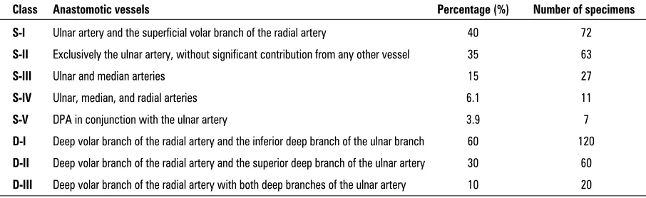
Table 1. A synoptic form of all the classes of both superficial and deep palmar arches with percentages and number of
specimens for each class
Class Anastomotic vessels Percentage (%) Number of specimens
S-I Ulnar artery and the superficial volar branch of the radial artery 40 72
S-II Exclusively the ulnar artery, without significant contribution from any other vessel 35 63
S-III Ulnar and median arteries 15 27
S-IV Ulnar, median, and radial arteries 6.1 11
S-V DPA in conjunction with the ulnar artery 3.9 7
D-I Deep volar branch of the radial artery and the inferior deep branch of the ulnar branch 60 120
D-II Deep volar branch of the radial artery and the superior deep branch of the ulnar artery 30 60
The extent of radial and ulnar contribution to the blood supply of the hand can be determined through a variety of invasive and non-invasive methods, in-cluding angiography, ultrasonography [18] and the standard Allen test. In fact a combination of the stan-dard Allen test and ultrasonography has been report-ed to be very successful [10, 18–20, 22]. Ruengsakul-rach et al. [20] have suggested the use of a modified Allen test (Allen-LEC) to determine whether variations exist such as a radial version of the persistent median artery and a type of high-take off the superficial dor-sal radial artery exist. Either of these exceptional situ-ations might yield misleading information during a standard Allen test (without confirmatory angiog-raphy or ultrasonogangiog-raphy) that could potentially re-sult in serious ischaemia following radial artery har-vest such as that described by Parks et al. [17]. The findings of Bianchi and Leiro support our assertion that in most cases a complete SPA is usually sufficient to provide adequate blood flow to the thumb in con-junction with a complete DPA [2]. We found that these requirements were fulfilled in 90% of our cases.
With the recent advances in the field of endoscopic surgical removal of the radial artery reported by Con-nelly et al. [4] and their promising results, it is apparent that the use of the radial artery as a CABG is being met with some degree of approval. Connelly et al. [4] reported the use of the radial artery in 60% of coro-nary bypass cases. They reported a reduction in infec-tion rate, discomfort, scarring and possible neurolog-ical deficiency, even in patients with such complicat-ing factors as diabetes or peripheral vascular disease. The removal of the radial artery endoscopically can be performed by a properly trained physician’s assis-tant in as little as 15 min. The reported poor long-term performance of saphenous vein grafts [8] allows one to remain cautiously optimistic regarding the use of this novel technique in the future.
It is interesting to note that many authors [3, 5–7, 9, 15, 19, 21, 23] have tried to explore the anatomy and morphology of the superficial and deep palmar arches and very few have reported similar results and percentages. Ethnic and gender differences, sample size and different classification interpretation could be some of the reasons that the results are not uni-form. However, it is important to note that this study is representative of a small subset of the human population, and only serves to illustrate the many variations in the anatomy of the palmar arch. We hope that our findings will help to promote more positive outcomes in surgical procedures requiring
REFERENCES
1. Al-Turk M, Metcalf WK (1984) A study of the super-ficial palmar arteries using the Doppler Ultrasonic Flow meter. J Anat, 138: 27–32.
2. Bianchi H, Leiro R (1987) The arterial trunk of the thumb-index digital collaterals. Surg Radiol Anat, 9: 63–67. 3. Coleman S, Anson B (1961) Arterial patterns in the
hand based upon a study of 650 specimens. Surg Gy-necol Obst, 113: 409–424.
4. Connelly MW, Torrillo LD, Stauder MJ, Patel NU, McCabe JC, Loulmet DF, Subramanian VA (2002) En-doscopic radial artery harvesting: Results of first 300 patients. Ann Thor Surg, 74: 502–506.
5. Gajisin S, Zbrodowski A (1993) Local vascular contri-bution of the superficial palmar arch. Acta Anat, 147: 248–251.
6. Gellman H, Botte MJ, Shankwiler J, Gelberman RH (2001) Arterial patterns of the deep and superficial palmar arches. Clin Orthop, 383: 41–46.
7. Jelicic N, Gajisin S, Zbrodowski A (1988) Arcus palmaris superficialis. Acta Anat, 132: 187–190.
8. Johnson WH 3rd, Cromartie RS 3rd, Arrants JE, Wuamett JD, Holt JB (1998) Simplified method for can-didate selection for radial artery harvesting. Ann Tho-rac Surg, 65: 1167.
9. Ikeda A, Ugawa A, Kazihara Y, Hamada N (1988) Arterial patterns in the hand based on three-dimen-sional analysis of 220 cadaver hands. J Hand Surg, 13A: 501–509.
10. Kochi K, Sueda T, Orihashi K, Matsuura Y (1999) New noninvasive test alternative to Allen’s test: snuff-box technique. J Thorac Cardiovasc Surg, 118: 756–758. 11. Mezzogiorno A, Passiatore M, Mezzogiorno V (1994)
Anatomic variations of the deep palmar arteries in man. Acta Anat, 149: 221–224.
12. Murakami T (1969) On the position and course of the deep palmar arteries, with special reference to the so-called palmar metacarpal arteries. Okajimas Fol Anat, 46: 177–199.
13. Olave E, Prates JC (1999) Deep palmar arch patterns in Brazilian individuals. Surg Radiol Anat, 21: 267–271. 14. Olave E, Gabrielli C, Del Sol N, Rodriques CF, Prates JC
(1998) A biometric study on the relationships between the deep palmar arch and the superficial palmar arch, the distal wrist and palmar creases. Folia Morphol, 57: 383–388. 15. Olave E, Prates JC, Del Sol M, Gabrielli C (1997) Ana-tomical relationships between the deep palmar arch and the deep branch of the ulnar nerve. Folia Mor-phol, 56: 187–193.
16. Olave E, Prates JC, Gabrielli C, Pardi P (1997) Median artery and superficial palmar branch of the radial artery in the carpal tunnel. Scand J Plast Reconstr Surg Hand Surg, 31: 13–16.
17. Parks BJ, Arbelaez J, Horner RL (1978) Medical and surgical importance of the arterial blood supply of the thumb. J Hand Surg, 3: 383–385.
Car-19. Rodriguez-Niedenfuhr M, Sanudo JR, Vazquez T, Nearn L, Logan B, Parkin I (1999) Median artery revised. J Anat, 195: 57–63.
20. Ruengsakulrach P, Buxton BF, Eizenberg N, Fahrer M (2001) Anatomic assessment of hand circulation in harvesting the radial artery. J Thor Cardio Surg, 122: 178–180.
21. Ruengsakulrach P, Eizenberg N, Fahrer C, Fahrer M, Buxton BF (2001) Surgical implications of variations in hand collateral circulation: Anatomy revisited. J Thor Cardio Surg, 122: 682–686.
22. Starnes SL, Wolk SW, Lampman RM, Shanley CJ, Prager RL, Kong BK, Fowler JJ, Page JM, Babcock SL, Lange LA, Erlandson EE, Whitehouse WM Jr (1999) Noninvasive evaluation of hand circulation before ra-dial artery harvest for coronary artery bypass grafting. J Thor Cardio Surg, 117: 261–266.