R E S E A R C H
Open Access
Pattern and process in the evolution of the sole
dioecious member of Brassicaceae
Valerie L Soza, Vietnam Le Huynh and Verónica S Di Stilio
*Abstract
Background:Lepidium sisymbrioides, a polyploid New Zealand endemic, is the sole dioecious species in
Brassicaceae and therefore the closest dioecious relative of the model plantArabidopsis thaliana.The attractiveness
of developing this system for future studies on the genetics of sex determination prompted us to investigate historical and developmental factors surrounding the evolution of its unisexual flowers. Our goal was to determine
the evolutionary pattern of polyploidization ofL. sisymbrioidesand the timing and process of flower reproductive
organ abortion. To that end, we used a combination of phylogenetics to place this species within the complex
history of polyploidization events inLepidiumand histology to compare its floral ontogeny to that of its closest
hermaphroditic relatives and toA. thaliana.
Results:Using a nuclear locus (PISTILLATA), we reconstructed the gene tree amongLepidiumtaxa and applied a
phylogenetic network analysis to identify ancestral genomes that contributed to the evolution ofL. sisymbrioides.
Combining this phylogenetic framework with cytological and genome size data, we estimatedL. sisymbrioidesas
an allo-octoploid resulting from three hybridization events. Our investigations of flower development showed that unisexual flowers appear to abort reproductive organs by programmed cell death in female flowers and by developmental arrest in male flowers. This selective abortion occurs at the same floral developmental stage in both
males and females, corresponding toArabidopsisstage nine.
Conclusions:Dioecy in Brassicaceae evolved once inL. sisymbrioidesfollowing several allopolyploidization events, by a process of selective abortion of reproductive organs at intermediate stages of flower development. Different developmental processes, but similar timing of abortions, affect male versus female flower development. An increased understanding of how and when reproductive organs abort in this species, combined with our estimates of ancestral genome contributions, ploidy and genome size, lay the foundation for future efforts to examine the genetic mechanisms involved in the evolution of unisexual flowers in the closest dioecious relative of the best studied model plant.
Keywords:allopolyploidy, programmed cell death, dioecy, floral ontogeny, genome size, organ arrest, phylogenetic
network,PISTILLATA, sex differentiation, unisexual flowers
Background
The family Brassicaceae contains approximately 338 gen-era and 3,709 species [1,2] and includes the model plant
Arabidopsis thaliana, which has a floral morphology rep-resentative of the vast majority of the family. Only 5% of genera within Brassicaceae show deviations from the basic floral plan of four sepals, four petals, six stamens (four medial and two lateral), and two fused carpels [3]. One of these genera that diverge from the basic Brassicaceae
floral morphology, Lepidium (230 species [1]), is widely
distributed in temperate and subtropical areas [4]. Early-diverging lineages in the genus comprise outcrossing dip-loid species from the Old World that exhibit the basic floral plan of the family, whereas derived lineages tend to be selfing allopolyploids from the New World, Australia, and New Zealand with reduced flowers (that is, fewer sta-mens and/or reduced petals) [5]. Among the latter, the
New Zealand endemicLepidium sisymbrioidesis the only
dioecious species in the whole Brassicaceae [4,6-10]. Staminate flowers ofL. sisymbrioidesconsist of four to six stamens and a reduced ovary [10,11], whereas carpellate
* Correspondence:[email protected]
Department of Biology, University of Washington, Box 351800, Seattle, WA 98195-1800, USA
flowers have three to seven staminodes and a functional pistil with style and stigma [10]. Nonfunctional
reproduct-ive organs of unisexual flowers of L. sisymbrioides have
been loosely described as ‘abortive’ [8,11], but the exact timing and process of the abortions remain unknown.
Incongruence between phylogenetic trees using nuclear versus chloroplast DNA regions suggests reticulate evolu-tion within the genus [12]. It appears that hybridizaevolu-tion, followed by whole genome duplication, resulted in
predom-inantly allopolyploid Lepidium species in the Americas,
Australia, and New Zealand [5]. These hybridization events may have contributed to the reduced stamens and petals observed in these species [5], in which case dioecy could represent another example of organ reduction.
In angiosperms, dioecy often follows polyploidization, presumably due to chromosomal rearrangements that fa-cilitate the evolution of sex chromosomes or the break-down of gametophytic self-incompatibility, followed by inbreeding depression [13-15]. Correlations between is-land habitat and dioecy are also common, through selec-tion for outcrossing in small, colonizing hermaphroditic populations, [16]. In fact, New Zealand taxa in general show a higher incidence of gender dimorphism com-pared to their continental sister taxa [17,18]. Combined evidence for reticulate evolution and polyploidy in New
Zealand Lepidium [5,12] suggests that L. sisymbrioides
may also be an allopolyploid. We therefore hypothesize that this species represents another case of dioecy evolving
in an island species, following hybridization and
polyploidization.
The lineages containingLepidium and Arabidopsis
di-verged from each other relatively recently, approximately
35 million years ago (mya) [19].Lepidium sisymbrioides,
therefore, offers the potential to uncover the genetic mechanisms involved in the evolution of unisexual flowers by being the closest dioecious relative to the most thor-oughly investigated model plant. Determining the develop-mental stage and process of reproductive organ abortion should facilitate the identification of candidate genes in-volved in the evolution of unisexual flowers in this species as genes involved in sporogenesis and gametogenesis have
been identified inArabidopsis [20,21]. Six developmental
processes leading to reproductive organ abortion in uni-sexual flowers are recognized: cell death, programmed cell death, parenchymatization, arrest of development, change in timing of otherwise normal developmental events, and inviable pollen [22]. Identifying which of these processes contributes to the development of male and female flowers inL. sisymbrioides, as well as estimating this spe-cies’genome size and ploidy history, will facilitate future efforts to uncover the genetic mechanisms involved in the evolution of dioecy.
The overall goal of this study was to investigate the pat-tern of polyploidization and the developmental processes
underlying the evolution of separate sexes in L.
sisym-brioides, the sole dioecious member of Brassicaceae. To
that end, we 1) identified ancestral genomes within L.
sisymbrioides and close relatives, 2) estimated ploidy and
genome size forL. sisymbrioidesand close relatives and 3)
investigated the timing and process of organ abortion by comparing its floral development to that of its close herm-aphroditic relatives and toArabidopsis thaliana.
Methods
Sampling methods
Three subspecies ofL. sisymbrioideswere originally
recog-nized:kawarau(Petrie) Thell.,matau(Petrie) Thell., and
sisymbrioides. All have been listed as nationally endan-gered because of a steep reduction in their distribution and abundance [23] and are difficult to sample. We sam-pled L. sisymbrioides subsp. sisymbrioides only because, despite reported habitat and morphological differences, all three subspecies are closely related [10].
In addition to published DNA sequences of Lepidium
available from GenBank (Appendix 1), we sampled L.
sisymbrioidessubsp.sisymbrioidesand three
hermaphro-ditic close relatives endemic to New Zealand,L. kirkii, L.
naufragorumand L. tenuicaule[9,10,12,24,25]. We sam-pledL. kirkiifrom an herbarium specimen (Appendix 1) and the remaining three species from cultivated acces-sions at the University of Washington (UW) greenhouse from wild-collected seed provided by P. Heenan (Landcare Research, Lincoln, New Zealand). Voucher specimens are listed in Appendix 1.
Molecular methods
Because we were primarily interested in the
polyploidi-zation history of L. sisymbrioides and its close relatives
(L. kirkii, L. naufragorum,and L. tenuicaule), we investi-gated reticulation events using the single-copy nuclear gene PISTILLATA (PI). PI had been previously used to
detect reticulation among other Lepidium taxa [5] and
therefore sequences were readily available (Appendix 1). Genomic DNA was extracted from one to two accessions for each of our four study species using the FastDNA Kit (MP Biomedicals, Solon, OH, USA) for cultivated
acces-sions or following the protocol of Hughey et al. [26] for
the herbarium specimen. We amplified and sequenced the
first intron of PI using PI-ITF and PI-ITR primers [5].
Polymerase chain reaction conditions were 95°C for 2 min, followed by 35 cycles of 94°C for 30 s, 60°C for 1 min, and 72°C for 1 min, with a final extension step at 72°C for 5 min. Amplified DNA was purified using ExoSAP-IT (USB Corporation, Cleveland, OH, USA).
Mini Kit (5 Prime Inc., Gaithersburg, MD, USA). Three to 27 positive clones were sequenced per accession (UW Biochemistry DNA Sequencing Facility or GENEWIZ, Seattle, WA, USA) for a total of 20 to 35 clones per taxon.
Phylogenetic analyses
PISTILLATA sequences were edited in Sequencher 4.9 (Gene Codes Corporation, Ann Arbor, MI, USA). We in-corporated sequences we generated from our four study species plus available sequences from other taxa in
Gen-Bank to the entire alignment of the PI first intron,
pro-vided by J. L. Bowman [5]. We then aligned all sequences manually using MacClade 4.08 [27]. Ambiguously aligned regions were excluded from subsequent analyses. Our data set is available through TreeBASE (http://purl.org/phylo/ treebase/phylows/study/TB2:S11886).
In order to detect whether PCR-mediated
recombin-ation had occurred among thePIcopies within a species,
we checked for recombination using RDP4 [28]. The PI
alignment was analyzed by the automated exploratory recombination analysis, which employs eight different recombination methods: RDP [29], BootScan [30], GEN-ECONV [31], MaxChi [32], Chimaera [33], SiScan [34], 3Seq [35], and LARD [36]. Analyses were run under the default general settings but as linear sequences and dis-entangling overlapping signals. The default settings for each method were used except for the following models: Felsenstein 1984 for BootScan and reversible process for LARD. Five recombinant sequences were identified and
removed from thePIalignment before subsequent
phylo-genetic analyses.
We reconstructed the phylogeny for the Lepidium PI
data set using Bayesian and likelihood analyses. For taxa
that had multiple clonal PI sequences, we chose one
clonal sequence from each monophyletic group of se-quences representing a given taxon that was recovered in a 50% majority rule consensus tree from preliminary Bayesian analyses of all clones and that was representa-tive of the majority of clones from a group. All other clonal sequences not forming monophyletic groups with other clones from the same taxon were included in the final analyses (Appendix 1).
For Bayesian and likelihood analyses, the model of
evo-lution for thePI data set was determined by jModelTest
2.1 [37,38]. The model selected under the Akaike
Informa-tion Criterion [39] was TVM + I +Γ. We specifiedL.
phle-bopetalumandL. perfoliatumas outgroups, as previously identified in various studies [5,9,25,40].
Bayesian analyses were conducted in MrBayes 3.2.2 [41,42] via the CIPRES Science Gateway 3.3 [43]. We used default priors of no prior knowledge for the parameters of the model. Bayesian analyses were conducted with three independent Markov Chain Monte Carlo [44] analyses of 10 million generations each. Metropolis coupling for each
analysis was conducted under the default settings. Conver-gence was determined when the average standard devi-ation of split frequencies remained less than 0.01. The first 10% of trees was discarded before convergence. The remaining trees from each run were pooled to construct a 50% majority rule consensus tree to obtain posterior prob-abilities (pp) and visualized with FigTree 1.4 [45].
Likelihood analyses were conducted in GARLI 2.0 [46]. Analyses were run under the default settings and included five search replicates to determine the maximum likeli-hood tree. To assess the reliability of clades in the result-ing likelihood tree, we conducted 1,000 nonparametric bootstrap (bs) replicates [47] in GARLI. Bootstrap repli-cates were conducted under the above settings, but in-cluded one search replicate and 10,000 generations as the first part of the termination condition. Bootstrap trees were summarized with SumTrees 3.3.1 [48] and visualized with FigTree.
Since multiplePIcopies in polyploidLepidiumtaxa had
been previously ascribed to allopolypoidy [5], we wanted to identify potential hybridization events leading to the evolu-tion of dioecy inL. sisymbrioides. To facilitate visualization of potential ancestral genomes that contributed to the evo-lution ofL. sisymbrioidesand its close relatives (L. kirkii,L. naufragorum, L. tenuicaule), we conducted network ana-lyses using the 50% majority rule consensus tree from
Bayesian analyses of the PIdata set as a multilabeled tree
(MUL tree). The MUL tree was imported into Dendro-scope 3.2.10 [49] and transformed into a phylogenetic
network using the Huber et al. [50] algorithm, which
minimizes the number of hybridization nodes.
Cytology
Chromosome counts were obtained from pollen mother cells (PMCs) from freshly collected floral buds from one
to two cultivated accessions of L. sisymbrioides and L.
tenuicauleusing a modified protocol of Kato [51].Floral buds were treated according to the protocol of Matsushita
et al. [52] and Wright et al. [53], modified with an N2O
treatment for 3 hours at 206 PSI and an enzyme digestion for 3 hours. PMCs were mounted in VECTASHIELD with DAPI (Vector Laboratories, Burlingame, CA, USA), ob-served and photographed using a Nikon Microphot-FX microscope (Nikon Instruments, Inc., Melville, NY, USA) and a Retiga 1300 monochrome camera (QImaging, Surrey, British Columbia, Canada).
Genome size estimation
Valley, CA, USA) before staining with propidium iodide and analyzed with a flow cytometer, as outlined in Davison
et al. [55]. CEN, with a 1C-value of 1223 Mbp or 1.25 pg [56], were used as an internal calibration standard. Animal standards have been discouraged by some authors for plant studies because (1) they cannot account for the huge range of plant genome sizes, (2) their nuclei structure may be dif-ferent from plant nuclei, and (3) their precise genome size is unknown [57]. However, for our purposes, the genome
size of CEN falls within the range ofLepidiumgenome size
estimates. Additionally, our goal was to produce relative holoploid genome size estimates, since absolute estimates are not feasible due to the lack of complete genome cover-age in most model taxa because of repetitive regions in the genome [55,57].
Samples were analyzed on a FACScan flow cytometer (Becton, Dickinson and Company, Franklin Lakes, NJ, USA) with FlowJo software (Tree Star, Ashland, OR, USA) at the UW Department of Immunology Cell Ana-lysis Facility. The 2C median nuclear peak of propidium
iodide fluorescence in Lepidium samples was compared
to that of the CEN standard to estimate the 2C nuclear
DNA content ofLepidiumin Mbp, then converted to pg
using the equation from Dolezelet al. [58].
Floral development observations
Floral tissue of L. naufragorum, L. sisymbrioides, and
L. tenuicaule was dissected and fixed overnight in formaldehyde-acetic acid-alcohol (FAA). Prior to scan-ning electron microscopy (SEM), tissue was dehydrated through an ethanol series (30 min each of 50%, 60%, 70%, 85%, 95% and 100%), critical-point dried, mounted, and sputter coated at the UW Department of Biology Imaging Facility. Observations were made with a JSM-840A scanning electron microscope (JEOL, Peabody, MA, USA).
The relative timing of floral organ initiation and
growth in Lepidium taxa had been previously shown to
be comparable to that of its model Brassicaceae relatives
(that is, Arabidopsis thalianaand Brassica napus), with
the exception of petal growth and stamen number [59,60]. Therefore, floral developmental stages for our
threeLepidiumstudy species were designated by the 13
characterized stages of the closely related model
Arabi-dopsis thaliana[20,61,62].
For histological observations, inflorescences were fixed in FAA, then dehydrated through an ethanol series end-ing in Citrisolv (Fisher Scientific, Kent, WA, USA), em-bedded in Paraplast Plus (McCormick Scientific, LLC,
St. Louis, MO, USA), and sectioned (5 or 8μm)
accord-ing to the protocol of Kramer [63]. Slides were deparaffi-nized with CitriSolv, hydrated through an ethanol series, stained in 1% Safranin O for 24 hr [64] and counter-stained with 0.5% Fast Green FCF for 30 sec or counter-stained
in 0.05% Toluidine Blue O in dH2O for 1 to 2 min, and dehydrated through an ethanol series ending in
Citri-Solv. Histological sections were mounted in Cytoseal™
60 (Richard-Allan Scientific, Kalamazoo, MI, USA) and observed using a Leica TCS SP5 II laser scanning confocal microscope (Leica Microsystems Inc., Buffalo Grove, IL, USA) with an excitation of 488 nm and an emission of 500 to 560 nm for Safranin O and an excitation of 561 nm and an emission of 625 to 690 nm for Fast Green FCF or using a Leitz Orthoplan 2 microscope (Ernst Leitz, Midland, Ontario, Canada) and photographed with a MicroPub-lisher 3.3 Real-Time Viewing camera (QImaging, Surrey, British Columbia, Canada).
TUNEL assays
We conducted TUNEL assays to determine whether pro-grammed cell death (PCD) was occurring in aborted
stamens from L. sisymbrioides female flowers.
Paraffin-embedded tissue sections were prepared as outlined above
from femaleL. sisymbrioidesand maleL. sisymbrioidesand
hermaphroditicL. tenuicaule for comparison. We used
the DeadEnd Fluorometric TUNEL System (Promega Corporation, Madison, WI, USA) according to manufac-turer’s instructions, including positive controls, and washed slides in PBS containing 0.1% Triton® X-100 and 5 mg/ml of BSA after terminating reactions to reduce background as recommended. Slides were mounted in VECTASHIELD with DAPI, except for negative control slides that were
un-treated and mounted in Cytoseal™60. Slides were observed
using a Leica TCS SP5 II laser scanning confocal micro-scope using an excitation of 405 nm and an emission of 430 to 550 nm for DAPI and an excitation of 488 nm and an emission of 500 to 535 nm for fluorescein.
Results
PISTILLATAgene duplication history suggests allopolyploidy inLepidium sisymbrioidesand relatives
In order to identify hybridization events leading to the
evolution of the dioecious speciesL. sisymbrioides and its
closest hermaphroditic relatives, L. kirkii, L. naufragorum
andL. tenuicaule, we amplified and sequenced the first
in-tron of the single-copy nuclear genePIfrom these species
and aligned them to other Lepidium sequences available
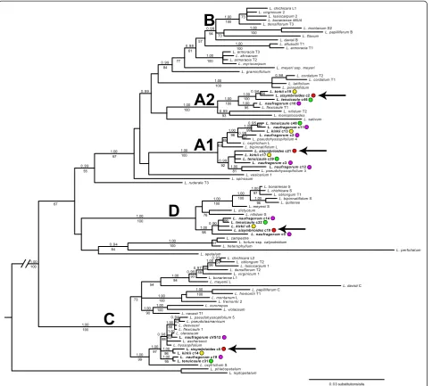
in GenBank or unpublished (provided by J. L. Bowman; Appendix 1). Phylogenetic analyses recovered five, strongly
supported clades (A1-D; pp ≥0.99, bs ≥94%; Figure 1)
representing five major copies of the PI intron from
American, Australian, and New Zealand (AANZ) taxa.
Multiple copies of the PI intron within a taxon were
as distinct (pp = 1.00, bs = 100%) and indicative of two separate genomes, as evidenced by sequences from our four study species in both clades (Figure 1, colored dots).
Therefore, we found at least four distinct copies of thePI
intron in L. kirkii [GenBank:KJ648155-KJ648159], L.
naufragorum [GenBank: JN119859-JN119860/JN11986
2/KJ648160-KJ648165], L. sisymbrioides[GenBank: KJ6
48166-KJ648169], and L. tenuicaule [GenBank:KJ6481
70-KJ648174], representing clades A1, A2, C, and D (Figure 1). In addition, multiple sequences of L. kirkii, L. naufragorum,L. pseudohyssopifolium,and/orL. tenuicaule
within clades A1, C, and D (Figure 1) suggest that hybridization, gene duplication and/or allelic divergence are at play.None of the New Zealand taxa studied fell into the fifth clade B, which consists entirely of American taxa. Our results therefore suggest that all four New Zealand species are allopolyploids (and potentially allo-octoploids at minimum), originating from at least four divergent ge-nomes (represented by clades A1, A2, C, and D).
We further used the Bayesian majority rule consensus tree from thePIdata set (Figure 1) to estimate a phylogen-etic network to aid in the identification of hybridization
0.03 substitutions/site L. sativum
L. flavum
L. pseudotasmanicum
L. naufragorum c14 L. bonariense S
L. montanum L
L. papilliferum B L. montanum S2
L. nitidum S
L. bonariense 46U4
L. sisymbrioides c21
L. ruderale T3
L. naufragorum c12
L. densiflorum T2
L. naufragorum c3
L. dictyotum
L. violaceum
L. naufragorum c10 L. monoplocoides
L. hyssopifolium
L. phlebopetalum
L. kirkii c17
L. tenuicaule c46 L. alluaudii T1
L. myriocarpum
L. tenuicaule c31 L. desvauxii
L. sisymbrioides c2
L. chichicara S L. armoracia T2
L. vesicarium 1 L. spinosum
L. oblongum T2
L. aschersonii
L. davisii B
L. davisii C L. fremontii T1
L. cordatum T1
L. fremontii 2
L. oleraceum
L. virginicum 1
L. latifolium
L. pseudohyssopifolium 4
L. pseudohyssopifolium 5
L. naufragorum c11 L. chichicara L1
L. cordatum T2
L. bipinnatifidum S
L. kirkii c14
L. kirkii c8
L. oxytrichum S
L. densiflorum T3
L. flexicaule T1
L. tenuicaule c39 L. armoracia T3
L. bonariense L1 L. heterophyllum
L. graminifolium
L. oxytrichum L
L. kirkii c13
L. campestre
L. hirtum ssp. calycotrichum
L. kirkii c19
L. tenuicaule c48
L. naufragorum c5 L. virginicum 2
L. nitidum T2
L. naufragorum c2
L. naufragorum c19
L. flexicaule 1
L. meyeri ssp. meyeri
L. lasiocarpum 1
L. meyenii L
L. pinnatifidum
L. bipinnatifidum L
L. meyenii S
L. armoracia T1
L. tenuicaule c32
L. lasiocarpum 2
L. pseudohyssopifolium 3 L. africanum
L. quitense
L. chichicara L2
L. leptopetalum
L. naufragorum cVS12
L. sisymbrioides c5
L. papilliferum C L. oblongum T1
L. sisymbrioides c19
L. coronopus L. apetalum
L. perfoliatum
L. navasii T1
1.00
1.00 0.99
0 . 90
1.00 0.95 1.00 0.94 0.97 1.00 0.99 1.00 1.00 0.94 1.00 0.94 1.00 1.00 1.00 0.98 1.00 0.98 1.00 1.00 1.00 0.99 1.00 0.98 0.93 0.99 1.00 0.99 1.00 0.95 0.98 1.00 1.00 1.00 1.00 1.00 1.00 1.00 1.00 1.00 1.00 1.00 1.00 1.00 1.00 1.00 1.00 0.99 1.00
B
D
C
A1
A2
73 100 73 100 94 100 57 100 61 77 84 64 100 95 100 93 100 100 67 99 97 98 85 63 100 92 81 97 55 67 84 100 57 100 76 98 66 100 100 96 97 100 100 59 96 56 59 93 84 54 73 90 100 100 100 99 97 95 96 88 63 88 62Figure 1Bayesian majority rule consensus tree of the first intron of the nuclearPISTILLATA(PI) gene inLepidium.FourPIclades previously identified from a number of taxa within the genus are denoted asA-D(after [5]). Clade‘A’is not monophyletic in our study, and new clades identified by our study are denoted as A1 to A2. Sequences fromLepidiumtaxa that were generated in our study are in bold and indicated by a colored dot:L. kirkii(yellow),L. naufragorum(purple),L. sisymbrioides(red),L. tenuicaule(green). FourPIcopies fromL. sisymbrioides
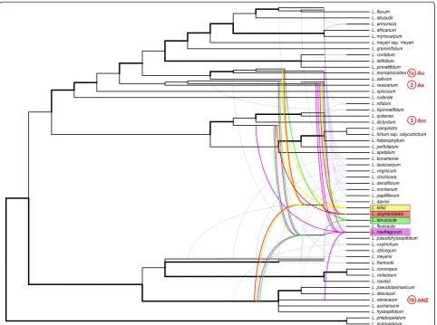
nodes and potential ancestral genomes contributing to our study species (Figure 2). According to the network,
our sampling includes 21 allopolyploid Lepidium taxa
(Figure 2, tree branches originating from curved lines), which are confirmed polyploids from the literature [5] and this study (L. sisymbrioides,L. tenuicaule). The remaining 31 taxa in our analyses do not show evidence of reticula-tion, and this may be due to diploidy, autopolyploidy, or gene loss.
The evidence suggests that Lepidium sisymbrioides is
derived from four distinct ancestral genomes (Figure 2, red lines): (1) a hybrid between (a) a descendant from
the common ancestor of theL. monoplocoidesgroup and
(b) the common ancestor of the group that includes L.
pseudotasmanicumandL. hyssopifolium(strong support),
(2) a descendant from the common ancestor of theL.
vesi-carium group (low support), and (3) a descendant from
the common ancestor of theL. dictyotumandL. quitense
group (strong support). Biogeographically, the
contri-bution of these genomes to L. sisymbrioides implies
hybridization among Australian and New Zealand (ANZ) taxa (1a and 1b, above), followed by hybridization with American (3) and potentially (with low support) Asian (2)
species. The other three New Zealand study species, L.
kirkii,L. naufragorum, and L. tenuicaule, show contribu-tions from four, five, and four distinct ancestral genomes, respectively (Figure 2). Of these three close hermaphro-ditic relatives,L. sisymbrioidesshares the most reticulation history with L. kirkii, followed by L. tenuicaule, then L. naufragorum(Figure 2).
Cytological observations reveal octoploidy in dioecious
L. sisymbrioidesand its hermaphroditic relative
L. tenuicaule
In order to confirm our results from thePIdata set, we
conducted chromosome counts in PMCs. Seed ofL.
kir-kii was not available, so its chromosome number
re-mains unknown.
Both L. sisymbrioides and L. tenuicaule had 2n = 64 chromosomes (Figure 3), corresponding to a ploidy of
8x(x= 8 is the base chromosome number for the genus
[2,4]). Octoploidy in these two species is consistent
with having four distinct copies of the PI intron as
shown by our phylogenetic and network analyses (A1, A2, C, D, Figures 1, 2). These four copies would there-fore represent four distinct diploid genomes in these
species’ history of hybridization and polyploidization
events.
Genome size estimations confirm ploidy estimates in
Lepidium sisymbrioidesand relatives
We examined holoploid genome size by calculating
1C-values for three of our New Zealand Lepidium
study species to confirm our estimates of ploidy and
to inform future genomic sequencing plans. Lepidium
sisymbrioides and L. tenuicaule had similar holoploid genome sizes of 0.63 and 0.66 pg, respectively,
whereas L. naufragorum’s holoploid genome size was
slightly over double that of the other two species at 1.41 pg (Table 1). Material from which the holoploid
genome size of L. naufragorum was obtained had
published chromosome counts from the same
popula-tion, indicating a ploidy of approximately 18x [65].
In conclusion, our holoploid genome size estimations
are consistent with L. sisymbrioides and L. tenuicaule
both being 8x and with L. naufragorum being 18x,
more than double the ploidy of the former two species.
Female and male flowers ofLepidium sisymbrioidesabort reproductive organs at comparable developmental stages but due to different processes
In order to assess the developmental stage and process
of abortion of reproductive organs in L. sisymbrioides,
we examined floral morphology and ontogeny of this species in comparison to the two closest hermaphroditic relatives available,L. naufragorumandL. tenuicaule. Since
floral developmental stages of hermaphroditic Lepidium
species are comparable to theA. thalianaontogenetic
sta-ging [20,61,62], we cross-referenced to this system for
convenience and reproducibility. Flower morphology ofL.
naufragorum andL. tenuicaulediffered fromA. thaliana
in petal size, number and arrangement of stamens, and
ovule number. Lepidium naufragorumflowers had petals
approximately as long as sepals and two lateral and two
medial stamens (Figure 4A), whereasL. tenuicauleflowers
had highly reduced petals, unnoticeable to the naked eye,
and four medial stamens (Figure 4B). All Lepidium taxa
produced a single ovule per locule.
In contrast toL. naufragorum andL. tenuicaule, both
male and female flowers of L. sisymbrioides generally
exhibited six stamens (two lateral and four medial; Figure 4C-F) with a few exceptions where only four medial (Figure 5O) or five stamens were found [see Additional file 1]. Petals were reduced (that is, shorter than sepals; Figure 4C-F, arrowheads), and four to six nec-taries were present among the stamen filaments in both sexes (Figure 4D, F, asterisks; [see Additional file 1]). In staminate flowers, the gynoecium arrested its develop-ment at intermediate stages, after differentiation of the anther locules (Figure 4C) and remained as a pistillode
while stamens expanded normally (Figure 4D), as in L.
naufragorumandL. tenuicaule(Figure 4A-B). In young carpellate flowers, stamens and carpels looked normal (Figure 4E). In later stages, however, stamen development was visibly arrested resulting in staminodia, whereas the
A
B
gynoecium developed normally (Figure 4F) as in L. nau-fragorum and L. tenuicaule (Figure 4A-B). From these morphological observations, both carpels and stamens
from male and female flowers ofL. sisymbrioides,
respect-ively, appeared to abort at intermediate stages of flower development.
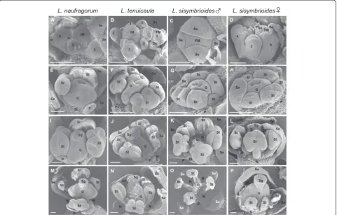
SEM of flower development in all three species showed flower meristems that initiated from the inflorescence
meri-stem in a similar fashion to Arabidopsis(Figure 5A-B, D,
Arabidopsis stages 1 to 2). As expected, sepal primordia developed first (Figure 5A-D, stages 3 to 4), followed by petals (Figure 5B), then presumably stamen and gynoecium primordia. Stamen filaments and anther locules differenti-ated within the androecium and the gynoecium developed as a tube through postgenital fusion of two carpels
(Figure 5E-F, stages 7 to 8). Subsequently, L.
naufra-gorum started to show more petal expansion than the other two species (compare Figure 5E-H). The gynoe-cial tube then closed at completion of postgenital fu-sion and began to differentiate a stigma with papillae (Figure 5I-J, L, stage 11). In staminate flowers of
L. sisymbrioides, after filaments and anther locules of the androecium had differentiated from one another, the gynoecium was arrested in its development (Figure 5K). The carpels of functional gynoecia expanded laterally, elongating and reaching full ma-turity with a clearly differentiated style and stigma
(Figure 5M-N, P, stage 12). In L. sisymbrioides
stam-inate flowers, the gynoecium remained aborted at
ma-turity (Figure 5O) in comparison to functional
gynoecia described above. Stamen filaments continued to elongate (Figure 5M-O, stage 12), except in
carpel-late flowers of L. sisymbrioides where they remained
much shorter than the gynoecium (Figure 5P). In L.
naufragorum, the only species with noticeable petals when mature, petals continued to expand, reaching the length of stamens (Figure 5M). In the other two species, petal primordia were initiated (Figure 5F-H) but never expanded, remaining small throughout development (Figure 5J-L) and not visible at maturity (Figure 5N-P).
Histological sections were performed to further inves-tigate the anatomical development of stamens and
car-pels. Lepidium naufragorum [see Additional file 2] and
L. tenuicaule revealed comparable reproductive organ development with no evidence of loss of organ function.
Therefore, only data from L. tenuicaule is compared
here againstL. sisymbrioides(Figure 6).
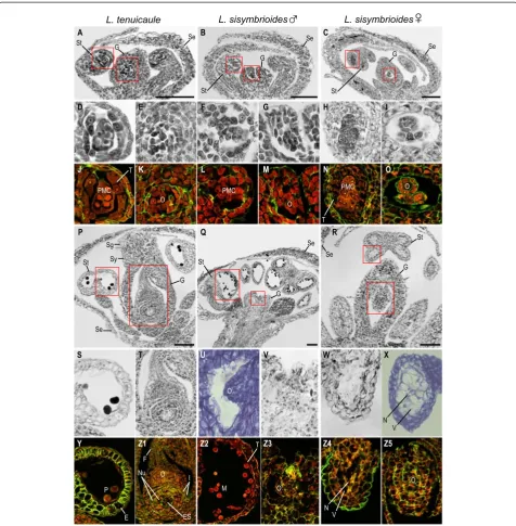
In hermaphroditic flowers ofL. tenuicaule, after stamen
filaments and anther locules differentiated (Figure 6A), anthers consisted of PMCs, tapetum and two outer anther
Table 1 Mean holoploid genome size (1C-value) and ploidy estimates for threeLepidiumspecies investigated Species 1C-value
(Mbp +/−s.d.)
1C-value (pg) Estimated ploidy levela
L. sisymbrioides 621 +/−7.84 0.635 8xb
L. tenuicaule 645 +/−29.41 0.660 8xb
L. naufragorum 1379 +/−61.56 1.410 18xc a
Ploidy estimates resulting from this study in bold.
b
This study, estimated based on chromosome counts (Figure3).
c
[65].
wall layers (middle layer and endothecium; Figure 6D, J, stage 9) and ovules began to develop in gynoecia (Figure 6E, K, stage 9). After meiosis of PMCs, anther wall layers degenerated, microspores underwent mitosis, and integuments enclosed the ovule [see Additional file 2, stage 12]. Subsequently, the androecium and gynoecium matured (Figure 6P). At this stage, the stamen filaments elongated (Figure 6P) and pollen sacs were composed of a single endothecium layer with secondary wall thicken-ings (Figure 6S, Y, stage 13). Pollen grains could be visu-alized with evident exine and the tapetum had degraded (Figure 6S, Y, stage 13). By this stage, the gynoecium had fused, elongated, and differentiated a style and stigma (Figure 6P, stage 13), and ovules had differentiated within each carpel (Figure 6T, Z1). Apical ovules consisted of an elongated funiculus and an embryo sac, surrounded by the nucellus and two integuments (Figure 6T, Z1, stage 13).
In staminate flowers of L. sisymbrioides, histological
sections revealed that after initiation of the gynoecium
(Figure 6B), sporogenous tissue (PMCs) was present in stamen locules (Figure 6F, L) and ovules had been initiated (Figure 6G, M, [see Additional file 2, stage 9]). However at later stages (Figure 6Q, stage 12; [see Additional file 2, stage 11]), as microspores matured within the anthers and the tapetum degenerated (Figure 6 Z2), the gynoecium failed to elongate and differentiate a style and stigma, and ovules did not grow nor differentiate (Figure 6U-V, Z3, [see Additional file 2]). Since the gynoecium arrest occurs before microsporogenesis (the production of tetrads from PMCs, stage 9), which normally precedes megasporogene-sis (stage 11) inArabidopsis, we conclude that the process for the loss of gynoecium function in male flowers is the arrest of development at a pre-meiotic, intermediate stage (stage 9).
In young carpellate flowers ofL. sisymbrioides(Figure 6C), sporogenous tissue (PMCs) inside the stamen locules (Figure 6H, N) and ovule initiation (Figure 6I, O) were evident at the same stage as in hermaphroditic flowers
L. naufragorum L. tenuicaule L. sisymbrioides
♂
L. sisymbrioides♀
A B C
J K L M N O
P Q R
Y Z1 Z2 Z3 Z4 Z5
Se
St G Se
St
G
Se
St
G
PMC
O
PMC
O
PMC O
Se St
G
Sg Se
St
G
Se
St
G
O
E P
ES
T
M O O
V N T
T
F
Sy
I Nu
D E F G H I
S T U V W X
L. tenuicaule L. sisymbrioides
♂
L. sisymbrioides♀
O
V N
Figure 6Histological sections of hermaphroditic and dioeciousLepidiumspecies.Floral developmental stages noted below follow
(Figure 6A, D-E, J-K, stage 9). By the time the gynoecium closed and a stigma and ovule began to dif-ferentiate (Figure 6R, Z5, [see Additional file 2, stage 11]), vacuolated cells pervaded anthers and PMCs had degenerated (Figure 6W-X, Z4). Stamen filaments did not elongate and neither tetrads, microspores, nor pollen were produced; pollen sacs appeared shrunken, filled with vacuolated cells, and no endothecium layer developed (Figure 6W-X, Z4). Based on the above observations, we propose that the developmental process for loss of androe-cium function in female flowers is likely cell death, as evi-denced by vacuolated cells (absence of stained cytoplasm) and nuclear degradation (Figure 6W-X, Z4) following the development of PMCs (Figure 6H, N). In conclusion, androecium abortion in female flowers occurs at a com-parable pre-meiotic stage to gynoecium abortion in male flowers (stage 9) but due to different processes, that is, cell death versus developmental arrest, respectively. Figure 7 summarizes our SEM and histological observations on
flower development inLepidiumstudy species (Figures 5, 6,
[see Additional file 2]) in reference to described develop-mental stages fromA. thaliana[20,61,62].
Programmed cell death in anther walls is involved in the degradation of pollen mother cells of femaleL.
sisymbrioidesflowers
Because we were finding evidence of cell death in
sta-mens of femaleL. sisymbrioides, we wanted to determine
whether PCD could be responsible for this abortion of stamens. To look for evidence of PCD, as characterized by DNA fragmentation, we conducted TUNEL assays on
histological sections of carpellate L. sisymbrioides and,
for comparison, staminate L. sisymbrioidesand L.
tenui-caule flowers. The TUNEL assay attaches fluorescein to fragmented DNA, eliciting a green fluorescent signal in
nuclei undergoing DNA degradation. Lepidium tissue
autofluoresced in the absence of staining under both DAPI and fluorescein excitation and emission ranges: cell walls, chloroplasts, nuclei, and pollen grains showed background signal (compare Figure 8A-B to C-D, G-H to I-J, and M-N to O-P). This autofluorescence contrib-uted additional histological evidence that cell death was
occurring in stamens of female L. sisymbrioides, as
evi-denced by the absence or degradation of cell walls, nu-clei, and pollen in the center of anther locules, where sporogenous tissue leading to pollen normally develops (compare Figure 8I-J to C-F and O-R). In spite of this autofluorescence, the use of negative and positive con-trols allowed us to observe strong, above-background, fluorescein signal in certain tissues at certain stages that indicate DNA degradation. For example, all nuclei in the
endothecium of mature, functional anther sacs ofL.
tenui-caule at stage 13 showed a strong, above-background, fluorescein signal indicating PCD (compare Figure 8B to D,
red arrow denotes one exemplary nucleus). More import-antly, we observed strong, above-background, fluorescein signal in all nuclei throughout all anther wall layers of
mature anthers from female L. sisymbrioides (compare
Figure 8H to J, red arrows denote exemplary nuclei from each layer). This was taken as evidence that these nuclei are undergoing DNA degradation, as observed in positive controls (treated with DNase) showing higher than above-background signal (compare Figure 8B to L, red arrow de-notes one exemplary nucleus). When comparing anthers
from female L. sisymbrioides that abort at stage 9 to
functional anthers from L. tenuicauleand maleL.
sisym-brioidesat the same stage (compare Figure 8I-J to E-F and Q-R), it appeared that PCD in anther wall layers was
con-tributing to the degradation of PMCs in carpellate L.
sisymbrioides flowers, in which tapetum, endothecium, and tetrads do not develop as in functional anthers from
L. tenuicaule and male L. sisymbrioides at the same stage. In summary, using the TUNEL assay as a proxy for PCD, we find evidence for PCD in the anther wall
layers of carpellateL. sisymbrioidesflowers, which likely
contributes to the degradation of PMCs and abortion of anthers at stage 9.
Discussion
Lepidium sisymbrioides is the sole dioecious member of Brassicaceae, and our phylogenetic analyses show that it is closely related to three other New Zealand
hermaph-roditic species: L. kirkii, L. naufragorum and L.
tenui-caule (Figures 1, 2). Increased phylogenetic sampling of
the PIfirst intron among AANZ taxa allows us to
iden-tify reticulation events leading toL. sisymbrioides,which
resulted from three past hybridization events (Figure 2). Of the three close relatives, the shared reticulation
history with L. kirkiiis a novel finding. Molecular,
cyto-logical, and genome size analyses provide evidence that
L. sisymbrioides is an allo-octoploid (Figures 1, 2 and 3, Table 1), with 64 chromosomes and an average holoploid genome size (1C-value) of 621 Mbp. By comparing the
floral ontogeny of unisexual flowers in L. sisymbrioides
to that of its close relatives and toArabidopsis thaliana, we show that unisexual flowers in this species arose from selective abortion of reproductive organs at a simi-lar floral developmental stage (Figure 7 stage 9) but by different processes in males and females. Differential abortion of the gynoecium in males appears to result from developmental arrest, while in females anther sterility re-sults from programmed cell death (Figure 6, 8).
Evolution of dioecy and unisexual flowers within Brassicaceae
The evolution of dioecy in Brassicaceae occurred only once
in the genus Lepidium. We infer that in L. sisymbrioides,
abortion of reproductive organs, as in type I flowers [66],
as all other members ofLepidiumare hermaphroditic. Our
floral ontogeny observations confirmed that reproductive organs are initiated and differentially aborted at the same floral developmental stage in male and female flowers of
L. sisymbrioides. The timing of abortion corresponds to
Arabidopsisstage 9 (Figures 4, 5, 6 and 7), which is broadly
considered an ‘intermediate’ stage of floral development
[22], after primordia initiation but before meiosis. Our
ontogeny shows that L. sisymbrioides is representative of
the majority of angiosperms with type I unisexual flowers that selectively abort reproductive organs at significantly correlated developmental stages between the two sexes [22]. This evidence suggests that similar regulatory switch points underlie male and female developmental pathways
as proposed by Diggleet al. [22] and comparable selective
forces are at play in the two sexes. However, the develop-mental stage and process of reproductive organ abortion in unisexual flowers across angiosperms vary widely with dif-ferent stages and processes occurring at equal frequencies [22].
With regard to the developmental process of organ
abortion in L. sisymbrioides, while sporogenous tissue
(PMCs) in stamen locules differentiates (Figure 6C, H, N), it quickly degenerates during development of carpellate flowers and becomes vacuolated with degraded nuclei (Figure 6R, W-X, Z4). Programmed cell death, which is in-volved throughout normal flower development [67], is pri-marily due to endogenous factors and is evidenced by cell death at a predictable time and location during tissue differentiation [68]. During normal flower development, the tapetum degenerates during microgametogenesis via PCD for proper microspore development and differen-tiation of pollen, [67]. Other studies have shown that premature tapetal degeneration can lead to male sterility [reviewed in 67]. Therefore, because we observe PCD in anther wall layers before microgametogenesis, this prema-ture tapetal degeneration is likely leading to male sterility in L. sisymbrioides females (Figures 6R, W-X, Z4, 8I-J, [see Additional file 2]).
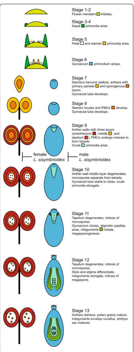
Two types of PCD occur in plants: autolytic and non-autolytic. The former generally occurs during normal plant development, whereas the latter occurs during plant-pathogen interactions [69,70]. Moreover, since loss of cell Stage 10
Anther wall middle layer degenerates, microspores separate from tetrads. Gynoecial tube starts to close, ovule primordia elongate.
Stage 12
Tapetum degenerates, mitosis of microspores.
Style and stigma differentiate, integuments elongate, mitosis of megaspore.
Stage 13
Anthers dehisce, pollen grains mature. Integuments envelop nucellus, embryo sac matures.
male
L. sisymbrioides
Stage 6
Gynoecium primordium arises.
Stage 9
Anther walls with three layers (endothecium , middle , and tapetum ), PMCs undergo meiosis to form tetrads.
Ovule primordia arise.
Stage 11
Tapetum degenerates, mitosis of microspores
Gynoecium closes, stigmatic papillae arise, integuments initiate, megasporogenesis
Stage 3-4
Sepal primordia arise.
Stage 1-2
Flower meristem initiates.
Stage 5
Petal and stamen primordia arise.
Stage 7
Stamens become stalked, anthers with primary parietal and sporogenous layers.
Gynoecial tube develops.
Stage 8
Stamen locules and PMCs develop. Gynoecial tube develops.
female
L. sisymbrioides
walls and cytoplasm, nuclear condensation, and increase in vacuolar volume are characteristic of autolytic PCD [70], this type of cell death is also likely involved in the degener-ation of PMCs inL. sisymbrioidesfemales (Figures 6R, 8I-J, [see Additional file 2]).
In male flowers of L. sisymbrioides, on the other
hand, the development of ovules and gynoecia is arrested shortly after initiation of ovule primordia. We found no evidence of cell death, parenchymatization, or change in timing of otherwise normal developmental events in arrested gynoecia. Ovule primordia remain evident in mature male flowers (Figure 6Q, U-V, Z3, Additional file 2). Therefore, of the six developmental pro-cesses reviewed in Diggleet al. [22], arrest of development best characterizes the abortion of the gynoecium in
L. sisymbrioidesmales.
Whole genome duplication events via hybridization in the evolution of dioecy inLepidium
Two different copies of thePIfirst intron were previously
identified among the ANZ taxa (clades A, C); only one
copy (clade C) was strongly supported [5]. Our PI
phyl-ogeny recovered at least four divergent copies of the first intron inL. sisymbrioidesand its close relatives (Figure 1), suggesting ancient allopolyploidization events, followed by
divergence of PI alleles. Based on our phylogenetic
network analyses, L. sisymbrioides has a history of three
allopolyploidization events: hybridization (1) between an Australian and Australian or New Zealand species, (2) with an Asian species, and (3) with an American species (Figure 2). Our study provides new evidence of an add-itional genome within the‘A’clade ofPI[5] and shows an additionalPIcopy in taxa from this clade (as in,L. kirkii,
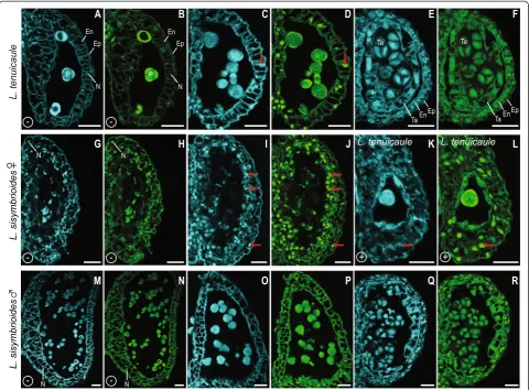
C D E F
G H
A B
I J K L
M N O P Q R
+ +
-
--
--
-En
P N
Ep Te
En Ta En
P N
Ep Te
En Ta
N N
N N
Ep Ep
L. tenuicaule L. tenuicaule
Figure 8Confocal microscopy of longitudinal sections of anther sacs from hermaphroditic, male and female flowers ofLepidium treated with TUNEL assay and controls. (A-F, K-L)HermaphroditicL. tenuicaule.(G-J)CarpellateL. sisymbrioides.(M-R)StaminateL. sisymbrioides.
L. naufragorum, L. sisymbrioides and L. tenuicaule; A1-A2, Figure 1), which would be expected of four divergent genomes contributing to several allopolyploidization events in our study species. Based on our cytological
and genome size estimates, L. sisymbrioides is an
octo-ploid, which would require several whole genome
duplica-tions. Together with our PI data, this evidence suggests
thatL. sisymbrioidesis an allo-octoploid composed of four different genomes.
AustralianLepidiumappear to have undergone a rapid
radiation during the Pliocene and Pleistocene, when the arid and cooler regions of the southeastern temperate bi-omes were expanding [71]. Previous studies suggested at least one dispersal event each from California and South Africa to Australia or New Zealand; most likely coloniz-ing Australia first, with at least two subsequent dispersal
events to New Zealand [12]. The majority of Lepidium
species produce mucilaginous seeds that adhere to birds [4], which may have facilitated long-distance dispersal among the Americas, Australia, New Zealand and the Old World [72-75]. Our results suggest that a hybridization event occurred either within Australia or between an Australian and a New Zealand ancestor, followed by hybridization with an Asian colonist and an American
colonist, resulting in the evolution of L. sisymbrioides
(Figures 1, 2). Colonization by an Asian ancestor is not well supported by our data and conflicts with previous studies indicating colonization by an African ancestor [12]; this contradictory evidence could be due to the use of different nuclear DNA regions. In spite of this, our results confirm at least two dispersal events to Australia or New Zealand from the New World and Old World that resulted in allopolyploidization, but exact New and Old World ancestry is uncertain. Additionally, we infer a hybridization between ANZ taxa not previously suggested.
Our 1C-value estimates forL. sisymbrioidesandL.
tenui-caulefall within the reported range for the family (0.15 to
2.43 pg [76-78]). Lepidium naufragorum lies outside the
high end of the range, consistent with it being highly
poly-ploid (18x [65]). Even though the 1C-value of L.
sisym-brioides (0.635 pg) is almost fourfold that of Arabidopsis thaliana(0.16 pg [79]), it is comparable to the size of other model plants such as rice (0.5 pg [80]), making it a likely candidate for whole genome sequencing. Genomic re-sources for this species would facilitate the investigation of sex determination and of the putative chromosomal rear-rangements that contributed to the evolution of dioecy after polyploidization. As new technologies and approaches are being developed [81,82], sequencing this octoploid will become more feasible in the near future.
Conclusions
The developmental process leading to the evolution of
dioecy in Lepidium sisymbrioides was placed in the
broader context of the historical patterns conditioning the evolution of separate sexes in this unique dioecious
relative ofArabidopsis. We have characterized the
devel-opmental stage and process of its unisexual flowers, pav-ing the way for future studies aimed at unravelpav-ing the genetic basis underlying reproductive organ abortion.
Having placedL. sisymbrioidesin a phylogenetic context,
determined its ploidy, hybridization history, and genome
size, and compared it toArabidopsis thalianaflower
de-velopment will facilitate the investigation of the role of polyploidy and of potential candidate genes in the evolu-tion of dioecy in Brassicaceae.
Appendix 1
Voucher information and GenBank accessions for
Lepi-diumtaxa (Brassicaceae) sampled in this study. Voucher
information provided only for taxa with sequences not downloaded from GenBank. Taxon, collector and
collec-tion number, origin, herbarium,PISTILLATAintron.
Lepidium africanum (Burm.f.) DC., AY114216; Lepi-dium alluaudii Maire, AY114221; Lepidium apetalum
Willd., AY114217; Lepidium armoracia Fisch. & C.A.
Mey., AY114218-AY114220;Lepidium aschersoniiThell.,
AY114222; Lepidium bipinnatifidum Desv.,
AY114223-AY114224; Lepidium bonariense L., AY114225-AY1
14227; Lepidium campestre (L.) R.Br., AY114228;
Lepi-dium chichicaraDesv., AY114229-AY114231; Lepidium cordatumWilld. ex Steven, AY114232-AY114233; Lepi-dium coronopus (L.) Al-Shehbaz, unvouchered, culti-vated from wild-collected seed from Madrid, Spain
(INIA seed accession 205-0261-68), JN119857;
Lepi-dium davisii Rollins, FJ541471/FJ541473; Lepidium densiflorum Schrad., AY114235-AY114236; Lepidium desvauxii Thell., AY114237; Lepidium dictyotum A.
Gray, AY114238; Lepidium flavum Torr., AY114239;
Lepidium flexicauleKirk, AY114240-AY114241;Lepidium fremontiiS. Watson, AY114243-AY114244;Lepidium gra-minifoliumL., AY114246;Lepidium heterophyllumBenth.,
AY114247; Lepidium hirtum (L.) Sm. ssp. calycotrichum
(Kunze) Thell., AY114248; Lepidium hyssopifoliumDesv.,
AY114249; Lepidium kirkiiPetrie, P. Heenan s.n.,
culti-vated from wild-collected seed from Galloway, New
Zealand, CHR, KJ648155-KJ648159; Lepidium
lasiocar-pum Nutt., AY114250-AY114251;Lepidium latifoliumL.,
AY114252; Lepidium leptopetalum F. Muell., AY114215;
Lepidium meyeniiWalp., AY114254-AY114255;Lepidium meyeri Claus ssp.meyeri, AY114269; Lepidium monoplo-coides F. Muell., AY114256; Lepidium montanum Nutt.,
AY114257/AY114259; Lepidium myriocarpum Sond.,
AY114260; Lepidium naufragorum Garn.-Jones & D.A.
Norton, V. Di Stilio 117, cultivated from wild-collected seed from Open Bay Islands, New Zealand, WTU,
JN119859-JN119860/JN119862/KJ648160-KJ648165;
from wild-collected seed from Gádor, Spain (INIA seed
accession 204-4472-76), JN119856; Lepidium nitidum
Nutt., AY114261-AY114262;Lepidium oblongumSmall,
AY114263-AY114264; Lepidium oleraceum Sparrm.,
AY114265; Lepidium oxytrichum Sprague,
AY114266-AY114267; Lepidium papilliferum (L.F. Hend.) A.
Nelson & J.F. Macbr., FJ541451/FJ541488; Lepidium
perfoliatum L., AY114268; Lepidium phlebopetalum (F.
Mull.) F. Mull., AY114214; Lepidium pinnatifidum
Ledeb., AY114270;Lepidium pseudohyssopifoliumHewson,
AY114271-AY114274;Lepidium pseudotasmanicumThell.,
AY114275;Lepidium quitenseTurcz., AY114276;Lepidium
ruderale L., AY114278; Lepidium sativum L., AY114279;
Lepidium sisymbrioides Hook. f., V. Soza 1924, cultivated from wild-collected seed from Twizel, South Canterbury,
New Zealand, WTU, KJ648166- KJ648169; Lepidium
spi-nosum Ard., AY114280; Lepidium tenuicaule Kirk, V. Di Stilio 116, cultivated from wild-collected seed from Shag
Point, New Zealand, WTU, KJ648170- KJ648174;Lepidium
vesicarium L., AY114281; Lepidium violaceum (Munby) Al-Shehbaz, unvouchered, cultivated from wild-collected seed from N. Azrou, Morocco (INIA seed accession
206-4096-84), JN119858;Lepidium virginicumL., AY114285.
Notes: INIA = Instituto Nacional de Investigaciones Agrarias, Madrid, Spain.
Additional files
Additional file 1:Staminate flowers ofLepidium sisymbrioides. A) Young staminate flower ofL. sisymbrioides, showing sepals (Se), five stamens (St), and aborted gynoecium (G). (B) Staminate flower ofL. sisymbrioides, showing sepals (Se), six nectaries (N) among stamen (St) filaments, and aborted gynoecium (G). Scale bar = 0.25 mm.
Additional file 2:Histological sections of hermaphroditic and dioeciousLepidiumspecies stained with Toluidine Blue O.(A-C) Flower at the pre-meiotic stage of microsporogenesis, stage 9; from left to right, (A) hermaphroditicL. naufragorum, (B) staminateL. sisymbrioides, and (C) carpellateL. sisymbrioides. (D, F, H) Anther locule from respective flower above. (E, G, I) Ovule from respective flower above. (J-L) Flowers later on in development, at the microgametogenesis stage, stages 11–12; from left to right, (J) hermaphroditicL. naufragorum, (K) staminateL. sisymbrioides, and (L) carpellateL. sisymbrioides. (M, O, Q) Anther locule from respective flower above. (N, P, R) Ovule from respective flower above. (S-U) Mature flowers, stage 13; from left to right, (S) hermaphroditic
L. naufragorum, (T) staminateL. sisymbrioides, and (U) carpellateL. sisymbrioides. (V, X, Z1) Anther locule from respective flower above. (W, Y, Z2) Ovule from respective flower above. Scale bar = 50μm in A-C; 100μm in J-U. E, endothecium; ES, embryo sac; G, gynoecium; I, integuments; M, microspores; N, nucleus; O, ovule; P, pollen; PMC, pollen mother cells; Se, sepals; Sg, stigmatic papillae; St, stamen; Sy = style; T, tapetum; V = vacuolated cell.
Abbreviations
AANZ:American, Australian, and New Zealand; ANZ: Australian and New Zealand; bs: bootstrap; BSA: bovine serum albumin; CEN: chicken erythrocyte nuclei; DAPI: 4’,6-diamidino-2-phenylindole; FAA: formaldehyde-acetic acid-alcohol; Mbp: millions of base pairs; PBS: phosphate buffered solution; PCD: programmed cell death; pg: picogram;PI:PISTILLATA; PMCs: pollen mother cells; pp: posterior probabilities; SEM: scanning electron microscopy.
Competing interests
The authors declare that they have no competing interests.
Authors’contributions
VLH conducted SEM observations of flower development and contributed to figures. VLS conducted DNA extractions, PCR, cloning, and sequencing, preparedPIalignments, conducted phylogenetic and network analyses, performed cytological and histological observations, and drafted the manuscript. VSD conceived, designed, and coordinated the study, collected genome size data, and participated in drafting and editing the manuscript. All authors read and approved the final manuscript.
Acknowledgements
The authors thank Peter Heenan for field collection of seeds fromL. naufragorum,L. sisymbrioides, andL. tenuicaule; John Bowman forPI
alignments; CHR for the herbarium specimen ofL. kirkii; Luca Comai and Jerry Davison for training and assistance with flow cytometry; Parisa Aalami-Monelli, Nadya Ali, Wai Pang Chan, Caitlin Connelly, Brittany Ng, and Patricia Salles Smith for assistance with data collection; Doug Ewing and Nora Kozlov for seed germination and plant care; Starr Matsushita and Delene Oldenberg for assistance with cytology; Cindy Skema for histological advice; University of Washington, Department of Biology funds to VSD; and anonymous reviewers for helpful suggestions.
Received: 21 June 2014 Accepted: 7 October 2014 Published: 12 November 2014
References
1. Al-Shehbaz IA, Beilstein MA, Kellogg EA:Systematics and phylogeny of the Brassicaceae (Cruciferae): an overview.Plant Syst Evol2006,259:89–120. 2. Warwick SI, Al-Shehbaz IA:Brassicaceae: chromosome number index and
database on CD-Rom.Plant Syst Evol2006,259:237–248.
3. Endress PK:Evolution and floral diversity: the phylogenetic surroundings ofArabidopsisandAntirrhinum.Int J Plant Sci1992,153:S106–S122. 4. Al-Shehbaz IA:The genera of Lepidieae (Cruciferae, Brassicaceae) in the
Southeastern United States.J Arnold Arboretum1986,67:265–311. 5. Lee JY, Mummenhoff K, Bowman JL:Allopolyploidization and evolution of
species with reduced floral structures inLepidiumL. (Brassicaceae). Proc Natl Acad Sci2002,99:16835–16840.
6. Kirk T:The Students’Flora of New Zealand and the Outlying Islands.
Wellington, New Zealand: John Mackay, Government Printer; 1899. 7. Bateman AJ:Note on dioecy in the Cruciferae.Heredity1955,9:415–415. 8. Webb CJ, Sykes WR, Garnock-Jones PJ:Flora of New Zealand: Naturalised
Pteridophytes, Gymnosperms, Dicotyledons, Volume IV.Botany Division D. S. I. R.: Christchurch, New Zealand; 1988.
9. Mummenhoff K, Brueggemann H, Bowman JL:Chloroplast DNA phylogeny and biogeography ofLepidium(Brassicaceae).Am J Bot2001,88:2051–2063. 10. Heenan PB, Mitchell AD, McLenachan PA, Lockhart PJ, de Lange PJ:Natural
variation and conservation ofLepidium sisymbrioidesHook, f. andL. solandriKirk (Brassicaceae) in South Island, New Zealand, based on morphological and DNA sequence data.N Z J Bot2007,45:237–264. 11. Allan HH:Flora of New Zealand. Volume I. Indigenous Tracheophyta
(Psilopsida, Lycopsida, Filicopsida, Gymnospermae, Dicotyledones).R. E. Owen, Government Printer: Wellington, New Zealand; 1961.
12. Mummenhoff K, Linder P, Friesen N, Bowman JL, Lee J-Y, Franzke A:
Molecular evidence for bicontinental hybridogenous genomic constitution inLepidiumsensu stricto (Brassicaceae) species from Australia and New Zealand.Am J Bot2004,91:254–261.
13. Miller JS, Venable DL:Polyploidy and the evolution of gender dimorphism in plants.Science2000,289:2335–2338.
14. Miller JS, Venable DL:The transition to gender dimorphism on an evolutionary background of self-incompatibilty: an example fromLycium (Solanaceae).Am J Bot2002,89:1907–1915.
15. Spigler RB, Lewers KS, Johnson AL, Ashman TL:Comparative mapping reveals autosomal origin of sex chromosome in octoploidFragaria virginiana.J Hered2010,101:S107–S117.
16. Sakai AK, Weller SG:Gender and sexual dimorphism in flowering plants: a review of terminology, biogeographic patterns, ecological correlates, and phylogenetic approaches.InGender and Sexual Dimorphism in Flowering Plants.Berlin, Germany: Springer; 1999:1–32.
18. Jesson LK:Ecological correlates of diversification in New Zealand angiosperm lineages.N Z J Bot2007,45:35–51.
19. Beilstein MA, Nagalingum NS, Clements MD, Manchester SR, Mathews S:
Dated molecular phylogenies indicate a Miocene origin forArabidopsis thaliana.Proc Natl Acad Sci2010,107:18724–18728.
20. Bowman JL:Arabidopsis: An Atlas of Morphology and Development.New York: Springer; 1994.
21. Ma H:Molecular genetic analyses of microsporogenesis and microgametogenesis in flowering plants.Annu Rev Plant Biol2005,
56:393–434.
22. Diggle PK, Di Stilio VS, Gschwend AR, Golenberg EM, Moore RC, Russell JRW, Sinclair JP:Multiple developmental processes underlie sex differentiation in angiosperms.Trends Genet2011,27:368–376.
23. De Lange PJ, Norton DA, Courtney SP, Heenan PB, Barkla JW, Cameron EK, Hitchmough R, Townsend AJ:Threatened and uncommon plants of New Zealand (2008 revision).N Z J Bot2009,47:61–96.
24. Mitchell AD, Heenan PB:Systematic relationships of New Zealand endemic Brassicaceae inferred from nrDNA ITS sequence data.Syst Bot
2000,25:98.
25. Mummenhoff K, Polster A, Muehlhausen A, Theissen G:Lepidiumas a model system for studying the evolution of fruit development in Brassicaceae.J Exp Bot2009,60:1503–1513.
26. Hughey JR, Silva PC, Hommersand MH:Solving taxonomic and nomenclatural problems in Pacific Gigartinaceae (Rhodophyta) using DNA from type material.J Phycol2001,37:1091–1109.
27. Maddison DR, Maddison WP:MacClade 4: Analysis of Phylogeny and Character Evolution.Sunderland, MA: Sinauer Associates; 2005. 28. Martin DP, Lemey P, Lott M, Moulton V, Posada D, Lefeuvre P:RDP3: a
flexible and fast computer program for analyzing recombination. Bioinformatics2010,26:2462–2463.
29. Martin D, Rybicki E:RDP: detection of recombination amongst aligned sequences.Bioinformatics2000,16:562–563.
30. Martin DP, Posada D, Crandall KA, Williamson C:A modified bootscan algorithm for automated identification of recombinant sequences and recombination breakpoints.AIDS Res Hum Retroviruses2005,21:98–102. 31. Padidam M, Sawyer S, Fauquet CM:Possible emergence of new
geminiviruses by frequent recombination.Virology1999,265:218–225. 32. Smith JM:Analyzing the mosaic structure of genes.J Mol Evol1992,
34:126–129.
33. Posada D, Crandall KA:Evaluation of methods for detecting recombination from DNA sequences: computer simulations.Proc Natl Acad Sci U S A2001,98:13757–13762.
34. Gibbs MJ, Armstrong JS, Gibbs AJ:Sister-Scanning: a Monte Carlo procedure for assessing signals in recombinant sequences.Bioinformatics
2000,16:573–582.
35. Boni MF, Posada D, Feldman MW:An exact nonparametric method for inferring mosaic structure in sequence triplets.Genetics2007,176:1035–1047. 36. Holmes EC, Worobey M, Rambaut A:Phylogenetic evidence for
recombination in dengue virus.Mol Biol Evol1999,16:405–409. 37. Guindon S, Gascuel O:A simple, fast, and accurate algorithm to estimate
large phylogenies by maximum likelihood.Syst Biol2003,52:696–704. 38. Darriba D, Taboada GL, Doallo R, Posada D:jModelTest 2: more models,
new heuristics and parallel computing.Nat Methods2012,9:772–772. 39. Akaike H:A new look at the statistical model identification.IEEE Trans
Autom Control1974,AC19:716–723.
40. Smith JF, Stillman AJ, Larson SR, Culumber CM, Robertson IC, Novak SJ:
Phylogenetic relationships amongLepidium papilliferum(L. Henderson) A. Nels. & J. F. Macbr.,L. montanumNutt., andL. davisiiRollins (Brassicaceae).J Torrey Botanical Society2009,136:149–163.
41. Huelsenbeck JP, Ronquist F:MRBAYES: Bayesian inference of phylogenetic trees.Bioinformatics2001,17:754–755.
42. Ronquist F, Huelsenbeck JP:MrBayes 3: Bayesian phylogenetic inference under mixed models.Bioinformatics2003,19:1572–1574.
43. Miller MA, Pfeiffer W, Schwartz T:Creating the CIPRES Science Gateway for inference of large phylogenetic trees.InProceedings of the Gateway Computing Environments Workshop (GCE), 2010.New Orleans, LA: Institute of Electrical and Electronics Engineers; 2010:1–8.
44. Yang ZH, Rannala B:Bayesian phylogenetic inference using DNA sequences: a Markov Chain Monte Carlo method.Mol Biol Evol1997,
14:717–724.
45. Rambaut A:FigTree Version 1.4.2012. [http://tree.bio.ed.ac.uk/software/figtree/]
46. Zwickl DJ:Genetic algorithm approaches for the phylogenetic analysis of large biological sequence datasets under the maximum likelihood criterion.Austin: The University of Texas; 2006.
47. Felsenstein J:Confidence limits on phylogenies: an approach using the bootstrap.Evolution1985,39:783–791.
48. Sukumaran J, Holder MT:DendroPy: a Python library for phylogenetic computing.Bioinformatics2010,26:1569–1571.
49. Huson DH, Scornavacca C:Dendroscope 3: an interactive tool for rooted phylogenetic trees and networks.Syst Biol2012,61:1061–1067.
50. Huber KT, Oxelman B, Lott M, Moulton V:Reconstructing the evolutionary history of polyploids from multilabeled trees.Mol Biol Evol2006,
23:1784–1791.
51. Kato A:Air drying method using nitrous oxide for chromosome counting in maize.Biotech Histochem1999,74:160–166.
52. Matsushita SC, Tyagi AP, Thornton GM, Pires JC, Madlung A:Allopolyploidization lays the foundation for evolution of distinct populations: evidence from analysis of syntheticArabidopsisallohexaploids.Genetics2012,
191:535–547.
53. Wright KM, Pires JC, Madlung A:Mitotic instability in resynthesized and natural polyploids of the genusArabidopsis(Brassicaceae).Am J Bot2009,
96:1656–1664.
54. Greilhuber J, Dolezel J, Lysak MA, Bennett MD:The origin, evolution and proposed stabilization of the terms“genome size”and“C-value”to describe nuclear DNA contents.Ann Bot2005,95:255–260. 55. Davison J, Tyagi A, Comai L:Large-scale polymorphism of
heterochromatic repeats in the DNA ofArabidopsis thaliana.BMC Plant Biol2007,7:44.
56. Gregory TR:Animal Genome Size Database.2013 [www.genomesize.com] 57. Dolezel J, Greilhuber J:Nuclear genome size: are we getting closer?
Cytometry A2010,77:635–642.
58. Dolezel J, Bartos J, Voglmayr H, Greilhuber J:Nuclear DNA content and genome size of trout and human.Cytometry A2003,51A:127–128. 59. Bowman JL, Smyth DR:Patterns of petal and stamen reduction in Australian species ofLepidiumL. (Brassicaceae).Int J Plant Sci1998,
159:65–74.
60. Chehregani A, Sedaghat M:Pollen grain and ovule development in Lepidium vesicarium(Brassicaceae).Int J Agriculture & Biology2009,
11:601–605.
61. Muller A:Zur Charakterisierung der Bluten und Infloreszenzen von Arabidopsis thaliana(L.) Heynh.Kulturpflanze1961,9:364–393. 62. Smyth DR, Bowman JL, Meyerowitz EM:Early flower development in
Arabidopsis.Plant Cell1990,2:755–767.
63. Kramer EM:Methods for studying the evolution of plant reproductive structures: comparative gene expression techniques.InMolecular Evolution: Producing the Biochemical Data, Part B. Volume 395.Edited by Zimmer EA, Roalson EH. San Diego, CA: Elsevier Academic Press; 2005:617–636. 64. Johansen DA:Plant Microtechnique. New York.London: McGraw-Hill Book
Company, Inc.; 1940.
65. De Lange PJ, Murray BG:Contributions to a chromosome atlas of the New Zealand flora - 37.Miscellaneous families. N Z J Bot2002,40:1–23. 66. Mitchell CH, Diggle PK:The evolution of unisexual flowers: morphological
and functional convergence results from diverse developmental transitions.Am J Bot2005,92:1068–1076.
67. Wu HM, Cheung AY:Programmed cell death in plant reproduction.Plant MolBiol2000,44:267–281.
68. Noodén LD:Plant Cell Death Processes.San Diego, CA: Elsevier Academic Press; 2004.
69. Van Doorn WG, Beers EP, Dangl JL, Franklin-Tong VE, Gallois P, Hara-Nishimura I, Jones AM, Kawai-Yamada M, Lam E, Mundy J:Morphological classification of plant cell deaths.Cell Death & Differentiation2011,18:1241–1246.
70. Van Doorn WG:Classes of programmed cell death in plants, compared to those in animals.J Exp Bot2011,62:4749–4761.
71. Crisp M, Cook L, Steane D:Radiation of the Australian flora: what can comparisons of molecular phylogenies across multiple taxa tell us about the evolution of diversity in present–day communities?Philos Trans R Soc Lond B Biol Sci2004,359:1551–1571.
72. Carlquist S:The biota of long-distance dispersal.V. Plant dispersal to Pacific Islands. Bulletin of the Torrey Botanical Club1967,94:129–162.
73. Niemi A:Lepidium ruderaleL. on gull skerries in the archipelago SW of Helsingfors.Memoranda Societatis pro Fauna et Flora Fennica1968,
74. Mummenhoff K, Hurka H, Bandelt H:Systematics of AustralianLepidium species (Brassicaceae) and implications for their origin - evidence from IEF analysis of RUBISCO.Plant Syst Evol1992,183:99–112.
75. Garnock-Jones P, Norton D:Lepidium naufragorum(Brassicaceae), a new species from Westland, and notes on other New Zealand coastal species ofLepidium.N Z J Bot1995,33:43–51.
76. Johnston JS, Pepper AE, Hall AE, Chen ZJ, Hodnett G, Drabek J, Lopez R, Price HJ:
Evolution of genome size in Brassicaceae.Ann Bot2005,95:229–235. 77. Oyama RK, Clauss MJ, Formanová N, Kroymann J, Schmid KJ, Vogel H,
Weniger K, Windsor AJ, Mitchell-Olds T:The shrunken genome ofArabidopsis thaliana.Plant Syst Evol2008,273:257–271.
78. Lysak MA, Koch MA, Beaulieu JM, Meister A, Leitch IJ:The dynamic ups and downs of genome size evolution in Brassicaceae.Mol Biol Evol2009,
26:85–98.
79. Bennett MD, Leitch IJ, Price HJ, Johnston JS:Comparisons with Caenorhabditis(similar to 100 Mb) andDrosophila(similar to 175 Mb) using flow cytometry show genome size inArabidopsisto be similar to 157 Mb and thus similar to 25% larger than theArabidopsisgenome initiative estimate of similar to 125 Mb.Ann Bot2003,91:547–557. 80. Bennett MD, Smith JB:Nuclear DNA amounts in angiosperms.Philos Trans
R Soc Lond B Biol Sci1991,334:309–345.
81. Burton JN, Adey A, Patwardhan RP, Qiu R, Kitzman JO, Shendure J:
Chromosome-scale scaffolding of de novo genome assemblies based on chromatin interactions.Nat Biotechnol2013,31:1119–1125.
82. Krasileva K, Buffalo V, Bailey P, Pearce S, Ayling S, Tabbita F, Soria M, Wang S, Consortium I, Akhunov E, Uauy C, Dubcovsky J:Separating homeologs by phasing in the tetraploid wheat transcriptome.Genome Biol2013,14:R66.
doi:10.1186/2041-9139-5-42
Cite this article as:Sozaet al.:Pattern and process in the evolution of the sole dioecious member of Brassicaceae.EvoDevo20145:42.
Submit your next manuscript to BioMed Central and take full advantage of:
• Convenient online submission • Thorough peer review
• No space constraints or color figure charges • Immediate publication on acceptance
• Inclusion in PubMed, CAS, Scopus and Google Scholar • Research which is freely available for redistribution