Soukup
et al.
R E S E A R C H
Open Access
The Nodal signaling pathway controls left-right
asymmetric development in amphioxus
Vladimir Soukup
1, Luok Wen Yong
2, Tsai-Ming Lu
2, Song-Wei Huang
2, Zbynek Kozmik
1*and Jr-Kai Yu
2,3*Abstract
Background:Nodal is an important determinant of the left-right (LR) body axis in bilaterians, specifying the right side in protostomes and non-chordate deuterostomes as opposed to the left side in chordates. Amphioxus represents an early-branching chordate group, rendering it especially useful for studying the character states that predate the origin of vertebrates. However, its anatomy, involving offset arrangement of axial structures, marked asymmetry of the oropharyngeal region, and, most notably, a mouth positioned on the left side, contrasts with the symmetric arrangement of the corresponding regions in other chordates.
Results:We show that the Nodal signaling pathway acts to specify the LR axis in the cephalochordate amphioxus in a similar way as in vertebrates. At early neurula stages, Nodal switches from initial bilateral to the left-sided expression and subsequently specifies the left embryonic side. Perturbation of Nodal signaling with small chemical inhibitors (SB505124 and SB431542) alters expression of other members of the pathway and of left/right-sided, organ-specific genes. Upon inhibition, larvae display loss of the innate alternation of both somites and axons of peripheral nerves and loss of left-sided pharyngeal structures, such as the mouth, the preoral pit, and the duct of the club-shaped gland. Concomitantly, the left side displays ectopic expression of otherwise right-sided genes, and the larvae exhibit bilaterally symmetrical morphology, with duplicated endostyle and club-shaped gland structures.
Conclusions:We demonstrate that Nodal signaling is necessary for establishing the LR embryonic axis and for developing profound asymmetry in amphioxus. Our data suggest that initial symmetry breaking in amphioxus and propagation of the pathway on the left side correspond with the situation in vertebrates. However, the organs that become targets of the pathway differ between amphioxus and vertebrates, which may explain the pronounced asymmetry of its oropharyngeal and axial structures and the left-sided position of the mouth.
Keywords:Nodal signaling, Amphioxus, Left-right asymmetry, Mouth opening, Embryonic development
Background
Bilaterians exhibit varying degrees of asymmetry along the left-right (LR) body axis. Most animals exhibit a symmet-rical outward appearance with conserved directional asym-metry of the visceral organs, while others, such as snails and crabs, possess strikingly asymmetrical external features [1-3]. Many embryos usually undergo an initial symmetry-breaking event, which is followed by the asymmetrical propagation of signaling cues and gene expression, and eventually asymmetrical organ formation [4]. Although the
nature of the symmetry-breaking event varies between dif-ferent animals [5-7], the signaling cascade that patterns the LR axis seems to be highly conserved [1,2,8].
Nodal, a transforming growth factor beta (TGF-β) superfamily factor, has been previously identified to be central to determining LR asymmetry through unilateral activation of downstream genes. In mouse, Nodal is ini-tially expressed bilaterally around the node, where the encoded protein interacts with its co-ligand GDF1. The Nodal/GDF1 heterodimer exhibits higher activity than the Nodal homodimer and also acts at a longer range [9]. The action of the Nodal inhibitor Cerl2 on the right side en-sures that Nodal becomes preferentially active on the left side, and this activity is transferred to the left lateral plate mesoderm [10-12]. Here, Nodal activates its own expression and also triggers expression of the TGF-β * Correspondence:kozmik@img.cas.cz;jkyu@gate.sinica.edu.tw
1
Institute of Molecular Genetics, Academy of Sciences of the Czech Republic, Videnska 1083, Prague 14220, Czech Republic
2
Institute of Cellular and Organismic Biology, Academia Sinica, 128 Academia Road, Section 2, Nankang, Taipei 11529, Taiwan
Full list of author information is available at the end of the article
factor Lefty2 and transcription factor Pitx2. Lefty2, which diffuses at a high velocity and inhibits Nodal, en-sures that Nodal becomes restricted to the left side; here, Nodal further enhances its own expression and expression ofLefty2 and Pitx2[13]. Pitx2 subsequently triggers expression of downstream targets and promotes tissue-specific proliferation and differentiation, leading to asymmetrical development of the affected organs ([14] and literature therein).
Nodal signaling has been identified in most deutero-stomes, where it determines both internal organ asym-metry and asymmetric external development [14-18]. In protostomes, Nodal signaling directs shell coiling in snails and probably also LR asymmetry of internal or-gans in other lophotrochozoans; on the other hand, Nodal has not been identified in ecdysozoans, despite numerous examples of directional LR asymmetries in these organisms [19-21]. Interestingly, the recent finding that the Nodal-Pitx cascade is responsible for asymmet-ric budding and branching morphogenesis of polyps in
Hydra suggests that regulation of asymmetric morpho-genesis by the Nodal pathway is an ancient trait that originated prior to the split of cnidarians and bilaterians [22]. Despite its conserved use throughout eumetazoans, there is a key difference in the site of expression and function of Nodal: it defines the left side in vertebrates and vertebrate chordates, but the right side in non-chordate deuterostomes and lophotrochozoans [15,20]. This change of expression is likely related to the pro-posed inversion of the dorso-ventral axis in the common ancestor of chordates [23-26], which caused a concomi-tant flipping of the right and left sides.
Amphioxus is advantageous for studying the events that occurred just after the dorso-ventral inversion. This group of marine invertebrates shares many common features (in-cluding notochord, dorsal nerve cord, pharyngeal gill slits, and metameric somital segments) with vertebrates, but lacks the vertebral column and the elaborate head struc-tures derived from neural crest cells. Several vertebrate or-gans have identifiable homologs in amphioxus. The early developmental stages and adult stages of amphioxus are also highly similar to their counterparts in vertebrates. Upon sequencing the whole genome [27,28], the phylogen-etic positioning of amphioxus at the earliest diverging chordate clade is supported, while vertebrates and the highly derived tunicates are now placed together as a sister group [29,30]. Its phylogenetic position among the chor-dates and similarities to vertebrates have enabled amphi-oxus to provide crucial insights into the ancestral state of vertebrate traits [31].
Amphioxus LR asymmetry is a peculiarity among other chordates and represents an interesting area of study (Figure 1). During embryonic development, the somites are formed asymmetrically on the left and right sides [32];
furthermore, the arrangement of somites is staggered, with the left set of somites positioned slightly forward as com-pared to the right set (Figure 1A,B). As a consequence, in larval and adult amphioxus, the muscle segments and per-ipheral nerves running along the myomere boundaries are out of register, with the left side positioned half a segment anterior to the right side [33] (Figure 1C). Even more con-spicuously, the entire pharyngeal region displays a marked asymmetry (Figure 1D,E,F,G,H,I,J). The left-sided position-ing of the mouth durposition-ing the larval stages is considered unique to amphioxus (Figure 1F) and raises serious ques-tions regarding the proposed homology with the median mouths of other chordates. On the left side, the structure related to the vertebrate anterior pituitary, the preoral pit, develops from the left coelomic pouch that fuses with the epidermis anterior to the mouth [34] (Figure 1E,G). On the right side, the pharyngeal wall differentiates into the endostyle (Figure 1E,H) that represents the homolog of the vertebrate thyroid gland and into the club-shaped gland, an enigmatic structure with no clear counterpart in other animals. The club-shaped gland forms a transverse tube, the dorsal secretory part of which connects to the right pharyngeal wall, while the ventral non-secretory duct opens externally on the left side [35] (Figure 1E,H,I). The pharyngeal asymmetry is further exaggerated by the first few gill slits that develop ventrally on the right side behind the club-shaped gland (Figure 1E,J). Although the marked LR asymmetric features of amphioxus have been known since the late 19th century (reviewed in [1]), it is still not completely clear whether this asymmetry is also controlled by the conserved Nodal signaling cascade. During the last decade, the expression patterns of homologs of vertebrate
Nodal,Lefty2,Gdf1,Cerl2, andPitx2have been described, and the LR asymmetric expression patterns of those genes hint that Nodal signaling may play a role in patterning the LR axis in amphioxus [36-41]. However, functional studies of the Nodal pathway during LR patterning have not been carried out in the amphioxus system to date.
Methods
Identification of left-right asymmetric gene markers in amphioxus
The sequences ofBranchiostoma floridaegenes reported to be asymmetrically expressed along the left and right sides (including Gdf1/3 [38], Pitx [36,39], Nkx2.1 [42],
Hand [43], m-actin [44], and Hu/Elav [33,45]) were BLAST (Basic Logical Alignment Search Tool)-searched against the B. floridae expressed sequence tag (EST) database [46] (http://amphioxus.icob.sinica.edu.tw/) to iden-tify potential cDNA clones (Additional file 1: Table S1). The clones were amplified using EST-specific primers (Additional file 2: Table S2) to synthesize probes for RNAin situ hybridization. Clones ofNodal,Lefty, and FoxE4were described previously [40,41,47].B. floridae CerberuscDNA, which could not be identified in the EST database, was amplified by polymerase chain reaction (PCR) using a cDNA library constructed in the pBluescript vector [48]. PCR was performed using the Expand High FidelityPLUS PCR System (Roche, Basel, Switzerland). PCR products were then ligated into the pGEM®-T Easy vector (Promega, Madison, WI, USA), amplified, and sequenced.
Published sequences of amphioxus genes that were previously shown to display asymmetric expression in the pharynx (Pitx, Lhx3, Dkk1/2/4, FoxE4, FoxQ1, and
Nkx2.1 (TTF-1)[36,39,42,47,49-53]) were BLAST-searched for their potential Branchiostoma lanceolatum orthologs using the European amphioxus transcriptome database [54]. After confirmation using translated nucleotide BLAST (tBLASTx), the candidate sequences were amplified using AccuPrime Pfx DNA Polymerase (Life Technologies, Carlsbad, CA, USA) and sequence-specific primers (see Additional file 2: Table S2). The PCR products were cloned into the pCR-Blunt II-TOPO vector (Life Technologies, Carlsbad, CA, USA), and the identity of the clones was confirmed by sequencing. TheB. lanceolatum Krox clone was kindly provided by Stephanie Bertrand (Laboratoire Arago, Banyuls-sur-Mer, France).
Animal collection
Three species of amphioxus, Branchiostoma floridae,
Branchiostoma belcheri, andBranchiostoma lanceolatum, were used in this study. B. floridae adults were collected in Tampa Bay, FL, USA, during the summer breeding sea-son.B. belcheriadults were collected from Kinman Island near the Xiamen area in southeastern China [55]. Gametes were obtained by electric stimulation [56] or by spon-taneous spawning of gravid animals. Fertilization and culturing of the embryos were carried out as previously described [56]. B. lanceolatumadults were collected in Banyuls-sur-Mer, France, prior to the summer breeding season and raised in the lab until spawning. The spawn-ing of males and females was induced by temperature shift as described [57]. Embryos were staged according
to Hirakow and Kajita [58,59], and neurula-stage em-bryos were further divided into more defined stages ac-cording to Lu et al. [33].
In situhybridization, immunostaining, cryosection, and image acquisition
To synthesize riboprobes, cDNA fragments were ampli-fied as templates as previously described [60]. Antisense or sense digoxigenin (DIG)-labeled riboprobes were syn-thesized using DIG RNA labeling mix (Roche, Basel, Switzerland) with T7 or SP6 RNA polymerase (Promega, Madison, WI, USA), depending on the insert orientation. Whole-mount in situ hybridization on amphioxus em-bryos was performed as previously described [61] with slight modifications to improve the results. Probe incu-bation during the hybridization process was performed at 65°C overnight. Immunostaining of F-actin and acety-lated alpha-tubulin was carried out as previously described [33], with slight modifications. Embryos were de-ciliated by using a p1000 pipette to gently pipette seawater onto embryos placed in a 45-μm meshed basket. The streaming seawater pushes the embryo against the meshed surface repeatedly, which creates a force that makes the surface cilia fall off. After this procedure, embryos were fixed with 4% of PFA in MOPS buffer. The secondary anti-body used to detect acetylated alpha tubulin was chan-ged to Alexa-635-conjugated goat anti-mouse antibody. Hoechst 33342 (Invitrogen, Carlsbad, CA, USA) was used for nuclear staining. Cryosections were obtained using a Leica CM1950 cryostat (Leica Biosystems, Heidelberg, Germany) afterin situhybridization.
out by manipulating the intensity threshold and voxel value. The preoral pit, endostyle, club-shaped gland, and gill slits were contoured manually by selecting the out-line of the nuclear staining of each organ across each z-section from the dorso-ventral plane. To ensure that the manually contoured structures accurately depicted the relevant organs, an automated contour of the whole pharyngeal region was generated; the automatic and manual contours were cross-checked to ensure they overlapped. Further cross-checking with previous cryo-sections was conducted to ensure consistency of the model. The outline of the mouth was generated by re-moving the rest of the automated pharyngeal region model with several clipping planes, leaving the surface around the mouth opening. Snapshots and videos of the 3D model were also made in Imaris x64.
Pharmacological inhibition of Nodal signaling
B. floridae,B. belcheri, andB. lanceolatumembryos and lar-vae were treated with Nodal signaling inhibitors SB431542 (Tocris, Bristol, UK) and SB505124 (Sigma, St. Louis, MO, USA) at the indicated concentrations and for vari-ous lengths of time.
For B. floridae and B. belcheri treatments, SB505124 and SB431542 were dissolved in dimethyl sulfoxide (DMSO) to prepare a 50 mM stock. The stock was then diluted with filtered seawater to make working solutions of 5 and 10 μM for SB505124 and 10 and 20 μM for SB431542. The embryos were treated at the late gastrula stage (G5/6) by direct application. The control group was treated with equal amounts of DMSO. The embryos were then collected and fixed at early pre-hatching neurula (N0), early neurula (N1), mid-neurula (N2), late neurula (N3), and open mouth (L2) stages for further analysis.
For B. lanceolatum treatments, SB505124 was dis-solved in DMSO to prepare a 3 mM stock. This stock solution was diluted in filtered seawater to final concen-trations of 0.1, 0.5, 1.0, 5, 10, and 50μM and applied to amphioxus embryos at N0. The larvae were raised until the open mouth stage and then fixed. Control embryos were treated with filtered seawater containing an equal amount of DMSO. Larvae treated with a concentration of 0.1 μM displayed a wild-type phenotype while larvae treated with 0.5μM or higher showed a phenotype with altered morphology. After this pilot experiment, we per-formed two types of treatments. For long-term treat-ments, embryos were raised in 1 μM SB505124 from cap-shaped gastrula (G3), mid-gastrula (G4), late gas-trula (G5/G6), gasgas-trula/neurula (N0), hatching neurula (N1), mid-neurula (N2), or early larva (L1) until the open mouth stage (L2) and then fixed. For time-restricted treatments, embryos at G4, G5, N0, or N1 were treated with 0.5 μM SB505124; the concentration was adjusted to 0.1μM by adding the adequate amount
of filtered seawater at N0, N1, N2, N3, or L1. Embryos were then raised until L2 and fixed. After fixation, the control and treated larvae were used for morphological analysis andin situhybridization.
Results
Asymmetric expression of developmental regulatory genes across the left-right axis during amphioxus embryogenesis and larval development
To establish whether cephalochordates use a conserved genetic network for LR patterning, we first surveyed the expression patterns of amphioxus homologs of the Nodal signaling cascade and genes known to be responsible for vertebrate LR asymmetric development. Previously, it has been shown thatNodal,Gdf1/3,Lefty, andCerberusare all expressed symmetrically in the dorsal mesendoderm dur-ing amphioxus gastrulation [37,38,40,41]. To further de-termine the exact starting point of LR asymmetric expression of these important signaling molecules, we carefully selected finely staged B. floridae neurulae for examination. At the N0 stage (neurulae with zero so-mites), Nodaland Gdf1/3are expressed bilaterally in the dorsal paraxial mesoderm (Figure 2A,B). Interestingly,
Leftyexpression becomes localized to the left side up to the embryonic midline (Figure 2C). On the other hand, the Nodal antagonist Cerberus is expressed only on the right side at this stage (Figure 2D). Thus, left-sidedLefty
expression and right-sidedCerberusexpression appear to represent the earliest recognizable LR asymmetric gene expression during amphioxus embryogenesis. In addition,
Pitx expression cannot be detected in six of the ten N0-stage embryos examined (Figure 2E); in the remaining four N0-stage embryos, Pitx is expressed weakly on the left side (Figure 2F), suggesting that left-sidedPitx expres-sion has just begun to be established at the N0 stage.
At the N1 stage (neurulae with three somites), Nodal is still expressed in a bilateral fashion, but the expression in the left domain is stronger than that in the right do-main (Figure 2G). Similarly, the expression dodo-mains of
Gdf1/3 and Lefty are more restricted to the left side by this stage (Figure 2H,I); Lefty transcripts are also found medially up to the embryonic midline (Figure 2I, white arrowhead). Cerberus is expressed on the right side of the embryo (Figure 2J), and some Cerberus transcripts can be detected at the midline (Figure 2J, white arrow-head). At this stage,Pitxis strongly expressed on the left side in both the mesendoderm and the ectoderm (Figure 2K).
At the N2 stage (neurulae with eight somites), Nodal,
In addition to the aforementioned Nodal cascade genes, several transcription factor genes also display the LR asymmetric expression pattern during this stage and the subsequent N3 stage (neurulae with more than eight so-mites).FoxE4, a gene specifically expressed in the devel-oping club-shaped gland and the adult endostyle [47,62], is preferentially expressed on the right side (Figure 2Q).
Nkx2.1 is expressed in the anterior archenteron, with right-sided expression reaching slightly posterior to the expression on the left side (Figure 2R). The Handgene is expressed in the lateral/ventral mesoderm during amphioxus development [43], and its expression is con-sistently stronger on the right side as compared to that on the left side (Figure 2S).
Major organogenesis starts to take place during the subsequent larval stages. At the L1 stage, the larva
exhibits LR asymmetric expression of several genes in the pharyngeal region. Here, we show B. lanceolatum
images as representatives in Figure 3, because during the course of this study, we obtained more pharyngeal gene markers fromB. lanceolatum. Available gene expres-sion patterns from B. floridae show identical patterns (Additional file 3: Figure S1). On the left side, Pitx is expressed broadly in the anterior pharynx spanning the mouth and preoral pit areas (Figure 3A,A’), whereasLhx3
andDkk1/2/4 expression is more focused (Figure 3B,B’,C, C’). Aside from their expression in the neural tube,Lhx3
prospective mouth opening, with theKroxpattern nested within the FoxE4 pattern (Figure 3D,D’,E,E’). Nkx2.1 and
FoxQ1 are expressed slightly anterior to the FoxE4-Krox
pattern on the right side;FoxQ1 is also expressed poster-iorly along the length of the pharynx (Figure 3F,F’,G,G’).
At the L2 stage, the pharyngeal region differentiates into distinct organs, namely the preoral pit and mouth on the left side and the endostyle and club-shaped gland on the right side. On the left side, Pitx and Dkk1/2/4
continue to be expressed at the margins of the mouth opening and, together with Lhx3, are also expressed in the preoral pit (Figure 3H,I,J,H’,I’,J’). On the right side,
FoxE4 transcripts become confined to the whole club-shaped gland (Figure 3K,K’), while Krox is confined to
the dorsal part of this gland (Figure 3L,L’). Additionally, expression of both Nkx2.1 and FoxQ1 is largely re-stricted to the endostyle on the right side, and FoxQ1
expression is also observed in the pharyngeal bands (Figure 3M,M’,N,N’).
In summary, we observed gradual establishment of asymmetric gene expression of (i) signaling molecules in the Nodal signaling cascade and (ii) the putative downstream transcription factorPitxacross the LR axis during the early neurula stage (N0 to N1). We also characterized a number of marker genes that exhibited LR asymmetric expression patterns in the mid/late neu-rula stage or in the subsequent larval stages. In the fol-lowing sections, we will use the expression patterns of Figure 3Left-right asymmetric expression of developmental regulatory genes during larval stages.Images are ofB. lanceolatumat the early larval stage (L1) and the open mouth stage (L2) from either left lateral view or dorsal view.‘L’marks the left side and‘R’marks the right side; anterior is to the left. Black arrowheads indicate the preoral pit, black arrows mark the mouth region, white arrows point to the club-shaped gland, and white arrowheads mark the endostyle. Scale bar, 100μm.(A-G, A’-G’)At L1,Pitxis expressed broadly in the left anterior pharyngeal region spanning the area of the prospective preoral pit and the mouth.Lhx3expression is confined to the prospective preoral pit, whileDkk1/2/4
these marker genes to assess the effects of blocking Nodal signaling with small molecule inhibitors during amphioxus development.
Treatments with Nodal signaling inhibitors disturb the expression of early left-right regulatory genes, even before morphological asymmetry can be observed
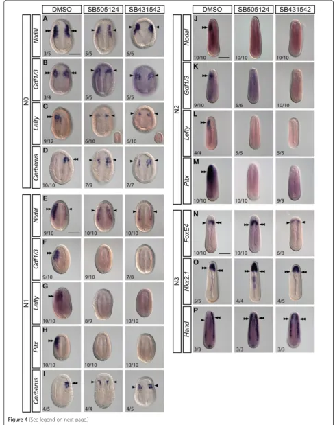
We next examined whether blocking Nodal signaling would affect the asymmetric expression of Nodal cascade genes and other LR marker genes. We treatedB. floridae
embryos with Nodal signaling inhibitors SB505124 and SB431542 starting from the late gastrula stage (G5/G6) and then analyzed these embryos at subsequent stages. Treated N0 embryos exhibit bilateral expression ofNodal
and Gdf1/3, as compared to controls (Figure 4A,B). Ex-pression ofLefty and Cerberus, which is restricted to ei-ther the left or right sides in control embryos, becomes bilateral for both genes upon the treatment (Figure 4C,D). The treated larvae exhibit slightly reduced expression levels of all the examined factors, and in the case ofLefty, the expression can even be reduced to an undetectable level (Figure 4C, inset images).
At the N1 stage, when expression ofNodalis normally biased to the left side, the treated embryos display weak
Nodalexpression on both sides, and the pattern appears to be symmetrical (Figure 4E). Similarly, expression of the other two left-sided genes,Gdf1/3andLefty, is abol-ished by the treatment (Figure 4F,G) and expression of the downstream transcription factor Pitx is also lost (Figure 4H). On the other hand, treatment changes the expression pattern ofCerberusfrom right-sided to bilat-eral (Figure 4I). At the subsequent N2 stage, treatment abolishes expression of Nodal (Figure 4J). Similarly, ex-pression of Gdf1/3, Lefty, and Pitx is no longer detect-able in the treated N2 embryos (Figure 4K,L,M).
Analysis of other genes displaying asymmetric expression at N3 revealed that their expression patterns are also af-fected by treatments with Nodal inhibitors.FoxE4, the ex-pression of which is biased towards the right pharyngeal wall in wild types, exhibits symmetric expression in a
horseshoe-shaped pattern upon either treatment
(Figure 4N). In a similar manner, the rightward-biased expression pattern of Nkx2.1 becomes symmetrical following treatment (Figure 4O); the expression of
Hand, which normally shows differential expression between the left and right lateral plate mesoderm, be-comes equally strong on both sides (Figure 4P).
Taken together, these data demonstrate that inhibition of Nodal signaling abolishes the asymmetrical expression of the Nodal cascade genes and results in symmetrical expression of other downstream organ-specific genes during the neurula stage; therefore, Nodal signaling is necessary for establishing the LR molecular asymmetry in developing amphioxus embryos.
Treatments with Nodal signaling inhibitors alter the left-right asymmetric arrangement of developing muscle blocks and the nervous system
Asymmetrical somite arrangement, which occurs at the mid-neurula stage, is the earliest recognizable morpho-logical feature of LR asymmetric development in amphi-oxus (reviewed in [1]). To investigate the effect of blocking Nodal signaling on the development of somite structures, we treated B. floridae and B. belcheri em-bryos with SB505124 and SB431542 from the late gastrula stage (G5/6) and then observed somite development in the neurula and myomere structure at larval stages using phalloidin staining of F-actin or usingin situhybridization of the muscle actin gene (m-actin). Phalloidin staining en-abled detection of mild asymmetrical arrangement of so-mites in N2-stage neurulae, with left-sided soso-mites positioned slightly more forward as compared to those on the right (Figure 5A, DMSO control); this asymmetrical pattern became more apparent at the N3 stage when em-bryos have more than eight somites (Figure 5B, DMSO control). After treatments with either Nodal inhibi-tors, somites become symmetrically aligned on both sides of the developing notochord at the N2 and N3 stages (Figure 5A,B). Consistently, m-actin in situ
hybridization showed the same effect of blocking Nodal signaling on somite asymmetry (Figure 5C), that is, the
m-actin-expressing muscle precursor cells became sym-metrically aligned in the treated N3 embryos. This ef-fect is especially apparent in the middle/posterior part of the body, where the normal asymmetric arrangement of somites is most easily discerned (Figure 5C, white dashed lines).
(See figure on previous page.)
Previous anatomical studies have revealed the asym-metrical arrangement of amphioxus somatic motoneu-rons in the central nervous system (CNS) and their axonal tract structures [63-65]. Several studies on devel-opmental gene expression patterns also uncovered such LR asymmetric patterns in putative CNS neurons during amphioxus embryogenesis [45,66-70], indicating that the developing CNS also exhibits LR asymmetry. More im-portantly, this CNS neuron asymmetry corresponds to the asymmetric somite arrangement in neurula-stage embryos, suggesting that their development might be controlled by a common developmental pathway. To de-termine whether Nodal signaling also controls the LR asymmetrical distribution of CNS neurons, we used the expression pattern of the pan-neuronal markerHu/Elav
to examine the distribution of post-mitotic neurons in the embryos treated with the two Nodal signaling inhibi-tors. Consistent with previous descriptions, we observed that CNS neurons form recognizable clusters in two lon-gitudinal columns along the rostral-caudal axis of the developing CNS in normal N3-stage embryos (Figure 5D, DMSO control). Much like the asymmetry of somites, the arrangement of these neuronal clusters is staggered between the left and right columns, with the left ones be-ing positioned slightly forward of the right ones (Figure 5D, white dashed lines in DMSO control). Nodal inhibitor treatments caused the arrangement of CNS neuronal clus-ters to become bilaterally symmetrical (Figure 5D), con-sistent with the change seen in the somite development. In addition, ERR, which stains both somites and certain neurons in the anterior CNS [66], also exhibits asymmet-rical expression along the LR axis (Figure 5E, DMSO con-trol). Upon treatment, expression of ERR loses its staggered arrangement along the anterior-posterior axis and becomes bilaterally symmetrical (Figure 5E). The arrangement of somatic musculature and neurons at the N3 stage becomes more pronounced with subse-quent differentiation of these tissues; by L1 and L2, the control larvae exhibit a clear alternating pattern of muscle blocks and axonal structures of spinal nerves (Figure 5F,G, DMSO controls), while the larvae treated with Nodal inhibitors display bilaterally symmetrical patterns of muscle blocks and the axon-innervated po-sitions of the CNS (Figure 5F,G).
Overall, these data suggest that the Nodal signaling pathway is necessary for the proper development of asymmetrical muscular and neural tissues in amphioxus and that inhibition of the Nodal pathway causes symme-trization of these tissues.
Inhibition of Nodal signaling results in the loss of the left-sided mouth opening and duplication of right-left-sided pharyngeal structures
To investigate the effect of inhibition of Nodal signaling on the development of the amphioxus pharynx, we treated B. lanceolatum and B. floridae embryos with SB505124 from N0 to L2. We analyzed the pharyngeal morphology of the control and treated larvae using im-munofluorescence and in situ hybridization (Figure 6 and Additional file 3: Figure S1).
Confocal microscopy analysis revealed that the phar-ynx of the treated larvae exhibits symmetrical morph-ology, with loss of left-sided structures such as the mouth and the preoral pit. Moreover, the left side of the treated larvae seems to be a mirror image of the right side, with duplication of the otherwise right-sided endo-style and club-shaped gland (Figure 6A,A’) To confirm the identity of individual pharyngeal structures, we per-formedin situhybridization against previously identified organ-specific marker genes (see Figure 3). In agreement with the morphological analysis, in situ hybridization demonstrated that genes expressed in the preoral pit and in the mouth in the control larvae (Pitx, Lhx3, and
Dkk1/2/4) become downregulated following treatment (Figure 6B,B’,C,C’,D,D’). On the other hand, Krox and
FoxE4 transcripts, which mark the right-sided club-shaped gland in wild type, exhibit ectopic expression on the left side of the treated larvae, thus presenting a bilat-erally symmetrical signal (Figure 6E,E’,F,F’). Detailed ana-lysis uncovered that theFoxE4-positive andKrox-negative region of the club-shaped gland (that is, the duct of the club-shaped gland) is lost in the treated larvae, suggesting that only the dorsal part of the club-shaped gland is du-plicated (Figure 6I,I’,J,J’). Similarly, FoxQ1 and Nkx2.1, which are expressed in the right-sided endostyle in wild type, display bilaterally symmetrical signals in the anterior portion of the pharynx upon treatment (Figure 6G,G’,H,H’, K,K’). To confirm the phenotype observed in whole
(See figure on previous page.)
Figure 5Inhibition of Nodal signaling alters the left-right asymmetric arrangement of muscle segments and nervous system.All images are taken from the dorsal view.‘L’marks the left side,‘R’marks the right side, and asterisks (*) mark the anterior. The notochord is marked with an ‘n’. Dashed lines mark somite borders and white arrows mark axons of the peripheral nerves. Scale bar, 50μm.(A, B)B. belcheriembryos were stained with phalloidin to mark somite outlines. Morphological asymmetry is barely visible at N2, but asymmetrical arrangement of the somites is apparent by the N3 stage. Treatment causes the staggered arrangement of somites to become symmetrical.(C-E)Expression ofB. floridae m-actin
Figure 6Inhibition of Nodal signaling disrupts formation of the left-sided mouth and causes duplication of the right-sided pharyngeal structures in amphioxus larvae. (A, A’)Dorsal views of the pharyngeal region ofB. lanceolatumL2 larvae stained forβ-catenin and acetylatedα-tubulin. ‘L’indicates the left side and‘R’indicates the right side; anterior is to the left. Following administration of SB505124, the larvae exhibit loss of the preoral pit (pp, black arrowhead), the mouth (m, black arrow), and the first gill slit (fgs, white double arrow) and ectopic development of the club-shaped gland (csg, white arrow) and the endostyle (en, white arrowhead).(B-H, B’-H’)In situhybridization of the treatedB. lanceolatumlarvae revealed loss of expression of
Pitx,Lhx3, andDkk1/2/4and bilateral expression ofKrox,FoxE4,FoxQ1, andNkx2.1concomitant with the duplication of the right side.(I-K, I’-K’)View from the left side displaying loss of the duct of the club-shaped gland (white double arrowhead) upon treatment with SB505124 as deduced from the expression ofKrox,FoxE4, andNkx2.1.(L-O, L’-O’)Transverse sections ofB. floridaelarvae through the pharynx showing the loss ofPitxexpression concomitant with the loss of both the preoral pit (black arrowhead) and the mouth (black arrow) and the ectopic left-sided expression ofFoxE4and
mounts, we sectioned the control and treated larvae upon hybridization. The treated larvae display loss of left-sided expression of Pitx concomitantly with loss of the mouth and preoral pit (Figure 6L,L’,M,M’), and ectopic expression ofFoxE4 and Nkx2.1 on the left side, suggesting that the endostyle and the club-shaped gland develop on both the left and the right sides (Figure 6N,N’,O,O’). To obtain more comprehensive images of the larval morphology, we used Imaris x64 to construct 3D organ models (Figure 6P,P’, Additional file 4: Video S1, and Additional file 5: Video S2). Snapshots of the 3D model display consistent changes of morphology for the treated larvae. The predominantly left-sided preoral pit and mouth are lost upon treatment while the predominantly right-sided endostyle and club-shaped gland form ectopically on the left side, almost forming a mirror image of their right-sided counterparts.
These data suggest that inhibition of Nodal signaling results in the loss of left-sided identity, leading to the ab-sence of the mouth and preoral pit and duplication of the right-sided identity, with ectopic development of the endostyle and club-shaped gland on the left side.
Morphological asymmetry is specified during early neurula stages through asymmetrical expression of downstream targets
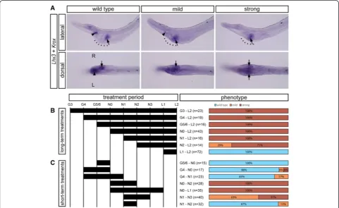
We took advantage of the aforementioned system to deter-mine the embryonic period during which LR asymmetry is specified. We performed a series of experiments in which SB505124 was applied to B. lanceolatum embryos for different time periods during development, and the resulting phenotypes at the open mouth stage (L2) were subsequently examined by in situ hybridization against the left-sided marker Lhx3 and the right-sided marker
Krox. Phenotypes were divided into three categories (Figure 7A):‘wild type’(asymmetrical pharyngeal morph-ology; expression of both Lhx3 and Krox present),‘mild’ (asymmetrical, but altered pharyngeal morphology; ex-pression of Lhx3 reduced or absent, expression of Krox
present on the right side), and ‘strong’ (symmetrical pharyngeal morphology; expression of Lhx3 lost, expres-sion ofKroxpresent on both left and right sides).
We first performed long-term treatments, in which SB505124 was administered to amphioxus embryos at
G3, G4, G5/6, N0, N1, N2, or L1 stages (Figure 7B). Ap-plication of the inhibitor at the N1 stage or earlier re-sults in larvae with the strong phenotype, application at N2 to L2 results in larvae with either strong or mild phenotype, and application at L1 to L2 results in larvae with a wild-type phenotype. This suggests that larval morphology is most susceptible to inhibition during early neurula stages. To confirm this hypothesis, we also performed short-term treatments (Figure 7C). Applica-tion of the inhibitor during late gastrula stages does not result in a significant increase of altered morphology, and the majority of the larvae display a wild-type pheno-type. On the other hand, application at early neurula stages, especially spanning N0 to N2, leads to symmetri-zation of the pharyngeal morphology.
These experiments are congruent with (i) the initial ob-servation of asymmetrical expression of Nkx2.1, FoxE4, and Handat N3 and (ii) the symmetrization of their ex-pression patterns upon treatment with Nodal inhibitors (Figure 4N,O,P). Together, these data suggest that mor-phological asymmetry in amphioxus is specified during the neurula stages through asymmetrical expression of downstream organ-specific target genes.
Discussion
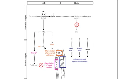
In this study, we demonstrate that expression of the Nodal signaling pathway members in amphioxus resembles that of their vertebrate orthologs and that Nodal is necessary for the left-sided expression of other downstream genes and for left-sided morphogenesis. Inhibition of Nodal results in (i) downregulation of the left-sided genes and bilateral expression of the otherwise right-sided genes and (ii) symmetrization of the larval body. Based on our de-tailed expression survey and time course experiments, we propose a scheme for LR axis establishment in amphioxus (Figure 8) and discuss it in an evolutionary context below.
1. The conservation of the role of Nodal signaling in left-right patterning in chordates
In vertebrates, the LR organizer directs the initial break of symmetry by induction of asymmetric gene expres-sion in its vicinity. The asymmetry at the LR organizer is then transmitted to induce asymmetry in the lateral plate mesoderm, where induction of asymmetric gene expression is followed by self-enhancement and amplifica-tion of the signals that result in polarizaamplifica-tion of the body, and establishment of left and right identities. Finally, these signals target organ-specific factors that are responsible for tissue-specific morphogenesis and differentiation of left- and right-sided organs. Our study shows that the Nodal signaling pathway patterns the LR axis and spe-cifies the left side during early amphioxus embryogen-esis principally in a similar way to that observed in vertebrates (Figure 8). In amphioxus, however, Nodal
acts at the place of its initial asymmetric induction, and thus, there is no transfer of asymmetrically induced sig-nals from the organizer to the lateral plate mesoderm.
We showed that Nodal and its co-ligand Gdf1/3 are expressed in a bilateral fashion at the onset of neurula-tion (N0) and become confined to the left side during subsequent stages, whereas Nodal inhibitors Cerberus
and Lefty display early asymmetric expression. Previous studies showed that Cerberus expression changes from bilateral to unilateral at the gastrula/neurula transition [37]. This change resembles that of its orthologs, Cerl2
in the mouse and CocoinXenopus, during specification of bilateral asymmetry at the LR organizer [71-73]. The organizer houses monociliated cells, the cilia of which beat in a simultaneous manner to generate a leftward fluid flow. The flow promotes decay of Cerl2mRNA on the left side of the node [74], and a similar mechanism may also affect Coco in Xenopus [73]. On the left side, lack of inhibition by Cerl2 results in local activation of Nodal that further suppresses expression of Cerl2, whereas on the right side, expression of Cerl2 inhibits action of Nodal [75]. Our data suggest that suppression ofCerberusby Nodal may also take place in amphioxus, given that inhibition of Nodal at the neurula stages re-sults in ectopic activation ofCerberus expression on the left side (Figure 4D,I). Thus, although amphioxusNodal
and Gdf1/3 are expressed symmetrically at N0, their right-sided suppression by Cerberus results in propaga-tion of the Nodal signal on the left side.
the midline expression of Cerberus at N2 can further block the potential transfer and ectopic activation of Nodal signaling on the right side (Figure 2O). Our time course experiments show that normal amphioxus larval morphology is susceptible to Nodal inhibition, espe-cially at N0 to N2, and we propose that the LR SELI system is active during this period.
The first signs of morphological asymmetry in amphi-oxus are observed after the LR SELI stages at N2 to N3, in which the embryos exhibit asymmetrical arrange-ment of somites and a unilaterally biased expression of organ-specific genes. In vertebrates, the LR morpho-logical asymmetry is conveyed by Pitx2 [82-87]. It is well understood that organs individually reply to the asymmetric Pitx2 signal, via differential proliferation, differentiation, cell shape changes, or apoptosis, but the
molecular mechanisms that linkPitx2to organ-specific morphogenesis are largely unknown and are only just being deciphered (see [14] and citations therein). In amphioxus, the Nodal target Pitx probably also acti-vates transcription of downstream factors in an organ-specific manner, thus triggering the offset placement of left-sided somites and peripheral nerves as opposed to their right-sided counterparts and promoting develop-ment of the preoral pit, mouth, and duct of the club-shaped gland (discussed later).
2. The significance of amphioxus left-right asymmetry and its implications for the development and evolution of the chordate characteristics
The discovery that the Nodal pathway acts to promote asymmetric development in amphioxus is in a good Figure 8Model of the Nodal signaling pathway during establishment of the LR asymmetry in amphioxus.Dashed lines mark proposed interactions based on the data from other chordates. Gray text denotes inhibition of expression of the respective factors. During neurulation,Nodal
accord with the situation in other chordates and even non-chordate deuterostomes and other bilaterians. How-ever, it is challenging to relate the extreme asymmetrical morphology in amphioxus to the almost perfect external symmetry in tunicates and vertebrates. Recent molecular studies support the hypothesis that a dorso-ventral inver-sion may have occurred in the common ancestor of chor-dates (reviewed in [25,26]), although the exact mechanism leading to this inversion is still debated [23,88-90]. Amphi-oxus resides within the chordate clade that split off from the lineage leading to tunicates and vertebrates just after this event. Therefore, it is difficult to determine whether the profound amphioxus asymmetry is somehow related to this event, that is, whether it reflects a recapitulation of the past evolutionary history, and thus represents a primi-tive chordate condition, or whether it exemplifies a peculi-arity of the cephalochordate lineage with no relation to tunicates or vertebrates. The mouth plays a pivotal role in this discussion, as its presence on the left side in amphi-oxus larvae casts doubts on its supposed homology with the median mouth of other chordates [91].
In tunicates and vertebrates, the mouth develops at the anterior neural boundary [92,93]. This boundary, referred to as the preplacodal ectoderm, is the location at which sensory placodes, cement organs, and the mouth originate and is demarcated by expression of members of the
Pax, Six, Eya, and Dach families of transcription factors [94]. Pitx factors define the anterior-most domain within the preplacodal ectoderm and are responsible for development of the mouth, cement organs, adeno-hypophysis, and lens [95]. Pitx factors are downstream of bone morphogenetic protein (BMP) signaling (an epidermal cue) and Otx2 (a neural cue) [96,97]. This initial anterior expression of Pitx factors is distinct from later left-sided Pitx2 expression that acts down-stream of Nodal signaling and is responsible for the LR asymmetry. Additionally, both the anterior and left-sided expression of Pitx factors is regulated at the transcrip-tional level by distinct BMP (Smad1/5)- and Nodal (FoxH1)-responsive elements [87,98,99].
In non-chordate deuterostomes, the mouth seems to be specified and regulated differently from that of verte-brates (Figure 9). In echinoderms, opposing Nodal/BMP signaling organizes the oral-aboral (dorso-ventral) axis, with Nodal signaling promoting, but BMP signaling inhi-biting, the oral fate [100-102]. Interestingly, Pitx2 does not seem to be part of the gene regulatory network pro-moting oral fate in echinoderms; if expression is ob-served in the oral ectoderm of an echinoderm species, it occurs only at later stages and in relation to LR asym-metry [103]. In hemichordates, the mouth develops on the non-Bmp side of the embryonic body (akin to mouth
development in echinoderms), whilePitxexpression can be found on the opposite side in the region of the pro-spective proboscis pore [104]. This structure has tradition-ally been proposed to be homologous to the anterior pituitary of vertebrates and the preoral pit of amphioxus [105]. The regulation ofPitxdownstream of BMP signal-ing in hemichordates [104] is reminiscent of the situation in tunicates and vertebrates and has been proposed as a key factor of the scenario for evolutionary repositioning of the mouth. According to this scenario, the position of the mouth in the chordate ancestor changed from echinoderm- and hemichordate-like anti-BMP and non-Pitx territory to tunicate- and vertebrate-like pro-BMP andPitx-expressing territory [92].
Amphioxus occupies a key position in this scenario and was previously proposed to exhibit a situation similar to that of vertebrates [92]. Yet, a bona fide vertebrate-like preplacodal ectoderm is not present at the anterior neural boundary [94,106], and members of the Six, Eya, Pax, and Dach families are expressed elsewhere throughout the body [107]. Yet,Pitxseems to be transiently expressed at the anterior neural boundary at the late gastrula stage (G6), and this expression coincides with the partially over-lapping bilateral expression of Nodal [39,40]. However, this anterior Pitx expression is diminished at the early pre-hatching neurula stage (N0), and a new left-sided do-main ofPitxexpression is induced at the N0 to N1 transi-tion (Figure 2E,F). Additransi-tionally, while treatments with Nodal inhibitors at neurula stages result in loss of the mouth, treatments spanning the late gastrula stages have only minor effects on mouth development (Figure 7). This suggests that eitherPitxexpression is not triggered by the Nodal signaling at these early stages or that it is not ne-cessary for the development of the mouth. Currently, it is unknown whether the anterior Pitx expression is downstream of neural Otx and epidermal BMP signal-ing and whether this circuitry plays any role in the de-velopment of the mouth, as in other chordates. Future research should therefore be aimed at uncovering the role of anteriorPitxexpression in amphioxus, to deter-mine whether this feature represents a shared charac-teristic between tunicates and vertebrates [108] or has a deeper phylogenetic significance.
The peculiar left-sided mouth in amphioxus has long been a matter of debate as to whether or not it is hom-ologous to the median mouth of vertebrates. Van Wijhe [109-111] argued that the amphioxus mouth is actually a modified first gill slit, which corresponds to the left spir-acle of vertebrates. The presence of this left-sided open-ing would be advantageous for filter feedopen-ing duropen-ing counter-clockwise rotation of the amphioxus larva; the original median mouth may have become greatly dimin-ished, with its remnants present in the preoral pit. This rather controversial assumption has been contested by
those who propose affiliation of the left-sided mouth of amphioxus with that of vertebrates. MacBride [112] suggested that the pharyngeal structures in amphioxus, including the mouth, are initiated in a primarily sym-metrical manner and that larval pharyngeal asymmetry can be explained by differential growth within the phar-ynx and the resultant displacement of organs. Willey [113], on the other hand, proposed that the develop-ment of the asymmetrical pharynx in amphioxus with its left-sided mouth could be explained as an evolution-ary consequence of the rostral prolongation of noto-chord. This prolongation would cause the pharynx to undergo a counter-clockwise torsion (when seen from behind) that would bring the anlagen of dorsal organs, like the mouth and preoral pit (see [114]), to the left side and those of ventral organs, like the endostyle and the club-shaped gland, to the right side [35,88,113]. Such morphogenesis could potentially be explained by differential growth or by cellular rearrangements of sur-rounding tissues, as proposed by MacBride [112], under the control of Nodal signaling. To date, cellular behav-ior during morphogenesis of the amphioxus pharynx has not been studied in depth. Holland and Holland [115] reported that cell proliferation is not correlated with potential pharyngeal torsion; rather, it is the dis-placed organs themselves, and not the surrounding tis-sues, that exhibit increased proliferation. However, if we propose that the pharyngeal organs are primarily established symmetrically and that the Nodal pathway controls the torsion, the anlagen of individual organs should remain at their supposed median position upon inhibition of this pathway (that is, the mouth and the preoral pit would be positioned dorsally and the endo-style and the club-shaped gland ventrally). In contrast, our results show that inhibition of the Nodal pathway results in the duplication of right-sided structures and does not result in the supposed development of medi-ally positioned mouth or endostyle (Figure 6). This im-plies that the primordia of the pharyngeal structures are established primarily asymmetrically and that their asymmetrical arrangement is probably not a secondary consequence of the torsion.
Another option is that the mouth in amphioxus is a derived cephalochordate characteristic with no counter-part in other chordates. Yasui and Kaji [91] argued that structures associated with the mouth during amphioxus metamorphosis as well as those associated with the mouth of the larval lamprey represent traits obtained independently due to similar feeding habits and thus demonstrate analogically derived characteristics. This hypothesis, together with the findings that mouth de-velopment is regulated by medially expressed Pitx
Nodal signaling) in amphioxus, raises serious questions regarding the a priori proposition of the homology of oral openings across chordates (Figure 9).
Despite the contested relationships among chordate mouths, the left-sidedPitx expression in amphioxus may serve similar functions as its vertebrate orthologs expressed medially at the anterior neural boundary. For example, mousePitx1andPitx2regulate both early mor-phogenesis of Rathke’s pouch and later proliferation of pi-tuitary cell precursors [116,117]. Pitx1 activates expression ofLhx3and acts in synergy with POU1F1/Pit1 to promote differentiation of pituitary cell types [118]. BothLhx3and
Pit1homologs are expressed in the amphioxus preoral pit [52,119], and thus, it is possible that they interact with up-streamPitx(Figure 8). Similarly, Pitx can regulate expres-sion ofDkk1/2/4in the amphioxus mouth and preoral pit. Previously, it was shown that Dkk1 is co-expressed with
Pitxfactors in the oral region ofXenopus[120,121]. More-over, microarray analysis demonstrated that activators of the Wnt/β-catenin pathway are downregulated, while in-hibitors of this pathway, includingDkk1, become upregu-lated, in the frog oral region; additionally, Dkk1
overexpression results in an enlarged mouth [122]. Local inhibition of the Wnt/β-catenin pathway in the mouth re-gion is then necessary for proper dissolution of the basal lamina between the ectoderm and the endoderm and for the subsequent break of the oral membrane. A direct link between Pitx factors and Dkk1 during vertebrate mouth development has not been identified at the time of writ-ing, but Pitx2 has been shown to activate expression of
Dkk2during development of the anterior segment of the eye in mouse [123]. The proposed regulation ofDkk1/2/4
expression by Pitx and local inhibition of Wnt/β-catenin signaling by Dkk1/2/4 may thus together form an ancient mechanism to promote fusion between ectoderm and endoderm epithelia, as a prerequisite for normal morpho-genesis of the amphioxus mouth and preoral pit.
We have shown here that Nodal signaling is respon-sible for establishment of the bilateral asymmetry of amphioxus paraxial structures, reminiscent of the role of Nodal in LR asymmetry on the pharyngeal region. The break of the initial symmetrical arrangement of paraxial structures starts at the mid-neurula stage (N2), when the fifth left somite displays a slightly anterior position and advanced differentiation to its right counterpart [124]. Shortly after (at early N3), this somite asymmetry is followed by asymmetrical development of axons of per-ipheral nerves [125]. During the course of development, the asymmetrical arrangement of somites and peripheral nerves is amplified, and new somites arise in a primarily asymmetric manner. BothNodalandPitxare expressed in the left anterior archenteron during outpocketing of the first somites (this study and [39,40]). We have shown that inhibition of Nodal signaling results in (i) the
symmetrical arrangement of somites by the late neurula stage (N3) and (ii) consequent development of bilaterally symmetrical muscle segments and peripheral nerves, resulting in the left side becoming aligned with the right side. The offset arrangement of somites regulated by Nodal signaling is specific to amphioxus development; however, the asymmetrical arrangement itself may also represent a basis for vertebrate somitogenesis. Previous experiments showed that depletion of retinoic acid in the zebrafish, chick, or mouse causes temporal acceler-ation of somite development on the left side [126-128], a situation reminiscent of normal development in amphi-oxus. Blum et al. [1] therefore proposed that retinoic acid signaling may act to shield vertebrate somites from Nodal signaling, which, in turn, may explain the verte-brate-specific transfer of the Nodal signal from the LR organizer to the lateral plate mesoderm without affecting the somites. Retinoic acid signaling thus seems to be im-portant for buffering the lateralizing effects on vertebrate somitogenesis, while the LR alternation of somites might be a primitive feature of chordate somitogenesis.
3. Symmetry breaking in amphioxus: hypotheses and prospects
tested by examining the effects of blocking ciliogenesis or cilia movement on LR asymmetric development in amphioxus embryos.
It is also unclear whether uneven distribution of ion channels influences symmetry breaking in amphioxus. Al-though there is clear evidence for uneven distribution of maternal transcripts of germline-related genes between the first two blastomeres of the amphioxus embryo, these maternal transcripts can be deposited into either the left or the right blastomere after the first cleavage [55,60]. It is unknown whether other maternal or early zygotic tran-scripts exhibit asymmetric distribution across the LR axis in cleavage-stage amphioxus embryos. Further surveys on early mRNA/protein distribution and pharmacological block of specific ion channels in cleavage-stage embryos are required to determine whether ion flux is involved in the symmetry breaking in amphioxus. We anticipate that the results obtained from amphioxus will provide import-ant information for understanding the evolution of LR patterning mechanisms in chordates.
Conclusions
We have shown that Nodal signaling is necessary for the establishment of LR asymmetry and for the determin-ation of the left side of the amphioxus body. Our expres-sion analysis and time course experiments on carefully staged neurulae allowed us to follow critical steps during the establishment of LR asymmetry; moreover, our ex-perimental design provides a basis for future research into the existence of leftward flow-generating cilia in amphioxus and their putative role in initial symmetry breaking [1]. Given the phylogenetic position of amphi-oxus, such studies promise to cast light on the shared and derived character states of LR asymmetry establish-ment among chordates.
Additional files
Additional file 1: Table S1.List of clones from the EST library used to synthesize probes ofB. floridaegenes.
Additional file 2: Table S2.List of PCR primers used for amplifying cDNA fragments ofB. floridaeandB. lanceolatumgenes.
Additional file 3: Figure S1.Branchiostoma floridae larvae display similar morphological changes upon treatment with SB505124. Asterisks (*) mark the anterior,‘L’marks the left side, and‘R’marks the right side. Scale bar, 25μm. (A, A’, B, B’)Pitxexpression marks the left-sided mouth (black arrow) and preoral pit (black arrowhead); both structures are lost upon treatment with SB505124. (C, C’, D, D’)FoxE4expression marks the whole club-shaped gland (white arrow), which resides mainly on the right side of the larva. Upon treatment, the club-shaped gland appears symmetrically on both the left and right sides. (E, E’, F, F’)Nkx2.1is expressed in the right-sided endostyle (white arrowhead). In the embryos treated with SB505124, the endostyle forms on both the left and right sides.
Additional file 4: Video S1.A 3D model reconstructed from z-stack images of pharyngeal structures in a controlB. floridaelarva. The anterior is pointed towards the left side. The initial view of the larva is a lateral view, which slightly tilts towards the left side. The larva is then rotated clockwise
when viewed from the posterior end. The preoral pit (green) opens on the left side, but the organ itself resides up to the midline. The position of the left-sided mouth has been indicated by highlighting the cells around it (yellow). The endostyle (blue) resides on the right side, anterior to the club-shaped gland (red); the dorsal part of the club-shaped gland is situated mainly on the right side, while the ventral part crosses over to the left. The first two gill slits (purple) reside behind the endostyle. The first gill slit appears tilted towards the right, and the opening emerges on the right side; the second gill slit forms in a more medial position.
Additional file 5: Video S2.A 3D model reconstructed from z-stack images of pharyngeal structures in aB. floridaelarva treated with SB505124. The anterior is pointed towards the left side. The initial view of the larva is a lateral view, which tilts slightly towards the left side. The larva is rotated clockwise when viewed from the posterior end. The preoral pit does not form in the treated larva, leaving a vacant region within the rostrum. The mouth does not open in the treated larva. The endostyle (blue) forms ectopically on the left side, resulting in a‘U’-shaped organ. The club-shaped gland (red), still posterior to the endostyle, also forms a‘U’-shaped structure, caused by the ectopic formation of the organ on the left. The first two gill slits (purple) are both located medially in the treated larva.
Abbreviations
BLAST:Basic Logical Alignment Search Tool; BMP: bone morphogenetic protein; CNS: central nervous system; DIG: digoxigenin; DMSO: dimethyl sulfoxide; EST: expressed sequence tag; LR: left-right; SELI: self-enhancement and lateral inhibition; tBLASTx: translated nucleotide BLAST; TGF-β: transforming growth factor beta.
Competing interests
The authors declare that they have no competing interests.
Authors’contributions
VS, ZK, and JKY conceived and designed the experiments. VS, LWY, TML, SWH, and JKY performed the experiments. VS, LWY, TML, and JKY analyzed the data. VS, LWY, and JKY wrote the paper. All authors read and approved the final manuscript.
Acknowledgements
We thank Linda Holland and Nicholas Holland at the Scripps Institution of Oceanography, UCSD, and Daniel Meulemans Medeiros at the University of Colorado, Boulder, for collecting theB. floridaeadults. We are grateful to Hector Escriva and Stephanie Bertrand at Laboratoire Arago, Banyuls-sur-Mer, France, for providing theB. lanceolatumadults and theB.l. Kroxclone. We also thank Cho-Fat Hui, Director of the ICOB Marine Research Station, and Che-Huang Tung, Meng-Yun Tang, Tzu-Kai Huang, Veronika Noskova, and Jindra Pohorela for culturing the amphioxus in our laboratories. We thank Dr. Shao-Chun Hsu in the ICOB core facility for the technical support with confocal microscopy. We thank Sarka Takacova and Dr. Duncan Wright for the English editing. We also thank the three anonymous reviewers for their helpful comments. VS is supported by the Grant Agency of the Czech Republic (14-20839P). The amphioxus work in the lab of ZK is supported in part by grant no. LH12047 from the Ministry of Education, Youth and Sports of the Czech Republic and by IMG institutional support RVO68378050. JKY is supported by the Ministry of Science and Technology, Taiwan (101-2923-B-001-004-MY2; 102-2311-B-001-011-MY3) and by a Career Development Award from Academia Sinica, Taiwan (AS-98-CDA-L06).
Author details 1
Institute of Molecular Genetics, Academy of Sciences of the Czech Republic, Videnska 1083, Prague 14220, Czech Republic.2Institute of Cellular and
Organismic Biology, Academia Sinica, 128 Academia Road, Section 2, Nankang, Taipei 11529, Taiwan.3Institute of Oceanography, National Taiwan
University, 1 Roosevelt Road, Section 4, Taipei 10617, Taiwan.
References
1. Blum M, Feistel K, Thumberger T, Schweickert A. The evolution and conservation of left-right patterning mechanisms. Development. 2014;141(8):1603–13.
2. Namigai EK, Kenny NJ, Shimeld SM. Right across the tree of life: the evolution of left-right asymmetry in the Bilateria. Genesis. 2014;52(6):458–70. 3. Palmer AR. Animal asymmetry. Curr Biol. 2009;19(12):R473–7.
4. Raya A, Izpisua Belmonte JC. Left-right asymmetry in the vertebrate embryo: from early information to higher-level integration. Nat Rev Genet. 2006;7(4):283–93.
5. Blum M, Schweickert A, Vick P, Wright CV, Danilchik MV. Symmetry breakage in the vertebrate embryo: when does it happen and how does it work? Dev Biol. 2014;393(1):109–23.
6. Shiratori H, Hamada H. The left-right axis in the mouse: from origin to morphology. Development. 2006;133(11):2095–104.
7. Vandenberg LN, Levin M. A unified model for left-right asymmetry? Comparison and synthesis of molecular models of embryonic laterality. Dev Biol.
2013;379(1):1–15.
8. Su YH. Telling left from right: left-right asymmetric controls in sea urchins. Genesis. 2014;52(3):269–78.
9. Tanaka C, Sakuma R, Nakamura T, Hamada H, Saijoh Y. Long-range action of Nodal requires interaction with GDF1. Genes Dev. 2007;21(24):3272–82. 10. Brennan J, Norris DP, Robertson EJ. Nodal activity in the node governs
left-right asymmetry. Genes Dev. 2002;16(18):2339–44.
11. Kawasumi A, Nakamura T, Iwai N, Yashiro K, Saijoh Y, Belo JA, et al. Left-right asymmetry in the level of active Nodal protein produced in the node is translated into left-right asymmetry in the lateral plate of mouse embryos. Dev Biol. 2011;353(2):321–30.
12. Saijoh Y, Oki S, Ohishi S, Hamada H. Left-right patterning of the mouse lateral plate requires nodal produced in the node. Dev Biol. 2003;256(1):160–72. 13. Nakamura T, Mine N, Nakaguchi E, Mochizuki A, Yamamoto M, Yashiro K,
et al. Generation of robust left-right asymmetry in the mouse embryo requires a self-enhancement and lateral-inhibition system. Dev Cell. 2006;11(4):495–504.
14. Nakamura T, Hamada H. Left-right patterning: conserved and divergent mechanisms. Development. 2012;139(18):3257–62.
15. Duboc V, Rottinger E, Lapraz F, Besnardeau L, Lepage T. Left-right asymmetry in the sea urchin embryo is regulated by nodal signaling on the right side. Dev Cell. 2005;9(1):147–58.
16. Luo YJ, Su YH. Opposing nodal and BMP signals regulate left-right asymmetry in the sea urchin larva. PLoS Biol. 2012;10(10):e1001402.
17. Morokuma J, Ueno M, Kawanishi H, Saiga H, Nishida H.HrNodal, the ascidian nodal-related gene, is expressed in the left side of the epidermis, and lies upstream ofHrPitx. Dev Genes Evol. 2002;212(9):439–46. 18. Yoshida K, Saiga H. Left-right asymmetric expression ofPitxis regulated by
the asymmetric Nodal signaling through an intronic enhancer in Ciona intestinalis. Dev Genes Evol. 2008;218(7):353–60.
19. Burdine RD, Caspary T. Left-right asymmetry: lessons from Cancun. Development. 2013;140(22):4465–70.
20. Grande C, Patel NH. Nodal signalling is involved in left-right asymmetry in snails. Nature. 2009;457(7232):1007–11.
21. Kuroda R, Endo B, Abe M, Shimizu M. Chiral blastomere arrangement dictates zygotic left-right asymmetry pathway in snails. Nature. 2009;462(7274):790–4. 22. Watanabe H, Schmidt HA, Kuhn A, Hoger SK, Kocagoz Y, Laumann-Lipp N,
et al. Nodal signalling determines biradial asymmetry in Hydra. Nature. 2014;515(7525):112–5.
23. Arendt D, Nubler-Jung K. Inversion of dorsoventral axis? Nature. 1994;371(6492):26.
24. De Robertis EM, Sasai Y. A common plan for dorsoventral patterning in Bilateria. Nature. 1996;380(6569):37–40.
25. Holland LZ, Carvalho JE, Escriva H, Laudet V, Schubert M, Shimeld SM, et al. Evolution of bilaterian central nervous systems: a single origin? EvoDevo. 2013;4(1):27.
26. De Robertis EM. Evo-devo: variations on ancestral themes. Cell. 2008;132(2):185–95.
27. Holland LZ, Albalat R, Azumi K, Benito-Gutierrez E, Blow MJ, Bronner-Fraser M, et al. The amphioxus genome illuminates vertebrate origins and cephalochordate biology. Genome Res. 2008;18(7):1100–11.
28. Putnam NH, Butts T, Ferrier DE, Furlong RF, Hellsten U, Kawashima T, et al. The amphioxus genome and the evolution of the chordate karyotype. Nature. 2008;453(7198):1064–71.
29. Bourlat SJ, Juliusdottir T, Lowe CJ, Freeman R, Aronowicz J, Kirschner M, et al. Deuterostome phylogeny reveals monophyletic chordates and the new phylum Xenoturbellida. Nature. 2006;444(7115):85–8.
30. Delsuc F, Tsagkogeorga G, Lartillot N, Philippe H. Additional molecular support for the new chordate phylogeny. Genesis. 2008;46(11):592–604. 31. Bertrand S, Escriva H. Evolutionary crossroads in developmental biology:
amphioxus. Development. 2011;138(22):4819–30.
32. Schubert M, Holland LZ, Stokes MD, Holland ND. Three amphioxusWntgenes (AmphiWnt3,AmphiWnt5, andAmphiWnt6) associated with the tail bud: the evolution of somitogenesis in chordates. Dev Biol. 2001;240(1):262–73. 33. Lu TM, Luo YJ, Yu JK. BMP and Delta/Notch signaling control the
development of amphioxus epidermal sensory neurons: insights into the evolution of the peripheral sensory system. Development. 2012;139 (11):2020–30.
34. Glardon S, Holland LZ, Gehring WJ, Holland ND. Isolation and developmental expression of the amphioxusPax-6gene (AmphiPax-6): insights into eye and photoreceptor evolution. Development. 1998;125(14):2701–10.
35. Lacalli T. Mucus secretion and transport in amphioxus larvae: organization and ultrastructure of the food trapping system, and implications for head evolution. Acta Zool. 2008;89(3):219–30.
36. Boorman CJ, Shimeld SM. Pitx homeobox genes inCionaand amphioxus show left-right asymmetry is a conserved chordate character and define the ascidian adenohypophysis. Evol Dev. 2002;4(5):354–65.
37. Le Petillon Y, Oulion S, Escande ML, Escriva H, Bertrand S. Identification and expression analysis of BMP signaling inhibitors genes of the DAN family in amphioxus. Gene Expr Patt. 2013;13(8):377–83.
38. Onai T, Yu JK, Blitz IL, Cho KW, Holland LZ. Opposing Nodal/Vg1 and BMP signals mediate axial patterning in embryos of the basal chordate amphioxus. Dev Biol. 2010;344(1):377–89.
39. Yasui K, Zhang S, Uemura M, Saiga H. Left-right asymmetric expression of
BbPtx, aPtx-related gene, in a lancelet species and the developmental left-sidedness in deuterostomes. Development. 2000;127(1):187–95. 40. Yu JK, Holland LZ, Holland ND. An amphioxusnodalgene (AmphiNodal)
with early symmetrical expression in the organizer and mesoderm and later asymmetrical expression associated with left-right axis formation. Evol Dev. 2002;4(6):418–25.
41. Yu JK, Satou Y, Holland ND, Shin IT, Kohara Y, Satoh N, et al. Axial patterning in cephalochordates and the evolution of the organizer. Nature. 2007;445(7128):613–7. 42. Venkatesh TV, Holland ND, Holland LZ, Su MT, Bodmer R. Sequence and
developmental expression of amphioxusAmphiNk2-1: insights into the evolutionary origin of the vertebrate thyroid gland and forebrain. Dev Genes Evol. 1999;209(4):254–9.
43. Onimaru K, Shoguchi E, Kuratani S, Tanaka M. Development and evolution of the lateral plate mesoderm: comparative analysis of amphioxus and lamprey with implications for the acquisition of paired fins. Dev Biol. 2011;359(1):124–36.
44. Kusakabe R, Kusakabe T, Satoh N, Holland ND, Holland LZ. Differential gene expression and intracellular mRNA localization of amphioxus actin isoforms throughout development: Implications for conserved mechanisms of chordate development. Dev Genes Evol. 1997;207(4):203–15. 45. Satoh G, Wang Y, Zhang P, Satoh N. Early development of amphioxus
nervous system with special reference to segmental cell organization and putative sensory cell precursors: a study based on the expression of pan-neuronal marker geneHu/elav. J Exp Zool. 2001;291(4):354–64. 46. Yu JK, Wang MC, Shin IT, Kohara Y, Holland LZ, Satoh N, et al. A cDNA
resource for the cephalochordate amphioxusBranchiostoma floridae. Dev Genes Evol. 2008;218(11–12):723–7.
47. Yu JK, Holland LZ, Jamrich M, Blitz IL, Hollan ND.AmphiFoxE4, an amphioxus winged helix/forkhead gene encoding a protein closely related to vertebrate thyroid transcription factor-2: expression during pharyngeal development. Evol Dev. 2002;4(1):9–15.
48. Langeland JA, Tomsa JM, Jackman Jr WR, Kimmel CB. An amphioxussnail
gene: expression in paraxial mesoderm and neural plate suggests a conserved role in patterning the chordate embryo. Dev Genes Evol. 1998;208(10):569–77.
49. Mazet F, Luke GN, Shimeld SM. The amphioxusFoxQ1gene is expressed in the developing endostyle. Gene Expr Patt. 2005;5(3):313–5.