PRIMARY RESEARCH
LINC01094 triggers radio-resistance in clear
cell renal cell carcinoma via miR-577/CHEK2/
FOXM1 axis
Yufeng Jiang
1†, Wei Li
2†, Yang Yan
2, Xudong Yao
2*, Wenyu Gu
2and Haimin Zhang
2*Abstract
Background: Radioresistance is an obstacle to limit efficacy of radiotherapy. Meanwhile, long non-coding RNAs (lncRNAs) have been reported to affect radioresistance. Here, we aimed to investigate lncRNAs involving radioresist-ance development of clear cell renal cell carcinoma (ccRCC), the most frequent type of renal cell carcinoma (RCC). Methods: The mRNA and protein expressions of genes were measured via qRT-PCR and western blot. The relation-ships among genes were verified by RIP and luciferase reporter assay. The radioresistance of ccRCC cells was evaluated through clonogenic survival assay, MTT assay and TUNEL assay.
Results: LINC01094 was over-expressed in ccRCC cell lines. LINC01094 expression was increased along with the radiation exposure time and the final stable level was 8 times of the initial level. Knockdown of LINC01094 resulted in enhanced radiosensitivity of ccRCC cells. Mechanically, LINC01094 was a ceRNA of CHEK2 by sponging miR-577. Also, the enhancement of LINC01094 on ccRCC radioresistance was mediated by CHEK2-stabilized FOXM1 protein. Conclusion: LINC01094 facilitates ccRCC radioresistance by targeting miR-577/CHEK2/FOXM1 axis, blazing a new trail for overcoming radioresistance in ccRCC.
Keywords: LINC01094, ccRCC radio-resistance, miR-577, CHEK2, FOXM1
© The Author(s) 2020. This article is licensed under a Creative Commons Attribution 4.0 International License, which permits use, sharing, adaptation, distribution and reproduction in any medium or format, as long as you give appropriate credit to the original author(s) and the source, provide a link to the Creative Commons licence, and indicate if changes were made. The images or other third party material in this article are included in the article’s Creative Commons licence, unless indicated otherwise in a credit line to the material. If material is not included in the article’s Creative Commons licence and your intended use is not permitted by statutory regulation or exceeds the permitted use, you will need to obtain permission directly from the copyright holder. To view a copy of this licence, visit http://creat iveco mmons .org/licen ses/by/4.0/. The Creative Commons Public Domain Dedication waiver (http://creat iveco mmons .org/publi cdoma in/ zero/1.0/) applies to the data made available in this article, unless otherwise stated in a credit line to the data.
Background
Renal cell carcinoma (RCC) is one of the most aggressive cancers and accounts for 3% of all adult malignancies [1]. Clear cell renal cell carcinoma (ccRCC), characterized by a high rate of metastasis and relapse [2], is the most common subtype of RCC and represents approximately 80–90% of all RCC cases [3, 4]. Importantly, patients with metastatic ccRCC make up over 30% of all ccRCC cases and the 5-year survival rate of them was lower than 20% due to the resistance to chemotherapy and radiotherapy,
[5, 6]. Although molecular characterization of ccRCC has got developed [7], the mechanism by which ccRCC patients obtain radioresistance or chemoresistance remains largely uncharted.
Radiotherapy is a commonly-applied cancer treatment as ionizing radiation (IR) damages cancer cell mainly via inducing DNA damage, especially DNA double strand breaks (DSBs) [8, 9]. The response of cancer cells to DNA damage is critical for tumor development, and enhanced repair on DNA DSBs results in resistance to IR [10]. In the past few decades, understanding of cellular signaling for DSBs repair has been gradually uncovered [11, 12]. Also, implication of non-coding RNAs (ncRNAs) in this process has been the focus on cancer research [13, 14].
Long non-coding RNAs (lncRNAs) are ncRNA tran-scripts with a length longer than 200 nts [15]. Recent studies indicated that lncRNAs play pivotal roles in the
Open Access
*Correspondence: yaoxudong67@sina.com; zhm1064@163.com †Yufeng Jiang and Wei Li are co-first author.
2 Department of Urology, Shanghai Tenth People’s Hospital, Tongji University School of Medicine, No.301 Yanchang Road, Jing’an District, Shanghai 200072, China
development of cancer radioresistance [16]. For example, SNHG18 improves radioresistance in glioma via inhibit-ing semaphorin 5A [17]. PCAT-1 regulates DSBs repair through repressing BRCA2 in prostate cancer [18]. Long intergenic non-protein coding RNA 1094 (LINC01094) is a lncRNA that has been scarcely explored before. Here, the TCGA data revealed that it was significantly upregu-lated in KIRC (Kidney renal clear cell carcinoma) tissues relative to the normal tissues. Therefore, we wondered whether LINC01094 was implicated in radioresistance development of ccRCC.
In the current study, we probed into the role and poten-tial mechanism of LINC01094 in ccRCC radioresistance.
Materials and methods
Cell culture
Human kidney proximal tubule cell (HK-2), human embryonic kidney cell (HEK-293T) and ccRCC cells (A-498, ACHN, 786-O, Caki-1) were purchased from American Type Culture Collection (ATCC; Manas-sas, VA, USA). All cells were cultivated in RPMI-1640 medium (Invitrogen, Carlsbad, CA, USA) adding 10% fetal bovine serum (FBS; Invitrogen) plus 1% penicillin/ streptomycin (Thermo Fisher Scientific, Grand Island, NY, USA) in a 5% CO2 atmosphere at 37 °C.
Quantitative real‑time PCR (qRT‑PCR)
RNA was extracted using TRIzol reagent (Thermo Fisher Scientific) and then reversely transcribed into cDNA. SYBR Green PCR Master Mix (Roche, Man-nheim, Germany) was applied on an Applied Biosystems 7900 Real-Time PCR System (Thermo Fisher Scientific) for real-time PCR. Relative RNA expression levels were assessed via 2−ΔΔCt method. GAPAH/U6 acted as
nor-malized gene.
Cell transfection
786-O or Caki-1 cells with irradiation treatment or not, were firstly added into six-well plates with non-serum culture medium for 1 day. The specific shRNAs for LINC01094 (shLINC01094#1/2) or non-specific control (shCtrl), miR-577 mimics, miR-NC were synthesized by RiboBio (Guangzhou, Guangdong, China). Besides, the pcDNA3.1 vector targeting LINC01094, CHEK2 or FOXM1 and empty vectors were constructed by Gene-chem (Shanghai, China). Lipofectamine 2000 (Invitro-gen) was used during transfection. Cells were collected after 48 h of transfection.
Colony formation assay
Cells were plated into 6-well culture plates with a con-centration of 800 cells per well. 14 days later, colonies
were fixed for 15 min in 100% methanol (Sigma-Aldrich, St. Louis, MO, USA) and then stained for 20 min using 0.1% crystal violet (Sigma-Aldrich) at room temperature.
MTT assay
Transfected 786-O or Caki-1 cells were seeded into 96-well plates with 4 Gy of irradiation treatment, cultur-ing for 0, 24, 48, 72 and 96 h. Proliferation of cells was tested via MTT assay. 10 µL of MTT reagent (Sigma-Aldrich) was added to each well for 4 h followed by DMSO incubation. Absorbance at 490 nm of cell lysates was detected via a plate reader (Bio-Tek Instruments, Winooski, VT, USA).
Immunofluorescence (IF) analysis
Cells were cultured on glass coverslips and fixed in in formaldehyde (Sigma-Aldrich), followed by permeabili-zation in cold methanol/acetone reaction. Afterwards, cell lines were treated with FITC-labeled phalloidin (Sigma-Aldrich) or primary antibody against phospho-Histone H2AX (ab2893; Abcam, Cambridge, USA), fol-lowing treatment with Alexa Fluor488-labeled or Alexa Fluor594-labeled (Thermo Fisher Scientific) second-ary antibody at room temperature for 1 h. Nuclei stain-ing was conducted with 4′,6-diamidino-2-phenylindole (DAPI; Thermo Fisher Scientific). Nikon Eclipse E600 microscope and ACT-1 software (Nikon) were finally utilized.
Nuclear‑cytoplasmic extraction assay
Nuclear and cytoplasmic proteins were extracted from Caki-1 or 786-O cells using the NE-PER kit (Thermo Fisher Scientific). The qRT-PCR was used for the detec-tion of LINC01094. GAPDH was used as an indicated reagent to detect the gene expression level in cytoplasm, while U6 was used to detect the expression level in nucleus.
RNA immunoprecipitation (RIP)
The Imprint RNA Immunoprecipitation Kit (Millipore, Bedford, OH, USA) was applied for RIP assay. Briefly, cell extracts were harvested after lysing in RIP lysis buffer (Solarbio, Beijing, China) and incubated with magnetic beads (Invitrogen) conjugated to antibodies of anti-Ago2 (Millipore) and anti-IgG (Millipore). RNA precipitates were extracted for qRT-PCR.
RNA pull down assay
binding RNAs were washed and eluted, qRT-PCR was conducted to analyze the expression of RNAs.
TUNEL staining assay
The Cell Death Detection Kit (Roche) was chosen to detect the apoptosis level. After TUNEL staining, cells were respectively dyed using DAPI (Thermo Fisher Sci-entific). Relative fluorescence intensity was observed by the confocal laser scanning microscope (CLSM; Leica Microsystems, Berlin, Germany).
Western blotting
Cells were lysed through RIPA buffer (Invitrogen). After centrifugation, the collected proteins were sepa-rated by SDS-PAGE (Bio-Rad, Hercules, CA, USA) and then transferred to polyvinylidene fluoride membranes (PVDF; Bio-Rad). The membranes were blocked with 5% nonfat milk and consecutively probed with primary antibodies including anti-γH2AX (ab2893, Abcam, Cambridge, USA), anti-cleaved PARP (ab32064, Abcam), anti-CHEK2 (ab109413, Abcam), anti-FOXM1 (ab180710, Abcam) and anti-GAPDH (ab245356, Abcam) as an internal control, followed by treated with secondary antibody. Finally, immunoreactivity was assessed via enhanced chemiluminescence (ECL; Bio-Rad).
Luciferase reporter assay
LINC01094-WT, CHEK2-WT and their respective mutations (LINC01094-MUT, CHEK2-MUT) were synthesized by Genepharma (Shanghai, China) and inserted into the pmirGLO dual-luciferase plasmid (Promega, Madison, WI, USA). Then, LINC01094-WT/MUT vector and CHEK2-LINC01094-WT/MUT vector were co-transfected into HEK-293T cells with indicated transfection plasmids, respectively. After 48 h of trans-fection, Dual Luciferase Report Assay System (Pro-mega) was applied to monitor luciferase activity.
Statistical analysis
Experiments mentioned in this study were conducted for three times or more. All data were shown as mean ± standard deviation (SD). Statistics analysis was performed on GraphPad 5.0 software (GraphPad Soft-ware, La Jolla, CA, USA). Differences were compared using Student’s t-test or one-way ANOVA, with P < 0.05 used as significance threshold.
Results
Depletion of LINC01094 improves the sensitivity of ccRCC cells to radiation
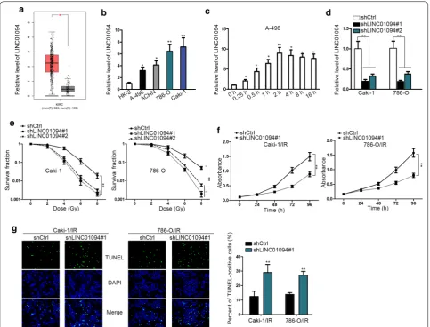
First of all, based on the TCGA data, we found that LINC01094 was obviously upregulated in KIRC tissues compared to the normal tissues (Fig. 1a). Also, it was demonstrated that LINC01094 was highly expressed in ccRCC cell lines (Fig. 1b). More importantly, LINC01094 was gradually upregulated in response to radiation exposure, and the final steady level was approximately 8 times of that under unexposed con-dition (Fig. 1c). On this basis, we suspected that LINC01094 might play a role in ccRCC radio-resistance development.
In order to verify above assertion, loss-of-func-tion assays were performed subsequently. As a result, LINC01094 was apparently silenced in both Caki-1 and 786-O cells by transfection of shRNAs targeting LINC01094 (Fig. 1d). Depletion of LINC01094 conse-quently resulted in enhanced sensitivity of both Caki-1 and 786-O cells responding to radiation, evidenced by impaired survival fraction in LINC01094-inhibited ccRCC cells under radiation (Fig. 1e). Besides, consid-ering Caki-1 and 786-O cells had worse survival frac-tion after being transfected with shLINC01094#1, we selected shLINC01094#1-transfected cells for fol-lowing assays. Moreover, through being exposed to 4 Gy of radiation, the proliferative ability was notably decreased in ccRCC cells upon LINC01094 depletion (Fig. 1f). Meanwhile, knockdown of LINC01094 led to markedly intensified apoptosis in these two ccRCC cells under radiation exposure (Fig. 1g). Therefore, we concluded that LINC01094 upregulation leads to strengthened resistance of ccRCC cells responding to irradiation.
LINC01094 confers radio‑resistance in ccRCC through enhancing DNA‑DSBs repair
checkpoint kinase 2 (CHEK2) protein, a pivotal factor involving in DNA damage repair, was verified to be dis-tinctly reduced under irradiated condition and was fur-ther downregulated by LINC01094 inhibition (Fig. 2d) Taken together, LINC01094 contributes to radiotoler-ance in ccRCC via improving DNA DSBs repair.
LINC01094 locates mainly in the cytoplasm and sponges miR‑577 in ccRCC
Particularly, we probed into the underlying mechanism by which LINC01094 affected ccRCC radioresistance. Firstly, we explored the distribution of LINC01094 in ccRCC cells, since the function of lncRNAs depends on their subcellular localization to some extent. As
a result, the online lncLocator suggested that most of LINC01094 located in the cytoplasm (Fig. 3a). Addi-tionally, subcellular fractionation confirmed that LINC01094 was largely a cytoplasmic lncRNA in ccRCC (Fig. 3b). Given that lncRNAs in the cytoplasm largely function as a competing endogenous RNA (ceRNA) in cancers, we wondered whether LINC01094 was an endogenous sponge of certain miRNAs. After bioinformatics analysis, miR-577, miR-545-3p and miR-330-3p were suggested as potential miRNAs that interacted with LINC01094. According to the results of RNA pull down assay, only miR-577 was significantly pulled down by biotinylated LINC01094 (Additional
file 1: Fig. S1A). Besides, miR-577 was downregulated in ccRCC cell lines in contrast to normal HK-2 cells (Fig. 3c). Moreover, we revealed that overexpression of miR-577 was stimulated by LINC01094 downregula-tion while the level of LINC01094 was mitigated under ectopic expression of miR-577 (Fig. 3d, e). Therefore, we speculated that LINC01094 might be a sponge of miR-577 in ccRCC.
To verify whether LINC01094 interacted with miR-577 in ccRCC cells, we constructed LINC01094-WT containing predicted binding sites of miR-577 and LINC01094-Mut containing mutated binding sites for luciferase reporter assay (Fig. 3f). As a conse-quence, the luciferase activity of LINC01094-WT was lessened under miR-577 upregulation, with that of
LINC0194-Mut unchanged all the way (Fig. 3g). Fur-thermore, the RIP result certified that both LINC01094 and miR-577 were harvested by anti-Ago2 (Fig. 3h), revealing the interaction between them in RNA-induced silencing complex (RISC). Based on these data, we concluded that LINC01094 locates in the cytoplasm and interacts with miR-577 in ccRCC.
LINC01094 increases FOXM1 protein in ccRCC by acting as a ceRNA of CHEK2 via absorbing miR‑577
Surprisingly, we discovered that CHEK2, a verified DNA damage-related gene, was predicted as one of the targets of miR-577 according to “ENCORI” database
(http://starb ase.sysu.edu.cn/). Importantly, the
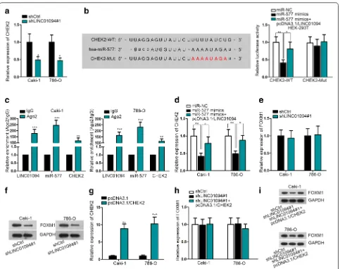
expres-sion of CHEK2 was distinctly decreased by LINC01094
suppression (Fig. 4a), further implying that CHEK2 might be the downstream effector of LINC01094 in ccRCC. Moreover, we mutated predicted binding sequences of CHEK2 and conducted the following RNA pull down assay and luciferase reporter assay. The RNA pull down assay validated the interaction between miR-577 and CHEK2 at predicted sites (Additional file 1: Fig. S1B). And luciferase reporter assay disclosed that lucif-erase activity of CHEK2-WT was reduced by miR-577 mimics but was partially recovered by LINC01094 co-overexpression while CHEK2-MuT showed no response under the same conditions (Fig. 4b). Also, the coexistence of LINC01094, miR-577 and CHEK2 mRNA in RISC was testified in RIP assays (Fig. 4c). Consistently, the expres-sion of CHEK2 was impeded by ectopic expresexpres-sion of miR-577, whereas such inhibition was attenuated under enforced LINC01094 expression (Fig. 4d). In other words,
LINC01094 prompts CHEK2 expression in ccRCC via competitively binding with miR-577.
Forkhead box M1 (FOXM1) is an oncogenic tran-scription factor that is always upregulated in various cancers including ccRCC [19, 20]. Recently, FOXM1 has been reported to contribute to radioresistance in several carcinomas [21, 22]. Meanwhile, it has also been demonstrated that FOXM1 was stabilized by CHEK2 and therefore enhanced the expression of FOXM1-targeted DNA repair genes [23]. In this regard, we presumed that FOXM1 might be the downstream gene of LINC01094/miR-577/CHEK2 axis involved in ccRCC radio-sensitivity regulation. As anticipated, inhibition of LINC01094 had no impact on the mRNA level of FOXM1 but led to a sharp reduction on the protein level of FOXM1 in both Caki-1 and 786-O cells (Fig. 4e, f). Moreover, LINC01094 silence-confined
FOXM1 protein level was recovered by CHEK2 upregu-lation, while the level of FOXM1 mRNA was scarcely influenced under the same condition (Fig. 4g–i). By and large, LINC01094 relies on CHEK2 to upregulate FOXM1 protein in ccRCC.
FOXM1 mediates LINC01094‑facilitated radioresistance in ccRCC
Subsequently, we attempted to make it clear that if FOXM1 was responsible for LINC01094-promoted DNA damage repair and consequent decreased radiosensitivity. As dem-onstrated in Fig. 5a, LINC01094 knockdown-restrained
FOXM1 protein was reversed under the FOXM1 overex-pression. In addition, upregulation of FOXM1 recovered the DNA DSBs repair controlled by LINC01094 suppres-sion in Caki-1 cells under irradiation (Fig. 5b). Moreover, lessened cell survival fraction, viability as well as acceler-ated cell apoptosis of 4 Gy radiation-exposed cells under LINC01094 silence were all normalized by FOXM1 upreg-ulation (Fig. 5c–e). All these assays proved that height-ened sensitivity of Caki-1 cells to radiation exposure under LINC01094 depletion was restored upon enforced FOXM1 expression. Hence, we drew a conclusion that LINC01094
Fig. 4 LINC01094 enhanced FOXM1 protein expression in ccRCC via miR-577/CHEK2 signaling. a qRT-PCR analysis of CHEK2 level in
LINC01094-inhibited ccRCC cells. b Luciferase reporter assay confirmed the competitive binding of LINC01094 and CHEK2 mRNA with miR-577. c
contributes to DNA DSBs repair to improve ccRCC radi-oresistance through FOXM1-responsible manner.
Discussion
Nowadays, radiotherapy has been commonly employed to treat more than half of human cancer types [24, 25], including renal cell carcinoma [26]. Nevertheless, some radioresistance-obtained cells, namely cancer stem-like cells, has become the major obstacle to control the effi-cacy of radiotherapy [27]. To overcome radioresistance, great efforts have been made over decades. Recently, lncRNAs have been recognized as novel regulators in radioresistance development [17, 28, 29]. Here, we stud-ied on a novel lncRNA LINC01094, which was suggested by TCGA to be highly-expressed in KIRC tissues Expres-sion of LINC01094 was ever-increasing till reaching a stable level (8 times of the inital level) in ccRCC cells during radiation exposure. On this basis, we assumed that LINC01094 might be associated with radioresistance
development in ccRCC. As expected, knockdown of LINC01094 led to increased sensitivity of ccRCC cells to radiation treatment, indicating the promotion effects of LINC01094 on ccRCC radioresistance. A recent study uncovered the implication of ncRNAs including lncR-NAs in DNA repair in radiation-exposed tumor develop-ment [13]. Ionizing radiation can induce a wide range of DNA lesions and clustered DNA lesions is the hallmark of IR. DNA double strand breaks (DSBs) play a key role during the process [30, 31]. Presently, we revealed that LINC01094 affected the radioresistance in ccRCC via promoting ability of ccRCC cells to repair DNA DSBs.
In recent years, lncRNAs in the cytoplasm have been emerged as competing endogenous RNAs in regulat-ing cancer development via spongregulat-ing certain miRNA to release such miRNAs-targeted mRNAs from deg-radation at post-transcriptional level [32, 33]. Herein, it was proved that LINC01094 was mainly a cytoplas-mic lncRNA in ccRCC cells. More importantly, we
demonstrated that miR-577, which was predominantly reported as an anti-tumor miRNA [34, 35], was directly sponged by LINC01094 in ccRCC. Further, CHEK2 (also called Chk2), a well-known factor that participated in DNA repair and radioresitance development [36–38], was recognized as the downstream target of miR-577 and had a competition with LINC01094 to interact with miR-577 in ccRCC cells. Besides, we validated that LINC01094 enhanced FOXM1 protein level in ccRCC at CHEK2-dependent manner, which was in consistent with a previous study that CHEK2 stabilized FOXM1 protein in stimulation of DNA repair-related genes [23]. Last but not least, rescue assays conducted in LINC01094-depleted ccRCC cells further certified that FOXM1 was the responsible effector underlying LINC01094-strength-ened radioresistance in ccRCC.
Conclusion
All in all, our work illustrated that LINC01094 contrib-utes to radioresistance development in ccRCC through modulating CHEK2-stabilized FOXM1 by acting as a ceRNA to up-regulate CHEK2, which might open up a new way to overcome the radioresistance in ccRCC.
Supplementary information
Supplementary information accompanies this paper at https ://doi. org/10.1186/s1293 5-020-01306 -8.
Additional file 1: Figure S1. (A-B) RNA pull down assay detected
rela-tive expression of miRNAs pulled down by different biotinylated RNAs. **P < 0.01.
Abbreviations
RCC : Renal cell carcinoma; lncRNAs: Long non-coding RNAs; ccRCC : Clear cell renal cell carcinoma; DSBs: DNA double-strand breaks; IR: Ionizing radiation; LINC01094: Long intergenic non-protein coding RNA 1094; KIRC: Kidney renal clear cell carcinoma; ATCC : American Type Culture Collection; FBS: Fetal bovine serum; qRT-PCR: Quantitative real-time PCR; IF: Immunofluorescence; DAPI: 4′,6-diamidino-2-phenylindole; RIP: RNA immunoprecipitation; CLSM: The confocal laser scanning microscope; PVDF: Polyvinylidene fluoride mem-branes; ECL: Enhanced chemiluminescence; SD: Standard deviation; CHEK2: Checkpoint kinase 2; ceRNA: Competing endogenous RNA; RISC: RNA-induced silencing complex; FOXM1: Forkhead box M1.
Acknowledgements
We sincerely thank all involvers for their support to our research.
Authors’ contributions
YJ and WL designed and revised this study. YY, XY, WG and HZ were responsi-ble for the collection of experimental materials. Each author conduced to this study and provided valuable advice for this manuscript. All authors read and approved the final manuscript.
Funding
The Project of Scientific Research Topic of Shanghai Municipal Health and Family Planning Commission: “The Effect of BX649059’s Interaction with micro-RNA on the Occurrence and Development of Renal Cancer and Its Molecular Mechanism” (201640146).
Availability of data and materials
Not applicable.
Ethics approval and consent to participate
Not applicable.
Consent for publication
Not applicable.
Competing interest
The authors declare that no competing interest exists in this study.
Author details
1 Department of Urology, Chongming Branch, Shanghai Tenth People’s Hospi-tal, Tongji University School of Medicine, No.66 Xiangyang Road, Chongming District, Shanghai 202157, China. 2 Department of Urology, Shanghai Tenth People’s Hospital, Tongji University School of Medicine, No.301 Yanchang Road, Jing’an District, Shanghai 200072, China.
Received: 11 July 2019 Accepted: 28 May 2020
References
1. Ricketts CJ, Crooks DR, Sourbier C, Schmidt LS, Srinivasan R, Linehan WM. SnapShot: renal cell carcinoma. Cancer Cell. 2016;29(4):610.e611. 2. Lokeshwar SD, Talukder A, Yates TJ, Hennig MJP, Garcia-Roig M,
Lahore-wala SS, Mullani NN, Klaassen Z, Kava BR, Manoharan M, et al. Molecular characterization of renal cell carcinoma: a potential three-microRNA prognostic signature. Cancer Epidemiol Biomark Prev. 2018;27(4):464–72. 3. Rini BI, Campbell SC, Escudier B. Renal cell carcinoma. Lancet.
2009;373(9669):1119–32.
4. Jemal A, Bray F, Center MM, Ferlay J, Ward E, Forman D. Global cancer statistics. CA Cancer J Clin. 2011;61(2):69–90.
5. Ficarra V, Guillè F, Schips L, de la Taille A, Galetti TP, Tostain J, Cindolo L, Novara G, Zigeuner R, Bratti E, et al. Proposal for revision of the TNM clas-sification system for renal cell carcinoma. Cancer. 2005;104(10):2116–23. 6. Dabestani S, Marconi L, Hofmann F, Stewart F, Lam TBL, Canfield SE,
Staehler M, Powles T, Ljungberg B, Bex A. Local treatments for metas-tases of renal cell carcinoma: a systematic review. Lancet Oncol. 2014;15(12):e549–61.
7. Cancer Genome Atlas Research N. Comprehensive molecular characteri-zation of clear cell renal cell carcinoma. Nature. 2013;499(7456):43–9. 8. Lichter AS, Lawrence TS. Recent advances in radiation oncology. N Engl J
Med. 1995;332(6):371–9.
9. Khanna KK, Jackson SP. DNA double-strand breaks: signaling, repair and the cancer connection. Nat Genet. 2001;27(3):247–54.
10. Sw L, Fen X. Ionizing radiation-induced DNA damage, response, and repair. Antioxid Redox Signal. 2014;21(2):251–9.
11. Chapman JR, Taylor Martin RG, Boulton Simon J. Playing the end game: DNA double-strand break repair pathway choice. Mol Cell. 2012;47(4):497–510.
12. Lieber MR. The mechanism of double-strand DNA break repair by the nonhomologous DNA end-joining pathway. Annu Rev Biochem. 2010;79:181–211.
13. Chowdhury D, Choi YE, Brault ME. Charity begins at home: non-coding RNA functions in DNA repair. Nat Rev Mol Cell Biol. 2013;14(3):181–9. 14. Thapar R. Regulation of DNA double-strand break repair by non-coding
RNAs. Molecules. 2018;23(11):2789.
15. Mercer TR, Dinger ME, Mattick JS. Long non-coding RNAs: insights into functions. Nat Rev Genet. 2009;10:155.
16. Michelini F, Pitchiaya S, Vitelli V, Sharma S, Gioia U, Pessina F, Cabrini M, Wang Y, Capozzo I, Iannelli F, et al. Damage-induced lncRNAs control the DNA damage response through interaction with DDRNAs at individual double-strand breaks. Nat Cell Biol. 2017;19(12):1400–11.
•fast, convenient online submission
•
thorough peer review by experienced researchers in your field
• rapid publication on acceptance
• support for research data, including large and complex data types
•
gold Open Access which fosters wider collaboration and increased citations maximum visibility for your research: over 100M website views per year
•
At BMC, research is always in progress.
Learn more biomedcentral.com/submissions
Ready to submit your research? Choose BMC and benefit from: 18. Prensner JR, Chen W, Iyer MK, Cao Q, Ma T, Han S, Sahu A, Malik R,
Wilder-Romans K, Navone N, et al. PCAT-1, a long noncoding RNA, regulates BRCA2 and controls homologous recombination in cancer. Cancer Res. 2014;74(6):1651–60.
19. Xue Y-J, Xiao R-H, Long D-Z, Zou X-F, Wang X-N, Zhang G-X, Yuan Y-H, Wu G-Q, Yang J, Wu Y-T, et al. Overexpression of FoxM1 is associated with tumor progression in patients with clear cell renal cell carcinoma. J Transl Med. 2012;10:200.
20. Wu X-R, Chen Y-H, Liu D-M, Sha J-J, Xuan H-Q, Bo J-J, Huang Y-R. Increased expression of forkhead box M1 protein is associated with poor prognosis in clear cell renal cell carcinoma. Med Oncol. 2012;30(1):346.
21. Kim S-H, Joshi K, Ezhilarasan R, Myers TR, Siu J, Gu C, Nakano-Okuno M, Taylor D, Minata M, Sulman EP, et al. EZH2 protects glioma stem cells from radiation-induced cell death in a MELK/FOXM1-dependent manner. Stem Cell Rep. 2015;4(2):226–38.
22. Liu N, Cui W, Jiang X, Zhang Z, Gnosa S, Ali Z, Jensen L, Jönsson J-I, Block-huys S, Lam EWF, et al. The critical role of dysregulated RhoB signaling pathway in radioresistance of colorectal cancer. Int J Radiat Oncol Biol Phys. 2019;104(5):1153–64. https ://doi.org/10.1016/j.ijrob p.2019.04.021. 23. Tan Y, Raychaudhuri P, Costa RH. Chk2 mediates stabilization of the FoxM1
transcription factor to stimulate expression of DNA repair genes. Mol Cell Biol. 2007;27(3):1007–16.
24. Schaue D, McBride WH. Opportunities and challenges of radiotherapy for treating cancer. Nat Rev Clin Oncol. 2015;12:527.
25. Allen C, Her S, Jaffray DA. Radiotherapy for cancer: present and future. Adv Drug Deliv Rev. 2017;109:1–2.
26. De Felice F, Tombolini V. Radiation therapy in renal cell carcinoma. Crit Rev Oncol Hematol. 2018;128:82–7.
27. Baumann M, Krause M, Hill R. Exploring the role of cancer stem cells in radioresistance. Nat Rev Cancer. 2008;8:545.
28. Li J, Ji X, Wang H. Targeting long noncoding RNA HMMR-AS1 suppresses and radiosensitizes glioblastoma. Neoplasia. 2018;20(5):456–66.
29. Tang T, Shan G, Zeng F. Knockdown of DGCR5 enhances the radiosen-sitivity of human laryngeal carcinoma cells via inducing miR-195. J Cell Physiol. 2019;234(8):12918–25.
30. Sage E, Shikazono N. Radiation-induced clustered DNA lesions: repair and mutagenesis. Free Radical Biol Med. 2017;107:125–35.
31. Kavanagh JN, Redmond KM, Schettino G, Prise KM. DNA double strand break repair: a radiation perspective. Antioxid Redox Signal. 2013;18(18):2458–72.
32. Noh JH, Kim KM, McClusky WG, Abdelmohsen K, Gorospe M. Cytoplasmic functions of long noncoding RNAs. Wiley Interdiscip Rev RNA IF4838. 2018;9(3):e1471.
33. Qi X, Zhang D-H, Wu N, Xiao J-H, Wang X, Ma W. ceRNA in cancer: pos-sible functions and clinical implications. J Med Genet. 2015;52(10):710–8. 34. Wang Y, Lu Z, Wang N, Feng J, Zhang J, Luan L, Zhao W, Zeng X. Long
noncoding RNA DANCR promotes colorectal cancer proliferation and metastasis via miR-577 sponging. Exp Mol Med. 2018;50(5):57. 35. Zhang W, Shen C, Li C, Yang G, Liu H, Chen X, Zhu D, Zou H, Zhen Y,
Zhang D, et al. miR-577 inhibits glioblastoma tumor growth via the Wnt signaling pathway. Mol Carcinog. 2016;55(5):575–85.
36. Zannini L, Delia D, Buscemi G. CHK2 kinase in the DNA damage response and beyond. J Mol Cell Biol. 2014;6(6):442–57.
37. Sancar A, Lindsey-Boltz LA, Unsal-Kaçmaz K, Linn S. Molecular mecha-nisms of mammalian DNA repair and the DNA damage checkpoints. Annu Rev Biochem. 2004;73:39–85.
38. Chen Y, Li Z, Dong Z, Beebe J, Yang K, Fu L, Zhang J-T. 14-3-3σ contributes to radioresistance by regulating dna repair and cell cycle via PARP1 and CHK2. Mol Cancer Res. 2017;15(4):418–28.
Publisher’s Note