P R I M A R Y R E S E A R C H
Open Access
The differential expression pattern of the
BMI-1,
SALL4
and
ABCA3
genes in myeloid leukemia
Qi Shen
1, Sichu Liu
1, Junyan Hu
1, Shaohua Chen
1, Lijian Yang
1, Bo Li
1, Xiuli Wu
1, Yu Ma
1, Jianchang Yang
2,
Yupo Ma
3and Yangqiu Li
1,4*Abstract
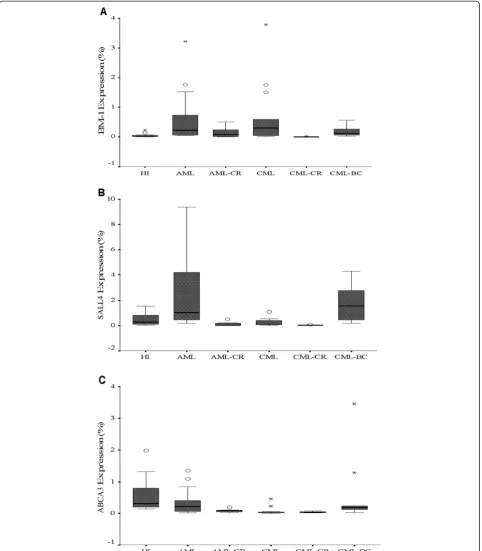
Background and methods:In order to characterize the expression pattern ofSALL4,BMI-1and ABCA3 genes in patients with myeloid leukemia and those who achieved complete remission (CR) after chemotherapy. Real-time PCR was used to determine the expression level of these genes in peripheral blood mononuclear cells from 24 patients with AML, eight patients with AML-CR, 13 patients with CML in the chronic phase (CML-CP), 12 patients with CML in blast crisis (CML-BC), 13 patients with CML-CR and 11 healthy individuals (HI).
Results:Overexpression of theBMI-1gene was found in the AML, CML-CP and CML-BC groups as compared with HI group, while the BMI-1 expression level was lower in patients who achieved CR. In contrast, significantly increasedSALL4expression was only found in AML group, additionally,SALL4expression was lower in the CML-CP and CML-CR groups compared with the HI group, while theSALL4expression level in the CML-BC group was higher and significantly greater than that in the CML-CP and CML-CR groups. Moreover, a positive correlation between the expression ofSALL4 and BMI-1genes was found in samples from most groups. There was no significant difference ofABCA3expression level in AML and CML-BC group in comparison with HI group. Interestingly, theABCA3expression level was significantly decreased in the CML-CP, AML-CR and CML-CR in comparison with the HI group. Moreover, theABCA3expression level in all of the CR groups was lower than that in their corresponding groups.
Conclusions:These results describe the alteredSALL4, ABCA3andBMI-1expression pattern in different phases of myeloid leukemia, which may relate to the development and progression to different diseases.SALL4expression was strongly correlated withBMI-1in most of the myeloid leukemia patient groups, providing a potential link betweenSALL4andBMI-1in leukemogenesis.
Keywords:SALL4gene,BMI-1gene, Real-time PCR, AML, CML
Background
The altered expression of genes, such asWT1,SCL, and Notch1, that play crucial roles in the regulation of hematopoietic progenitor cell proliferation is frequently found in leukemia [1-7]. Increasing data show that the genes involved in hematopoietic stem/progenitor cell (HSPC) proliferation change their expression pattern during leukemogenesis [8].
SALL4 (sal-like protein 4), a SALL gene family mem-ber that is a newly identified zinc-finger transcription
factor, was originally cloned based on its sequence hom-ology to Drosophila spalt (sal) [9-12]. Alternative spli-cing generates two variant forms of human SALL4 mRNA, SALL4A and SALL4B, and each has a different tissue distribution [9,13]. Recently, SALL4 has been shown to play an important role in maintaining ES cell (ESC) pluripotency and self-renewal properties.SALL4is involved in the self-renewal of leukemic initiation and HSPC [14]. Moreover, recent data have shown that SALL4plays an essential role in myeloid leukemogenesis. SALL4 is constitutively expressed in human leukemia cell lines and primary acute myeloid leukemia (AML) cells [9,13]. Transgenic mice that ubiquitously overexpress SALL4B exhibit myelodysplastic syndrome (MDS)-like symptoms and subsequently develop transplantable AML
* Correspondence:[email protected]
1Institute of Hematology, Jinan University, Guangzhou 510632, China 4
Key Laboratory for Regenerative Medicine of Ministry of Education, Jinan University, Guangzhou 510632, China
Full list of author information is available at the end of the article
[9,13], while SALL4 knockdown in leukemia cell lines triggers apoptosis [15].
BMI-1 is a member of the polycomb group of proteins, and it was initially identified inDrosophilaas a repressor of homeotic genes [9,16-18]. The BMI-1 gene was initially isolated as an oncogene that cooperates with c-myc in retroviral-induced B and T cell leukemia [19,20]. In humans,BMI-1is highly expressed in purified HSCs, and its expression declines with differentiation [9,21], and it plays an essential role in regulating adult, self-renewing HSPC and leukemia stem cells [9,21-27]. Knockout of theBMI-1 gene in mice results in the pro-gressive loss of all hematopoietic lineages [9,25]. BMI-1 expression appears to be important for the accumulation of leukemic cells. Interestingly, inhibiting tumor stem cell self renewal after BMI-1 deletion can prevent leukemic recurrence. Recently,BMI-1expression has been used as an important marker for predicting MDS develop-ment and the progression to AML [9,28]. BMI-1 overex-pression was also observed in a significant number of nasopharyngeal carcinoma tumors that correlated with advanced tumor progression, invasive stage and poor prognosis [19,29].
BMI-1was recently demonstrated to be a directSALL4 target gene. The induction of SALL4 expression is associated with increased levels of histone H3–K4 and H3–K79 methylation in theBMI-1promoter, indicating a novel connection between SALL4 and polycomb group proteins in leukemogenesis and a mechanism whereby aberrant SALL4 expression can directly alter BMI-1 expression [9].
Moreover,SALL4expression was higher in drug resist-ant primary acute myeloid leukemic patients than those from drug-responsive cases. In addition, SALL4 expression was enriched in the SP when compared to the non-SP counterpart. Recently, it is reported that SALL4could promote the expression of the ABC trans-porter genes, such as ATP binding cassette transtrans-porter A3 (ABCA3), suggesting that SALL4 can contribute to the SP phenotype by regulating the expression of
ABCA3andABCG2[15].
ABCA3 is a member of the ATP-binding cassette
(ABC) family of transport proteins and is required for perinatal respiratory adaptation. Mutations in ABCA3 resulted in fatal neonatal lung disease [30,31].ABCA3is highly expressed in AML and ALL patient samples and its expression is associated with unfavorable clinical treatment outcome. Furthermore, the expression of ABCA3 is enriched in leukemic SP cells and has been linked to multidrug resistance by facilitating lysosomal sequestration of drugs in AML primary cells and cell lines [15,32-35]. RNAi specific for ABCA3 led to a de-crease ofABCA3 expression in T-ALL cell line such as CCRF-CEM and Jurkat cells. Consequently, a significant
sensitization of cells to cytostatic drugs was achieved [35]. Moreover, both pharmacological blockade and the silencing of ABCA3 enhanced susceptibility of target B-cell lymphoma B-cells to anti-CD20 antibody-mediated lysis. Mechanisms of cancer cell resistance to drugs and antibodies are linked in an ABCA3-dependent pathway of exosome secretion [36].
Little is known about the expression pattern of the SALL4, ABCA3 and BMI-1 genes in patients with mye-loid leukemia and patients that achieved complete remis-sion after chemotherapy. In this study, we determined the expression characteristics of the SALL4, ABCA3and
BMI-1 genes in de novo AML and CML and complete
remission samples.
Results
The high amplification efficiency of the BMI-1 and SALL4genes was consistent with that of the β2M refer-ence gene. The PCR products from all of the genes of interest were confirmed using 2.5% agarose gel electro-phoresis followed by sequencing (data not shown). The BMI-1 and SALL4 genes were detected in all of the PBMC samples from the healthy individuals and those with myeloid leukemia.
Higher expression ofBMI-1in AML and CML
BMI-1 overexpression was found in the de novo AML (median: 0.303, p <0.001), CML-CP (median: 0.295, p=0.006), and CML-BC (median: 0.109, p=0.01) groups in comparison with the HI group (median: 0.027); the BMI-1 expression level in the AML-CR group (median: 0.078) was not significantly different compared with the HI group (p=0.322). Interestingly, the BMI-1 expression level in the CML-CR group (median: 0.003) was signifi-cantly lower than that in the HI (p< 0.0001) and CML groups (p< 0.0001).
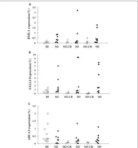
We next analyzed BMI-1 expression in different AML subtypes, including M2 (median: 0.400), M3 (median: 0.156) and M5 (median: 0.295), and all had a signifi-cantly higher expression level compared to the HI group (p=0.003, p=0.01 and p=0.004, respectively), while the BMI-1 expression level in the M2-CR (median: 0.070) and M3-CR (median: 0.099) groups was not significantly different in comparison to that of the HI group (p=0.514 and p=0.361, respectively) (Figures 1 and 2).
1.465; p=0.026), it was lower in the AML-CR (median: 0.026; p=0.026), M2-CR (median: 0.105; p=0.151) and M3-CR (median: 0.023; p=0.037) groups. Interestingly, the level ofSALL4 expression in the CML-CP (median: 0.093; p=0.213) and CML-CR groups (median: 0.025;
p<0.0001) was lower in comparison with the HI group, and the increasedSALL4expression level in the CML-BC group (median: 1.563) was significantly higher than that in the CML-CP (p=0.001) and CML-CR (p<0.0001) groups. The SALL4 expression level in all of the CR groups was
lower than that in their corresponding groups i.e., AML vs. AML-CR (p< 0.0001), M2 vs. M2-CR (p=0.017), M3 vs. M3-CR (p=0.007) and CML vs. CML-CR (p=0.011) (Figures 1 and 2).
Low expression ofABCA3in myeloid leukemia
The expression level ofABCA3seemed low in the differ-ent myeloid leukemia in comparison with the HI group.
There was no significant difference of ABCA3 expres-sion level in AML (median: 0.211; p=0.136), CML-BC (median: 0.174; p=0.097) and the different AML sub-types M2 (median: 0.242; p=0.215), M3 (median: 0.195; p = 0.186) and M5 (median: 0.221; p=0.364) in compari-son with the HI group (median: 0.313). While the ABCA3 expression level was significantly decreased in the CML-CP (median: 0.025; p <0.0001), AML-CR 0
0.5 1 1.5 2 2.5 3 3.5
HI M2 M2-CR M3 M3-CR M5
B
M
I-1
e
x
pr
essi
o
n
(%
)
A
0 1 2 3 4 5 6 7 8 9 10
HI M2 M2-CR M3 M3-CR M5
S
A
LL4
Ex
p
res
si
o
n
(%
)
B
0 0.5 1 1.5 2 2.5
HI M2 M2-CR M3 M3-CR M5
AB
CA3 e
x
p
re
ss
ion
(%
)
C
(median: 0.078; p=0.0011) and CML-CR (median: 0.037; p <0.0001) in comparison with the HI group. Moreover, the ABCA3 expression level in all of the CR groups was lower than that in their corresponding groups i.e., AML vs. AML-CR (p=0.042) (Figures 1 and 2).
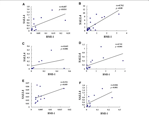
Correlation of relative expression ofBMI-1,SALL4and
ABCA3in myeloid leukemia
Correlation analysis of the relative expression levels of
BMI-1 and SALL4,SALL4 and ABCA3 was performed
using Spearman’s rank correlation analysis of the HI, AML and CML groups. A positive expression correl-ation level forBMI-1andSALL4genes was found in the HI (rs=0.687, p=0.014), AML (rs=0.762, p< 0.0001), M3 (rs=0.994, p< 0.0001), CML-CP (rs=0.742, p=0.004), CML-BC (rs=1=0.846, p=0.001), M2-CR (rs=1, p < 0.0001) and CML-CR (rs=0.534, p=0.049) groups. However, there was no significant correlation for both genes in
the M2 (rs=0.381, p=0.352), M5 (rs=0.643, p=0.086), M3-CR (rs=0.40, p= 0.060) and AML-CR (rs=0.643, p=0.086) groups (Figure 3). A similar result with a positive expression correlation level for genes SALL4 and ABCA3 was found in the HI (rs=0.783, p=0.004), while there was no significant correlation between the expression levels of both genes in all myeloid leukemia groups (Figure 4).
Discussion
BMI-1 and SALL4 are stem cell genes that modulate stem cell pluripotency and play a role in leukemogenesis. Dysregulated expression of both genes may have a cooperative effect in leukemogenesis [37]. Patients with RA and RARS who have a higher percentage of BMI-1+ cells showed disease progression to RAEB, suggesting thatBMI-1is a novel molecular marker that predicts the progression and prognosis of MDS [28]. In this study,
0 0.2 0.4 0.6 0.8 1 1.2 1.4 1.6 1.8
0 0.05 0.1 0.15 0.2 0.25
BMI-1
SALL4
rs=0.687 p=0.014
0 2 4 6 8 10 12 14
0 1 2 3 4
BMI-1
SALL4
rs=0.762 p=0.00
0 0.1 0.2 0.3 0.4 0.5 0.6
0 0.2 0.4 0.6
BMI-1
SALL4
rs=0.643 p=0.086
0 0.2 0.4 0.6 0.8 1 1.2
0 1 2 3 4
BMI-1
SALL4
rs=0.742 p=0.004
0 0.01 0.02 0.03 0.04 0.05 0.06 0.07
0 0.005 0.01 0.015 0.02
BMI-1
SALL4
rs=0.534 p=0.049
0 0.2 0.4 0.6 0.8 1 1.2 1.4 1.6 1.8
0 0.1 0.2 0.3
BMI-1
SALL4
rs=0.846 p=0.001
A B
C
F D
E
we analyzed BMI-1 and SALL4 expression in primary AML and CML at diagnosis and those in complete remission.
It has been shown that BMI-1 overexpression occurs in a variety of cancers including several types of leuke-mias and lymphomas [38]. In this study, BMI-1 was found to be overexpressed in AML and chronic phase CML patient groups; and its expression level was lower in patients who achieved complete remission. Similar results were reported by Sawa, M et al. who found that moderate to high BMI-1 expression was detected in AML patients, and the AML-M0 subtype showed higher relative expression of the BMI-1 transcript [39]. In addition, Merkerova, M et al. demonstrated that BMI-1 and its significantly higher BMI-1 transcript level in CML cells seem to play a secondary role in CML transformation [40]. Our results also indicate that a decreased BMI-1 expression level is associated with complete disease remission. Interestingly, the BMI-1 expression level in the CML-BC group appeared to be low in comparison with the de novo CML group, although the difference was not significant. Further investigation is needed using a larger patient cohort to extend our findings. Preliminary results indicate thatBMI-1may have potential as a therapeutic target for myeloid leukemia. It has been reported thatBMI-1 deple-tion by RNA interference leads to reduced U937 cell growth and proliferation and increased apoptosis [41],
and an antisense BMI-1 gene can inhibit the growth of K562 cells and upregulate p16 expression in K562 cells [42].
Using immunohistochemistry and real-time PCR, SALL4 was demonstrated to be constitutively expressed in human primary acute myeloid leukemia [13]. In this study, we found thatSALL4was overexpressed in differ-ent primary AML subtypes, and its expression was lower in the AML-CR patient group. These results are similar to the findings of Jeong, HW et al. who showed that AML patients who responded to treatment had decreas-ing SALL4 expression throughout the course of treat-ment, while AML patients with disease relapse or drug resistance had increasing SALL4 expression, which was correlated with disease progression [15]. Interestingly, unlike theSALL4expression characteristics in AML, the
SALL4 expression level in the CML-CP and CML-CR
groups was lower. Moreover, theSALL4expression level in patients with chronic phase CML was significantly lower than that in the CML-CR group. There is no dir-ect evidence demonstrating the SALL4 expression level in CML-CP and comparing the expression feature to healthy individuals; however, Lu and colleagues have found that the SALL4 protein was overexpressed in CML samples in blast crisis but not those in chronic phase by FACS [37]. Our results also demonstrated that SALL4 expression was higher in the CML-BC group in comparison with the CML-CP and CML-CR groups;
0 0.5 1 1.5 2 2.5
0 0.5 1 1.5 2
SALL4
ABCA3
rs=0.783
p=0.0004
0 0.2 0.4 0.6 0.8 1 1.2 1.4 1.6
0 2 4 6 8 10
SALL4
ABCA3
rs=-0.44
p=0.837
0 0.1 0.2 0.3 0.4 0.5
0 0.5 1 1.5
SALL4
ABCA3
rs=-0.121
p=0.694
0 0.5 1 1.5 2 2.5 3 3.5 4
0 2 4 6
SALL4
ABCA3
rs=-0.119
p=0.713
A B
C D
however, there was no significant difference in compari-son with the HI group. Is it possible thatSALL4 is pre-ferentially expressed in leukemic blasts? These results are similar to a report by Cui W et al. who demonstrated that only precursor B-cell lymphoblastic leukemias/ lymphomas and AML had detectableSALL4in neoplas-tic tissues [43]. The differentSALL4expression patterns in AML and CML suggest that these two disease entities may have different biological characteristics and/or mechanisms of leukemogenesis, at least for the associ-ation between SALL4and pathogenesis. However, there are not reports comparing the data of SALL4 expression level in CML-CP to healthy individuals, it is difficult to evaluate the significance of this finding. Recently, research from Zhu et al. showed that hematopoietic transcription factor PU.1 expression was significantly lower in newly diagnosed APL patient samples as com-pared to normal hematopoietic cells, which may relate to the expression level of PML-RARα, and they found that suppression of PU.1 expression occurred concur-rently with PML-RARα expression, the authors sug-gested that low PU.1 expression in APL patients is required for disease initiation and progression [44]. This finding might provide a direction in farther analysis the correlation ofSALL4with BCR-ABL in the pathogenesis of CML and to address this question.
In principle, the BMI-1 and SALL4 gene expression level should be positively correlated in stem cells [9]. Little is known about the expression pattern and differ-ences in the SALL4 and BMI-1 genes in patients with AML and CML. In this study, we analyzed the correl-ation between the relative expression levels of BMI-1 and SALL4. A positive expression level correlation was found for both genes in HI, AML, chronic phase CML, CML-BC and CML-CR patient groups; however, there was no significant correlation between these genes in patients with AML-CR, leaving their role in this group an open question. These results indicate that a positively correlated expression pattern is a common feature in patients with myeloid leukemia and healthy individuals, and both genes may cooperate during cell proliferation and differentiation.
Based on the different expression features ofSALL4in AML and CML, we further analyzed its regulating gene ABCA3,which is a member of the ATP-binding cassette (ABC) family of transport proteins [30,31]. Unlike the description by Wult, Norwood and Steinbach groups, who showed that ABCA3 is highly expressed in acute meyloid leukemia samples and is associated with unfavorable clinical treatment outcome [24,33,45], in the present study, lower expression level of ABCA3 was found not only in AML but also in CML groups, espe-cially in CML-CP and CR groups. Moreover, the expres-sion level of ABCA3 lost the correlation with SALL4
expression in leukemia patients. To determine whether these results relate to favorable clinical outcome, fur-ther investigation is needed. Additionally, detection of ABCA2, ABCB2 and ABCC10, which were found over-expressed in childhood AML, may be worthy to build the gene regulation network in proliferation of myeloid leukemia cells.
In conclusion, we determined the expression charac-teristics of the SALL4, ABCA3 and BMI-1genes in dif-ferent phases of AML and CML. Further studies will be needed to determine whether BMI-1 and SALL4 are novel therapeutic targets for leukemic stem/initiation cells in primary myeloid leukemia.
Methods
Samples
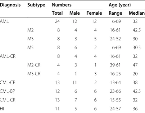
Twenty-four newly diagnosed and untreated patients with AML, eight cases with AML in complete remission (AML-CR), thirteen newly diagnosed and untreated patients with CML in chronic phase, 13 cases with CML-CR, and 12 cases with CML in blast crisis (CML-BC),were recruited, the details of the samples was listed in Table 1. The diagnoses of all patients were based on cytomorphology, immunohistochemistry, and cytoimmu-nological and cytogenetic analysis. Peripheral blood mononuclear cells (PBMCs) from 11 healthy individuals (HI) served as controls. Peripheral blood was collected by heparin anticoagulation, and PBMCs were separ-ated using the Ficoll-Hypaque gradient centrifugation method. All procedures were conducted in accordance with the guidelines of the Medical Ethics committees of the health bureau of Guangdong Province, China.
RNA extraction and cDNA synthesis
RNA was extracted using the Trizol kit (Invitrogen, Carlsbad, CA, USA) and then reverse-transcribed into first-strand cDNA using random hexamer primers and
Table 1 The details of samples used in study Diagnosis Subtype Numbers Age (year)
Total Male Female Range Median
AML 24 12 12 6-69 32
M2 8 4 4 16-61 42.5
M3 8 3 5 24-52 30
M5 8 6 2 6-69 30.5
AML-CR 8 4 4 16-61 32
M2-CR 4 3 1 39-61 47
M3-CR 4 1 3 16-25 20
CML-CP 13 11 2 13-64 38
CML-BP 12 6 6 23-66 42.5
CML-CR 13 7 6 15-55 32
the Superscript II reverse transcriptase Kit (Invitrogen) according to the manufacturer’s instructions.
Real-time quantitative reverse transcription–polymerase chain reaction (qRT–PCR)
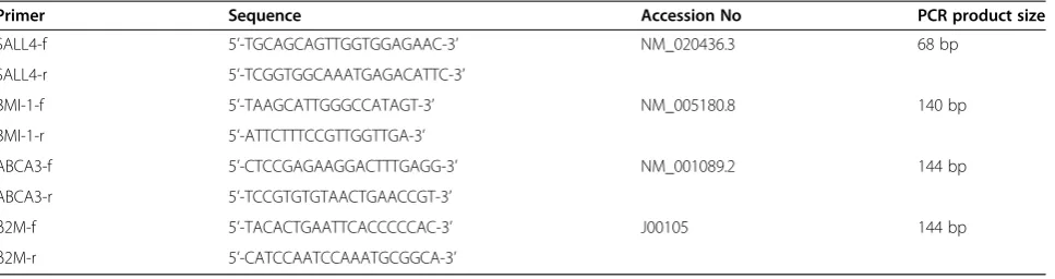
The expression levels ofBMI-1,SALL4,ABCA3 and the β2-microglobulin (β2-MG) reference gene were deter-mined by SYBR Green I real-time PCR. Briefly, PCR was performed in a 25 μL total volume containing 1 μL of cDNA, 9 μL of 2.5× SYBR Green mix (Tiangen, Beijing, China), and 10μmol/L primer pairs. After initial denaturation at 95°C for 2 min, 45 cycles consisting of the following procedure were performed using an MJ Research DNA Engine Opticon 2 PCR cycler (BIO-RAD, USA): 15 s at 95°C, 40 s at 64°C forβ2MandBMI-1, 60° C forSALL4, and 63°C forABCA3. The relative amounts of the genes of interest and the β2-MG reference gene were measured in two independent assays. The data are presented as the relative expression of the genes of inter-est relative to the internal control gene as determined by the 2(−ΔΔCT)method [1-3,5,46]. Additionally, the specific amplification of the PCR products was analyzed by melt-ing curve analysis and agarose gel electrophoresis. The primers used for real-time PCR for all gene amplifica-tions were synthesized by Shanghai Biological Engineer-ing Technology Services Co., Ltd. (Table 2). RT–PCR, for theBMI-1,SALL4andABCA3genes, was performed using the same primers described above, and the PCR products were sent to Shanghai Invitrogen Biotechnol-ogy Co. for DNA sequence analysis.
Statistical analyses
Differences in mRNA expression between two groups were analyzed using the Mann–WhitneyUtest. Data are presented as median. Spearman’s rank correlation ana-lysis was used to analyze theSALL4,BMI-1 andABCA3 mRNA levels in different samples using the SPSS 11.5 statistical software. Differences were considered statisti-cally significant atP< 0.05.
Competing interests
The authors declare that they have no potential conflicts of interest.
Authors’contributions
YQL and YPM contributed to concept development and study design. QS, SCL, SHC and YM performed the real-time PCR. JYH, LJY, BL, XLW, JCY were responsible for collection of clinical data. YQL and QS coordinated the study and helped drafting the manuscript. All authors read and approved the final manuscript.
Acknowledgements
This work was supported by Grants from National Natural Science Foundation of China (no. 81270604), the Fundamental Research Funds for the Central Universities (No. 21612116) and in part through NIH grant R01HL087948 (Y.M.).
Author details
1Institute of Hematology, Jinan University, Guangzhou 510632, China. 2
Department of Surgery,BST-9, School of Medicine, Stony Brook University, Stony Brook, NY 11794, USA.3Department of Pathology, BST-9, School of
Medicine, Stony Brook University, Stony Brook, NY 11794, USA.4Key Laboratory for Regenerative Medicine of Ministry of Education, Jinan University, Guangzhou 510632, China.
Received: 15 September 2012 Accepted: 10 October 2012 Published: 15 October 2012
References
1. Stams WAG, den Boer ML, Beverloo HB, Meijerink JPP, Stigter RL, van Wering ER, Janka-Schaub GE, Slater R, Pieters R:Sensitivity to L-asparaginase is not associated with expression levels of asparagine synthetase in t(12;21)+pediatric ALL.Blood2003,101(7):2743–2747. 2. Miglino M, Colombo N, Pica G, Grasso R, Clavio M, Bergamaschi M, Ballerini
F, Ghiso A, Ghiggi C, Mitscheunig L, Beltrami G, Cagnetta A, Vignolo L, Lucchetti MV, Aquino S, Pierri I, Sessarego M, Carella AM, Gobbi M:WT1 overexpression at diagnosis may predict favorable outcome in patients with de novo non-M3 acute myeloid leukemia.Leuk Lymphoma2011, 52(10):1961–1969.
3. Cardoso BA, de Almeida SF, Laranjeira AB, Carmo-Fonseca M, Yunes JA, Coffer PJ, Barata JT:TAL1/SCL is downregulated upon histone deacetylase inhibition in T-cell acute lymphoblastic leukemia cells.Leukemia2011, 25(10):1578–1586.
4. Aster JC, Blacklow SC, Pear WS:Notch signalling in T-cell lymphoblastic leukaemia/lymphoma and other haematological malignancies.J Pathol
2011,223(2):262–273.
5. Lin C, Zheng H, Wang C, Yang L, Chen S, Li B, Zhou Y, Tan H, Li Y: Mutations increased overexpression of Notch1 in T-cell acute lymphoblastic leukemia.Cancer Cell Int2012,12:13.
6. Takahashi S:Current findings for recurring mutations in acute myeloid leukemia.J Hematol Oncol2011,4:36.
7. Braoudaki M, Papathanassiou C, Katsibardi K, Tourkadoni N, Karamolegou K, Tzortzatou-Stathopoulou F:The frequency of NPM1 mutations in childhood acute myeloid leukemia.J Hemotol Oncol2010,3:41. Table 2 Primer sequences used for real-time PCR
Primer Sequence Accession No PCR product size
SALL4-f 5’-TGCAGCAGTTGGTGGAGAAC-3’ NM_020436.3 68 bp
SALL4-r 5’-TCGGTGGCAAATGAGACATTC-3’
BMI-1-f 5’-TAAGCATTGGGCCATAGT-3’ NM_005180.8 140 bp
BMI-1-r 5’-ATTCTTTCCGTTGGTTGA-3’
ABCA3-f 5’-CTCCGAGAAGGACTTTGAGG-3’ NM_001089.2 144 bp
ABCA3-r 5’-TCCGTGTGTAACTGAACCGT-3’
β2M-f 5’-TACACTGAATTCACCCCCAC-3’ J00105 144 bp
8. Yong AS, Stephens N, Weber G, Li Y, Savani BN, Eniafe R, Keyvanfar K, Kurlander R, Rezvani K, Barrett AJ:Improved outcome following allogeneic stem cell transplantation in chronic myeloid leukemia is associated with higher expression of BMI-1 and immune responses to BMI-1 protein.
Leukemia2011,25(4):629–637.
9. Yang JC, Chai L, Liu F, Fink LM, Lin P, Silberstein LE, Amin HM, Ward DC, Ma YP:Bmi-1 is a target gene for SALL4 in hematopoietic and leukemic cells.Proc Natl Acad Sci USA2007,104(25):10494–10499.
10. Kohlhase J, Hausmann S, Stojmenovic G, Dixkens C:Bink Schulz-Schaeffer W, Altmann M, Engel W: SALL3, a new member of the human spalt-like gene family, maps to 18q23.Genomics1999,62(2):216–222.
11. Al-Baradie R, Yamada K, St Hilaire C, Chan WM, Andrews C, McIntosh N, Nakano M, Martonyi EJ, Raymond WR, Okumura S, Okihiro MM, Engle EC: Duane radial ray syndrome (Okihiro syndrome) maps to 20q13 and results from mutations in SALL4, a new member of the SAL family.Am J Hum Genet2002,71(5):1195–1199.
12. Yang J, Aguila JR, Alipio Z, Lai R, Fink LM, Ma Y:Enhanced self-renewal of hematopoietic stem/progenitor cells mediated by the stem cell gene Sall4.J Hematol Oncol2011,4:38.
13. Ma YP, Cui W, Yang JC, Qu J, Di CH, Amin HM, Lai R, Ritz J, Krause DS, Chai L:SALL4, a novel oncogene, is constitutively expressed in human acute myeloid leukemia (AML) and induces AML in transgenic mice.
Blood2006,108(8):2726–2735.
14. Schuster JA, Stupnikov MR, Ma G, Liao WB, Lai R, Ma YP, Aguila JR: Expansion of hematopoietic stem cells for transplantation: current perspectives.Exp Hematol Oncol2012,1:12.
15. Jeong HW, Cui W, Yang YY, Lu JY, He J, Li AL, Song D, Guo Y, Liu BH, Chai L: SALL4, a stem cell factor, affects the side population by regulation of the ATP-binding cassette drug transport genes.PLoS One2011,6(4):e18372. 16. Zink D, Paro R:Drosophila Polycomb-group regulated chromatin inhibits
the accessibility of a trans-activator to its target DNA.EMBO J1995, 14(2):5660–5671.
17. Kennison JA:The Polycomb and trithorax group proteins of Drosophila: trans-regulators of homeotic gene function.Annu Rev Genet1995, 29:289–303.
18. Franke A, DeCamillis M, Zink D, Cheng N, Brock HW, Paro R:Polycomb and polyhomeotic are constituents of a multimeric protein complex in chromatin of Drosophila melanogaster.EMBO J1992,11(8):2941–2950. 19. Wu J, Hu D, Yang G, Zhou JY, Yang CF, Gao Y, Zhu ZY:Down-regulation of
BMI-1 cooperates with artemisinin on growth inhibition of nasopharyngeal carcinoma cells.J Cell Bochem2011,112(7):1938–1948.
20. Haupt Y, Alexander WS, Barri G, Klinken SP, Adams JM:Novel zinc finger gene implicated as myc collaborator by retrovirally accelerated lymphomagenesis in E mu-myc transgenic mice.Cell1991,65(5):753–763. 21. Park IK, Qian D, Kiel M, Becker MW, Pihalja M, Weissman IL, Morrison SJ,
Clarke MF:Bmi-1 is required for maintenance of adult self-renewing haematopoietic stem cells.Nature2003,423(6937):302–305.
22. Raaphorst FM:Self-renewal of hematopoietic and leukemic stem cells: a central role for the Polycomb-group gene Bmi-1.Trends Immunol2003, 24(10):522–524.
23. Dick JE:Stem cells: Self-renewal writ in blood.Nature2003,423(6937):231–233. 24. Ohta H, Sawada A, Kim JY, Tokimasa S, Nishiguchi S, Humphries RK, Hara J, Takihara Y:Polycomb group gene rae28 is required for sustaining activity of hematopoietic stem cells.J Exp Med2002,195(6):759–770.
25. Lessard J, Sauvageau G:Bmi-1 determines the proliferative capacity of normal and leukaemic stem cells.Nature2003,423(6937):255–260. 26. van der Lugt NM, Domen J, Linders K, van Roon M, Robanus-Maandag E,
te Riele H, van der Valk M, Deschamps J, Sofroniew M, van Lohuizen M, Bwrns A:Posterior transformation, neurological abnormalities, and severe hematopoietic defects in mice with a targeted deletion of the bmi-1 proto-oncogene.Genes Dev1994,8(7):757–769.
27. Iwama A, Oguro H, Negishi M, Kato Y, Morita Y, Tsukui H, Ema H, Kamijo T, Katoh-Fukui Y, Koseki H, van Lohuizen M, Nakauchi H:Enhanced self-renewal of hematopoietic stem cells mediated by the polycomb gene product Bmi-1.Immunity2004,21(6):843–851.
28. Mihara K, Chowdhury M, Nakaju N, Hidani S, Ihara A, Hyodo H, Yasunaga S, Takihara Y, Kimura A:Bmi-1 is useful as a novel molecular marker for predicting progression of myelodysplastic syndrome and patient prognosis.Blood2006,107(1):305–308.
29. Song LB, Zeng MS, Liao WT, Zhang L, Mo HY, Liu WL, Shao JY, Wu QL, Li MZ, Xia YF, Fu LW, Huang WL, Dimri GP, Band V, Zeng YX:Bmi-1 is a
novel molecular marker of nasopharyngeal carcinoma progression and immortalizes primary human nasopharyngeal epithelial cells.Cancer Res
2006,66(12):6225–6232.
30. Stahlman MT, Besnard V, Wert SE, Weaver TE, Dingle S, Xu Y, von Zychlin K, Olson SJ, Whitsett JA:Expression of ABCA3 in developing lung and other tissues.J Histochem Cytochem2007,55(1):71–83.
31. Saugstad OD, Hansen TW, Rønnestad A, Nakstad B, Tølløfsrud PA, Reinholt F, Hamvas A, Coles FS, Dean M, Wert SE, Whitsett JA, Nogee LM:Novel mutations in the gene encoding ATP binding cassette protein member A3 (ABCA3) resulting in fatal neonatal lung disease.Acta Paediatr2007, 96(2):185–190.
32. Chapuy B, Koch R, Radunski U, Corsham S, Cheong N, Inagaki N, Ban N, Wenzel D, Reinhardt D, Zapf A, Schweyer S, Kosari F, Klapper W, Truemper L, Wulf GG:Intracellular ABC transporter A3 confers multidrug resistance in leukemia cells by lysosomal drug sequestration.Leukemia2008, 22(8):1576–1586.
33. Wulf GG, Modlich S, Inagaki N, Reinhardt D, Schroers R, Griesinger F, Trümper L:ABC transporter ABCA3 is expressed in acute myeloid leukemia blast cells and participates in vesicular transport.
Haematologica2004,89(11):1395–1397.
34. Norwood K, Wang RY, Hirschmann-Jax C, Andreeff M, Brenner MK, Goodell MA, Wulf GG:An in vivo propagated human acute myeloid leukemia expressing ABCA3.Leuk Res2004,28(3):295–299. 35. Efferth T, Gillet JP, Sauerbrey A, Zintl F, Bertholet V, de Longueville F,
Remacle J, Steinbach D:Expression profiling of ATP-binding cassette transporters in childhood T-cell acute lymphoblastic leukemia.Mol Cancer Ther2006,5(8):1986–1994.
36. Aung T, Chapuy B, Vogel D, Wenzel D, Oppermann M, Lahmann M, Weinhage T, Menck K, Hupfeld T, Koch R, Trümper L, Wulf GG:Exosomal evasion of humoral immunotherapy in aggressive B-cell lymphoma modulated by ATP-binding cassette transporter A3.Proc Natl Acad Sci USA2011,108(37):15336–15341.
37. Lu J, Ma Y, Kong N, Alipio Z, Gao C, Krause DS, Silberstein LE, Chai L:Dissecting the role of SALL4, a newly identified stem cell factor, in chronic myelogenous leukemia.Leukemia2011,25(7):1211–1213. 38. Beà S, Tort F, Pinyol M, Puig X, Hernández L, Hernández S, Fernandez PL,
van Lohuizen M, Colomer D, Campo E:BMI-1 gene amplification and overexpression in hematological malignancies occur mainly in mantle cell lymphomas.Cancer Res2001,61(6):2409–2412.
39. Sawa M, Yamamoto K, Yokozawa T, Kiyoi H, Hishida A, Kajiguchi T, Seto M, Kohno A, Kitamura K, Itoh Y, Asou N, Hamajima N, Emi N, Naoe T:BMI-1 is highly expressed in M0-subtype acute myeloid leukemia.Int J Hematol
2005,82(1):42–47.
40. Merkerova M, Bruchova H, Kracmarova A, Klamova H, Brdicka R:Bmi-1 over-expression plays a secondary role in chronic myeloid leukemia transformation.Leuk Lymphoma2007,48(4):793–801.
41. Zhu W, Huang L, Xu XJ, Qian H, Xu WR:Anti-proliferation effect of BMI-1 in U937 cells with siRNA.Int J Mol Med2010,25(6):889–895. 42. Meng XX, Liu WH, Liu DD, Zhao XY, Su BL:Construction of antisense
Bmi-1 expression plasmid and its inhibitory effect on K562 cells proliferation.Chin Med J2005,118(16):1346–1350.
43. Cui W, Kong NR, Ma YP, Amin HM, Lai R, Chai L:Differential expression of the novel oncogene, SALL4, in lymphoma, plasma cell myeloma, and acute lymphoblastic leukemia.Mod Pathol2006,19(12):1585–1592.
44. Zhu X, Zhang H, Qian M, Zhao X, Yang W, Wang P, Zhang J, Wang K:The significance of low PU.1 expression in patients with acute promyelocytic leukaemia.J Hematol Oncol2012,5:22.
45. Steinbach D, Gillet JP, Sauerbrey A, Gruhn B, Dawczynski K, Bertholet V, de Longueville F, Zintl F, Remacle J, Efferth T:ABCA3 as a possible cause of drug resistance in childhood acute myeloid leukemia.Clin Cancer Res
2006,12(14 Pt 1):4357–4363.
46. Huang X, Chen S, Shen Q, Yang L, Li B, Zhong L, Geng S, Du X, Li Y: Analysis of the expression pattern of theBCL11Bgene and its relatives in patients with T-cell acute lymphoblastic leukemia.J Hematol Oncol
2010,3:44.
doi:10.1186/1475-2867-12-42