P R I M A R Y R E S E A R C H
Open Access
Anti-tumor effect of ribavirin in combination with
interferon-
α
on renal cell carcinoma cell lines
in vitro
Lichen Teng
1†, Dexin Ding
1†, Yongsheng Chen
1, Hongshuang Dai
1, Guobin Liu
1, Zhongjie Qiao
1*and Ruihua An
2*Abstract
Background:Ribavirin is an anti-viral drug; however, recent data suggest that it may also be effective in cancer therapy. This study investigated the effect of ribavirin alone or in combination with IFN-αon biological processes: proliferation, apoptosis, and migration of murine (Renca) and human renal carcinoma (RCC) cells (786–0)in vitro. Methods:Renca and 786–0 cells were treated with IFN-α, ribavirin, or a combination of IFN-αand ribavirin at varying concentrations. Cell proliferation was evaluated using CCK-8 assay. Induction of apoptosis and distribution of cell cycle were determined by flow cytometry. The migratory capacity of cells was quantified using a transwell migration assay. The toxic effect of these drugs was examined using MTT assay in HEK-293 cells. ELISA was used to measure IL-10 and TGF-βcontent in the culture supernatants.
Results:Our results showed that both ribavirin alone and in combination with IFN-αcould significantly inhibit the cell proliferation and arrest the cell cycle progress at the G2/M phase. These treatments also inhibited cell migration and IL-10 production, in a concentration-dependent manner, in 786–0 and Renca cells. Moreover, they significantly induced apoptosis of RCC cells and increased TGF-βproduction in concentration-dependent manner. No significant toxic effect was observed in HEK-293 cells. We also found that the effect of combined treatment was more pronounced than that of ribavirin or IFN-αalone. However, the combined effect of the two drugs was not synergistic.
Conclusion:Our findings suggest that ribavirin can negatively affect biological processes of RCC cells. This agent might become a new candidate for the treatment of RCC in the clinical setting.
Keywords:Ribavirin, RCC, Proliferation, Apoptosis, Migration
Background
Renal cell carcinoma (RCC) is one of the cancers most resistant to currently available chemotherapy, radiother-apy, and immunotherapy. Although nephrectomy and nephron-sparing surgery (NSS) are still the mainstream therapies in RCC, satisfactory outcomes are only achieved in patients with localized RCC. A Canadian study has demonstrated that at the time of first diagnosis, 25% of pa-tients with RCC are diagnosed with locally advanced RCC
with lymph node or local organ involvement, and 30% would present with metastasis [1]. Two decades ago, cyto-kine therapy, such as IL-2 or interferon alone or adminis-tered together, were typically used in the initial RCC therapy. The patients with the advanced or metastatic RCC would need a combination of surgery and immuno-therapy. Unfortunately, only a small proportion of such patients show significant and durable response to im-munotherapy. Since the advent of targeted therapy, tyro-sine kinase inhibitors, such as sorafenib and sunitinib, have attracted worldwide attention, bringing new hope to RCC patients. However, target therapy rarely gives a complete response in patients with localized or metastatic RCC. In addition, because of the high cost and severe side effects of such interventions, these drugs have not been widely used in China.
* Correspondence:zhongjieqiao@hotmail.com;504587957@qq.com †Equal contributors
1Department of Urology, The Affiliated Tumor Hospital, Harbin Medical
University, No. 150 Haping Road, Harbin city, Heilongjiang Province 150081, China
2
Department of Urology, The First Affiliated Hospital, Harbin Medical University, No. 31 Youzheng Street, Harbin city, Heilongjiang Province 150080, China
regulatory T cells and reverse the suppression of T effector cells [4]. The study suggests that ribavirin might exert a similar immunomodulatory effect in anti-cancer therapies. Some studies have shown that ribavirin can inhibit the ac-tivity of oncogenic eukaryotic translation-initiation factor eIF4E [5-8]. All these data have triggered our renewed interest in the potential effect of ribavirin on cancer cells.
Interferon-α(IFN-α) has shown anti-tumor activity in a variety of solid tumors and has been approved for RCC treatment. However, the available evidence demonstrates that IFN-α only produces modest benefits in unselected patients; randomized clinical trials show only a small sur-vival benefit [9,10]. This limited efficacy of IFN-α treat-ment has narrowed its application as a single agent in patients with RCC. Some investigators have even con-cluded that subcutaneous IFN-α should not be recom-mended for patients with metastatic RCC. However, it is worth considering combining IFN-α with other agents. This approach might enhance the anti-tumor effect of IFN-α in RCC. For example, combined ribavirin and IFN-α treatment might have a synergistic effect in the management of RCC. This study investigated the inhibi-tory effect of ribavirin and IFN-αon human and mouse RCC cell linesin vitro.
Results
The effect of IFN-α, ribavirin, or a combination of IFN-α and ribavirin on the proliferation of renca and 786–0 cells
Renca and 786–0 cells were treated with IFN-α, ribavi-rin, or a combination of IFN-α and ribavirin for 72 h. We observed significant decrease in OD values with increasing concentration of IFN-α (0, 20, 100, 500, 1000 IU/ml) or ribavirin (0, 100, 300, 500, 1000μM) in both cell lines (p < 0.05). These data demonstrate that the cell proliferation can be suppressed by IFN-αor riba-virin in a concentration-dependent manner. To investigate whether combined treatment can have a synergistic effect on the proliferation of these cells, the cell cultures were incubated with IFN-αand ribavirin at different concen-trations (IFN-α at 500 or 1000 IU/ml and ribavirin at 500 or 1000μM). We observed that, in comparison with ribavirin-only treatment, combined treatment signifi-cantly inhibited the proliferation of Renca and 786–0 cells in a concentration-dependent manner (p < 0.05). However, for both cell lines, the combined treatment
dence interval (CI) values showed an additive inhibitory effect when RCC cell lines were treated with IFN-αand ribavirin.
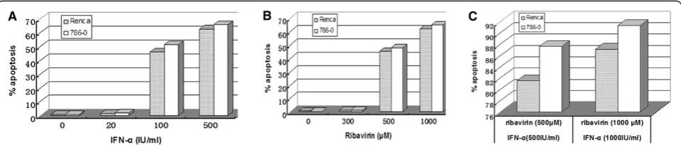
The effect of IFN-αalone, ribavirin alone, or a combination of IFN-αand ribavirin on apoptosis of human and murine RCC cells
Our results showed that IFN-αor ribavirin alone can in-duce apoptosis of RCC cells. However, the effect at lower concentrations of those agents was slight; only 1–2% apoptosis was observed in the cells treated with 20 IU/ml IFN-αor 300μM ribavirin. With higher concentrations of IFN-α or ribavirin, we observed significantly increasing apoptosis rate, to a maximum of 60–70% at 500 IU/ml of IFN-αor 1000μM ribavirin (Figure 2A and B). Thus, the effect of these agents on apoptosis was concentration-dependent. Interestingly, higher percentages of apoptotic cells (80–92%) were observed in RCC cells treated with a combination of IFN-αand ribavirin at high concentration levels (Figure 2C). CompuSyn analysis showed that com-bined treatment with IFN-αand ribavirin had an additive effect on apoptosis in RCC cells.
The effect of ribavirin and IFN-αon 786–0 cell cycle
Apart from the inhibition of proliferation and induction of apoptosis in RCC cells, we also investigated the effect of ribavirin and IFN-α on 786–0 cell cycle. Our results, shown in Figure 3A and B, demonstrate that the per-centages of 786–0 cells in the G0/G1 and S phase were similar in treated and untreated samples. With increasing concentration, however, the percentage of 786–0 cells in the G2/M phase decreased significantly, from 25.52% to 13.95%. The percentage of cells in the G2/M phase was lowest (13.95%) after treatment with 500μM of ribavirin and 500 IU/ml of IFN-α. These results suggest that riba-virin and IFN-αinduce the cell cycle arrest at the G2/M phase. CompuSyn analysis revealed an additive effect of the two agents on the cell cycle arrest.
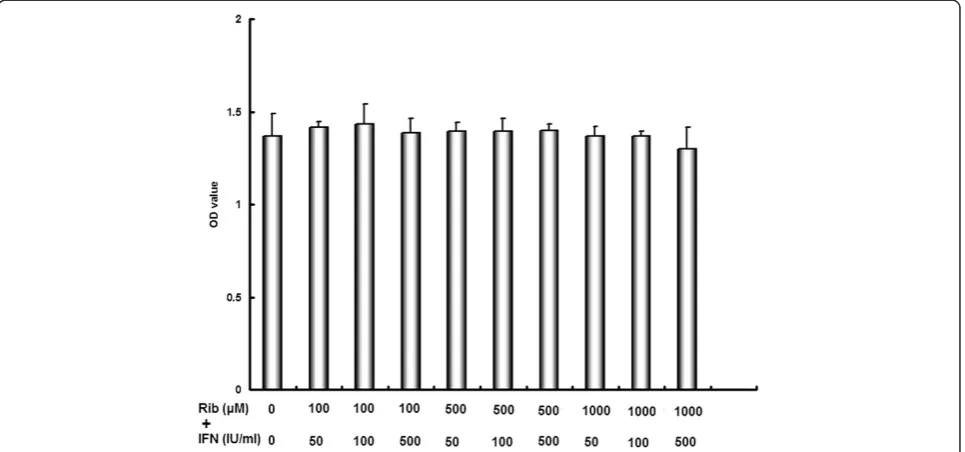
Cell cytotoxicity of ribavirin and IFN-αin the human embryonic kidney cell line (HEK-293)
HEK-293 cells was not significantly affected by com-bined treatment with different concentrations of riba-virin and IFN-α. The results, shown in the Figure 4, demonstrated that this treatment did not have signifi-cant cytotoxic effect in the normal kidney cell line.
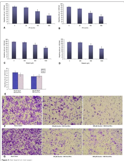
The effect of IFN-αalone, ribavirin alone, or a
combination of IFN-αand ribavirin on RCC cell migration
The transwell assay showed a significant decrease in cell migration for the cells incubated with IFN-αin compari-son with untreated cells, for both Renca and 786–0 cell lines. This effect was concentration-dependent for both cell lines (p < 0.05) (Figure 5A and B). The treatment with ribavirin significantly inhibited cell migration in both cell lines in a similar manner (p < 0.05) (Figure 5C and D), but the effect of ribavirin on cell migration was weaker than that of IFN-α. Surprisingly, after combined IFN-αand ribavirin treatment, fewer cells (50–70) under-went migration (p < 0.05) (Figure 5E-G). The results showed that both agents exerted a potent effect on cell migration. However, in comparison with the results for the combined treatment with 500 μM of ribavirin and 500 IU/ml of IFN-α, increasing IFN-α concentration
caused no further decrease in the number of migrating cells (in both cell lines, p > 0.05).
The effect of IFN-αalone, ribavirin alone, or combined treatment on IL-10 and TGF-βsecretion in supernatant of RCC cells
IL-10 concentrations in supernatants of Renca and 786– 0 cells treated with ribavirin at different concentrations were significantly lower (p < 0.05) than in the superna-tants from untreated cultures. These results demonstrate that ribavirin can inhibit IL-10 secretion in RCC cells in a concentration-dependent manner (Figure 6A). Simi-larly, IFN-α decreases IL-10 secretion in both treated RCC cell lines. However, the treatment with a combin-ation of IFN-α and ribavirin at higher concentration levels did not inhibit IL-10 secretion more than single-agent treatments (Figure 6A and B). In contrast, there was a trend towards increased TGF-βproduction. When TGF-β levels were measured with ELISA, we observed that both IFN-α and ribavirin on their own significantly increased TGF-βproduction in a concentration-dependent manner (in both Renca and 786–0 cells, p < 0.05). The combined treatment did not significantly increase TGF-β
Figure 1The effect of IFN-αand ribavirin alone and combined IFN-αand ribavirin treatment on the proliferation of the murine and human RCC cells. (A)Suppression of proliferation induced by IFN-αalone or in combination with ribavirin.(B)Suppression of proliferation induced by ribavirin alone or in combination with IFN-α. C1: combination of 500 IU/ml of IFN-αand 500μM ribavirin. C2: 1000 IU/ml of IFN-α and 1000μM ribavirin. The suppression was significant in the range of 20–1000 IU/ml of IFN-αor 100–1000μM ribavirin. (*indicates a significant difference in comparison with controls).
secretion. We also found that the increase in TGF-β pro-duction caused by IFN-αor ribavirin was more substantial in 786–0 cells than that in Renca cells (Figure 6C and D).
Discussion
The aim of this study was to evaluate the direct effect of ribavirin on RCC cells and the possibility that this agent might become a new candidate for RCC therapy. Ribavi-rin is a classic anti-viral agent and has been used for the treatment of various viral diseases for a long time. Previous studies have demonstrated that IFN-αalone or in combin-ation with other agents can directly inhibit the growth of cancer cells. However, up to now, ribavirin has not been used to treat RCC [11,12]. Ourin vitroexperiments revealed that ribavirin could directly inhibit proliferation
and migration of murine and human RCC cells, enhance their apoptosis, and arrest cell cycle progression at the G2/M phase. The effect of ribavirin on RCC cells was concentration-dependent. Our results also showed that ribavirin inhibited IL-10 production and increased TGF-β secretion. More importantly, combined treatment with IFN-αand ribavirin had an additive effect on the prolifera-tion, migraprolifera-tion, and induction of apoptosis. In this study, we did not observe significant toxic effects on normal cells. Thus, our results suggest that ribavirin might be a novel agent for the treatment of RCC.
The direct anti-tumor effect of ribavirin cells certainly deserves further investigation. Increasing ribavirin con-centration significantly increased the efficacy of its anti-cancer properties in vitro. We found that ribavirin (at
Figure 3Combined treatment with ribavirin and IFN-αaffected the RCC cell cycle. (A)Combination of 500 IU/ml of IFN-αand 500μM ribavirin blocked the G2/M phase cell cycle transition of 786–0 cells, in comparison with untreated control 786–0 cells. The percentage of 786–0 cells in the G0/G1 phase increased significantly to 58.32% but the percentage of cells in the G2/M phase decreased to 13.95%.(B)Quantitative analysis of the cell cycle distribution of 786–0 cells treated with ribavirin alone, IFN-αalone, or a combination of ribavirin and IFN-α(* < 0.05 versus control group).
cell migration. The level of transporter expression in RCC cells and intracellular levels of ribavirin and its metabolites are likely to be interrelated. It has been suggested that ri-bavirin is taken up by nitrobenzylthioinosine-sensitive (es)-nucleoside transporters and that the concentrative nucleoside transporters and sodium co-transport could be important at low ribavirin concentrations [13,14]. High intracellular levels of ribavirin and its metabolites also de-pend on the levels of ribavirin in the extracellular media [15,16]. Detailed studies of ribavirin metabolism and its transport processes in the RCC cells, which might modu-late intracellular levels of ribavirin, will be important for understanding the exact mechanisms of its action.
Ribavirin may be a pivotal combination partner in en-hancing the efficacy of IFN-α-based therapy. Here, we demonstrated the additive anti-tumor effect of ribavirin in combination with IFN-α. A study of an anti-viral therapy has concluded that ribavirin enhances specific interferon-sensitive gene expression by amplifying the IFN-αJAK/ STAT pathway; it has shown that STAT1 and STAT3
alone [17]. Another possible mechanism is that ribavirin stimulates ERK1/2 and subsequently promotes p53 ac-tivity [18]. In addition, ribavirin upregulates the expres-sion of IFN-a receptor in hepatocytes and augments interferon-stimulated gene induction [19,20]. These mechanisms are at least partly supported by the results of our study. However, further investigation should be performed to understand the mechanisms by which the combination of ribavirin and IFN-αexerts a potent anti-tumor effect.
It has been reported that ribavirin has anti-tumor properties in different types of malignant tumors, such as head and neck squamous cell carcinoma (HNSCC) cell line FaDu, in breast cancer and AML patients [15,20,21]. In our study, we demonstrated for the first time that both murine and human RCC cell lines can re-spond to the treatment with ribavirin alone or a com-bination of ribavirin and IFN-α. These results are consistent with previously reported data. As an onco-gene, eukaryotic translation-initiation factor 4E (eIF4E)
is widely overexpressed in various cancers [22]. eIF4E, which is an element of the mammalian target of rapamy-cin (mTOR) pathway, has been considered an important target for ribavirin anti-tumor activity [21,23]. Thus, the direct effect of ribavirin on RCC cells might be associated with eIF4E. Ribavirin might also enhance the efficacy of rapamycin analogues everolimus and temsirolimus in the treatment of renal cell carcinoma.
Apart from a direct effect on RCC cells, our study also confirmed that ribavirin reduced IL-10 production and increased TGF-β secretion, thus exerting an immuno-modulatory effect in RCC-cell microenvironment. Our previous studies have indicated that IL-10, a negatively immunomodulatory cytokine, had the ability to expand regulatory T cells (Tregs) and induce their conversion [24,25]. However, a recent clinical study has demon-strated that high TGF-β1 mRNA levels in peripheral blood in metastatic RCC patients are independently asso-ciated with favorable progression-free survival and overall survival. Thus, unlike IL-10, TGF-β appears to have an immune-promoting function [26]. More importantly, riba-virin has immunostimulatory effect (by multiple mechan-ism) in hepatitis C, such as enhancing proliferation of T effector cells, increasing production of IFN-Ɣin Th1 cells, and reversing Treg-mediated suppression of T effector cells [4]. It has been also suggested that ribavirin might reverse immunosuppression in malignant tumors, par-ticularly in RCC. All these data, in conjunction with our results, suggest that ribavirin alone or in combination with other agents might be useful in immunotherapy for RCC.
Conclusions
Our study provides the evidence that ribavirin in com-bination with IFN-αcan significantly inhibit the prolifer-ation and migrprolifer-ation, induce apoptosis, arrest the cell cycle at the G2/M phase, and decrease IL-10 production in the RCC cell lines. Moreover, this treatment has no toxic effects in normal cell line. We still need to conduct further investigation of the precise mechanism by which ribavirin directly suppresses RCC cells. Anti-tumor ef-fects of ribavirin should also be carefully examined in a model of RCC. These are some of the aspects of this complex system currently studied in our laboratory.
Materials and methods Cell line and cell culture
The study was approved by the Ethics Committee of Harbin Medical University (13016). This study used hu-man RCC cell lines 786–0 cells, which were donated by Dr. Shen of the Department of Urology at The first affili-ated hospital of Jilin University. Mice renal carcinoma cell lines Renca and HEK-293 cells purchased from American Type Culture Collection. Culture medium for
Renca consisted of complete growth medium RPMI-1640 (Hyclone, Irvine, CA, USA) supplemented with heat-inactivated (56°C, 30 min) 10% fetal bovine serum (FBS, Gibco), 100 U/ml penicillin and 100 μg/ml streptomycin (Gibco). 786–0 cells were grown in Dulbecco’s modified Eagle’s medium (DMEM, Hyclone) containing 10% FBS (Hyclone) and 100 U/ml penicillin and 100μg/ml strepto-mycin (Gibco). Above cells were cultured in an atmos-phere of 5% CO2 in air at 37°C.
Ribavirin and interferon α. Ribavirin and recombin-ant mouse and human Interferon α (IFN-α) were pur-chased Tokyo Chemical Industry (TCI) and eBioscience, respectively.
Effect of ribavirin alone, IFN-αalone or combination of ribavirin and interferonαon proliferation of renca and 786–0 cell lines
All cells were plated on 96-wells plates (5 × 10³cells/well) incubated in respectively serum overnight at 37°C in a 5% CO2 incubator. After removal of media, 100 μl/well
media containing different concentration of agents (ri-bavirin alone, IFN-αalone and combination of ribavirin and IFN-α) were added to each well. Plates were incu-bated for 72 h without subsequent media changed. There-after cells proliferation was measured using Cell Counting kit (CCK-8, Dojindo, Japan). Absorbance was measured at 450 nm. The OD values were measured to represent pro-liferation of cells. All the experiments were performed in triplicate.
Assessment apotosis by annexin-V and propidium iodide
To evaluate effect of ribavirin alone, IFN-α alone or combination ribavirin and interferon α on renal cell carcinoma cells, annexin-V and propidium iodide (PI) (Apoptosis Detection Kit, annexin-V-FITC, BD, Biosci-ences Pharmingen, San Diego, CA) were used double stain technique, which was also used to distinguish be-tween apoptosis (annexin-V positive, PI negative) and necrotic (annexin-V positive, PI positive) cells. 2 × 10⁵ cells under different culture condition were resuspended in 100μl binding buffer, incubated with 2μl Annexin-V-FITC (20 μg/ml) for 15 minutes on the ice in the dark, thereafter, each sample was incubated with 1 μl PI (50
μg/ml) for 2 minutes, and were then immediately analyzed by an Accuri C6 flow cytometer (BD Accuri Cytometers, MI). A minimum of 10,000 events was ac-quired for each sample.
Cell cycle analysis by flow cytometry
cells was incubated with or without ribavirin and IFN-α at different concentrations. After the cells were grown for 72 h, a volume 10 μl of the MTT reagent (Sigma, Hamburg, Germany) was added to each well. The plate was incubated for another 4 h at 37°C. After the MTT crystals were then solubilized with DMSO, absorbance of the cells was measured at 490 nm. Cytotoxity was expressed with OD value. All MTT experiments were performed in triplicates.
Transwell migration assay
The assay was performed using the 24 well conning chamber plate with 8-μm pore size polycarbornate mem-berane filters. 1 × 10⁵/well cells were placed in the upper chamber containing DMEM with 10% FBS, the lower chamber also contained DMEM with 10% FBS. The plates with cells were incubated at 37°C in a 5% CO2
in-cubator for 48 h. After incubation, the cells on the mem-berane filter were fixed with 4% (v/v) formaldehyde for 10 minutes at room temperature. The samples were washed with phosphate buffered solution (PBS) and were stained with 0.5% crystal violet for 30 minutes. The sam-ples were then washed (×3) with PBS. The cells on the upper surface of the filter removed with a cotton swab. The migratory cells were evaluated by counting the cells that migrated to the lower side of the filter by bright field microscopy at 200× magnification. Five random fields counted for each filter, and each sample was assayed in triplicate.
Detection of IL-10 and TGF-βconcentration in the supernatant by ELISA
To investigate effect IFN-α alone, ribavirin alone, and combination of IFN-αand ribavirin on production of IL-10 and TGF-β in the cell lines, we performed ELISA assay. The supernatant of each culture condition was collected. Thereafter all samples were measured using ELISA kit for IL-10 and TGF-β as per manufacturer’s instructions.
Statistical analysis
All experiments were repeated multiple and the results were expressed as the mean ± SD. Statistical analysis was performed using SSPS 13.0 software. One-way ANOVA with a multiple comparison was used for data
Authors’contributions
RA and LT conceived of the study, and ZQ carefully directed its progress with rich experience in research. LT drafted the manuscript. DD and YC carried out the cell biological studies and performed the statistical analysis. HD performed ELISA assay. GL helped to revise the manuscript. All authors read and approved the final manuscript.
Acknowledgements
The study was founded by Harbin Medical University cancer hospital research fund (JJ2010-16) and postdoctoral financial assistance of Heilongjiang province (LBH-Z11090). This study was supported in part by grants (670) from Heilongjiang Provincial Department of Public Health.
Received: 23 September 2013 Accepted: 25 May 2014 Published: 3 September 2014
References
1. Canadian Cancer Society, National Cancer Institute of Canada:Canadian Cancer Statistics.Canada, Toronto: 2009.
2. Wohnsland A, Hofmann WP, Sarrazin C:Viral determinants of resistance to treatment in patients with hepatitis C.Clin Microbiol Rev2007,20:23–38. 3. Dixit NM, Perelson AS:The metabolism, pharmacokinetics and
mechanisms of antiviral activity of ribavirin against hepatitis C virus.Cell Mol Life Sci2006,63:832–842.
4. Langhans B, Nischalke HD, Arndt S, Braunschweiger I, Nattermann J, Sauerbruch T, Spengler U:Ribavirin exerts differential effects on functions of CD4+Th1, Th2, and regulatory T cell clones in hepatitis C.PLoS One
2012,7:e42094.
5. Kentsis A, Topisirovic I, Culjkovic B, Shao L, Borden KL:Ribavirin suppresses eIF4E-mediated oncogenic transformation by physical mimicry of the 7-methyl guanosine mRNA cap.Proc Natl Acad Sci U S A2004,
101:18105–18110.
6. Kentsis A, Volpon L, Topisirovic I, Soll CE, Culjkovic B, Shao L, Borden KL:
Further evidence that ribavirin interacts with eIF4E.RNA2005,
11:1762–1766.
7. Assouline S, Culjkovic B, Cocolakis E, Rousseau C, Beslu N, Amri A, Caplan S, Leber B, Roy DC, Miller WH Jr, Borden KL:Molecular targeting of the oncogene eIF4E in acute myeloid leukemia (AML): a proof-of-principle clinical trial with ribavirin.Blood2009,114:257–260.
8. Borden KL:Pondering the puzzle of PML (promyelocytic leukemia) nuclear bodies: can we fit the pieces together using an RNA regulon? Biochim Biophys Acta2008,1783:2145–2154.
9. Parton M, Gore M, Eisen T:Role of cytokine therapy in 2006 and beyond for metastatic renal cell cancer.J Clin Oncol2006,24:5584–5592. 10. Coppin C, Porzsolt F, Awa A, Kumpf J, Coldman A, Wilt T:Immunotherapy
for advanced renal cell cancer [serial online].Cochrane Database Syst Rev
2005,1:CD001425.
11. Sayers TJ, Wiltrout TA, McCormick K, Husted C, Wiltrout RH:Antitumor effects ofα-interferon andγ-interferon on a murine renal cancer (renca)
in vitroandin vivo.Cancer Res1990,50:5414–5420. 12. Kusano H, Ogasawara S, Akiba J, Nakayama M, Ueda K, Yano H:
Antiproliferative effects of sorafenib and pegylated IFN-α2b on human liver cancer cellsin vitroandin vivo.Int J Oncol2013,42:1897–1903. 13. Jarvis SM, Thorn JA, Glue P:Ribavirin uptake by human erythrocytes and
the involvement of nitrobenzylthioinosine-sensitive (es)-nucleoside transporters.Br J Pharmacol1998,123:1587–1592.
laevis oocytes expressing recombinant plasma membrane human nucleoside transporters.Eur J Pharmacol2007,557:1–8.
15. Borden KL, Culjkovic-Kraljacic B:Ribavirin as an anti-cancer therapy: acute myeloid leukemia and beyond?Leuk Lymphoma2010,51:1805–1815. 16. Stevenson NJ, Murphy AG, Bourke NM, Keogh CA, Hegarty JE, O’Farrelly C:
Ribavirin enhances IFN-αsignalling and MxA expression: a novel im-mune modulation mechanism during treatment of HCV.PLoS One2011,
6:e27866.
17. Liu WL, Yang HC, Su WC, Wang CC, Chen HL, Wang HY, Huang WH, Chen DS, Lai MY:Ribavirin enhances the action of interferon-αagainst hepatitis C virus by promoting the p53 activity through the ERK1/2 pathway.PLoS One2012,7:e43824.
18. Feld JJ, Nanda S, Huang Y, Chen W, Cam M, Pusek SN, Schweigler LM, Theodore D, Zacks SL, Liang TJ, Fried MW:Hepatic gene expression during treatment with peginterferon and ribavirin: identifying molecular pathways for treatment response.Hepatology2007,46:1548–1563. 19. Thomas E, Feld JJ, Li Q, Hu Z, Fried MW, Liang TJ:Ribavirin potentiates
interferon action by augmenting interferon-stimulated gene induction in hepatitis C virus cell culture models.Hepatology2011,53:32–41. 20. Tan K, Culjkovic B, Amri A, Borden KL:Ribavirin targets eIF4E dependent
Akt survival signaling.Biochem Biophys Res Commun2008,375:341–345. 21. Pettersson F, Yau C, Dobocan MC, Culjkovic-Kraljacic B, Retrouvey H, Puckett
R, Flores LM, Krop IE, Rousseau C, Cocolakis E, Borden KL, Benz CC, Miller WH Jr:Ribavirin treatment effects on breast cancers overexpressing eIF4E, a biomarker with prognostic specificity for luminal B-type breast cancer.Clin Cancer Res2011,17:2874–2884.
22. Graff JR, Konicek BW, Carter JH, Marcusson EG:Targeting the eukaryotic translation initiation factor 4E for cancer therapy.Cancer Res2008,
68:631–634.
23. Pópulo H, Lopes JM, Soares P:The mTOR signalling pathway in human cancer.Int J Mol Sci2012,13:1886–1918.
24. Seo N, Hayakawa S, Takigawa M, Tokura Y:Interleukin-10 expressed at early tumour sites induces subsequent generation of CD4(+) T-regulatory cells and systemic collapse of antitumour immunity. Immunology2011,103:449–457.
25. Teng L, Liu L, Su Y, Yuan X, Li J, Fu Q, Chen S, Wang C:Suppression of alloimmunity in mice by regulatory T cells converted with conditioned media.J Surg Res2011,171:797–806.
26. Busse A, Asemissen A, Nonnenmacher A, Ochsenreither S, Fusi A, Braun F, Stather D, Schmittel A, Miller K, Thiel E, Keilholz U:Systemic immune tuning in renal cell carcinoma: favorable prognostic impact of TGF-β1 mRNA expression in peripheral blood mononuclear cells.J Immunother
2011,34:113–119. doi:10.1186/1475-2867-14-63
Cite this article as:Tenget al.:Anti-tumor effect of ribavirin in combination with interferon-αon renal cell carcinoma cell linesin vitro.
Cancer Cell International201414:63.
Submit your next manuscript to BioMed Central and take full advantage of:
• Convenient online submission
• Thorough peer review
• No space constraints or color figure charges
• Immediate publication on acceptance
• Inclusion in PubMed, CAS, Scopus and Google Scholar
• Research which is freely available for redistribution