R E S E A R C H A R T I C L E
Open Access
Influence of a 6-month physical training
program on serum and urinary
concentrations of trace metals in middle
distance elite runners
M. Maynar
1, I. Bartolomé
1, J. Alves
2, G. Barrientos
2, F. J. Grijota
3, M. C. Robles
1and D. Muñoz
1*Abstract
Background:The aim of this survey was to determine the effects of an aerobic physical training program of six months duration on the serum and urinary concentrations of essential trace elements among middle distance runners and untrained, non-sportsmen participants.
Methods:24 well-trained, middle-distance (1500 and 5000 m), aerobic male runners (AG) were recruited at the beginning of their training season and 26 untrained males formed the control group (CG). All participants were from the same region of Spain, and all of them had been living in this area for at least two years. Serum and urine of samples of Cobalt (Co), Copper (Cu), Manganese (Mn), Molybdenum (Mo), Selenium (Se), Vanadium (V) and Zinc (Zn) were obtained at the beginning of the training season, and six months later, from all participants. All samples were analyzed with inductively coupled plasma mass spectrometry (ICP-MS).
Results:Two-way ANOVA showed significant differences relative to group effect in serum concentrations of Co, Cu, Mn, Mo, Se and Zn. Attending to time effect, there were differences in Mn (p= 0.003) and Zn (p= 0.001). The group x time interaction revealed differences only in the case of Mn (p= 0.04). In urine, significant differences between group were obtained in Co, Cu, Mn, Se and V. Time effect showed changes in Co, Cy, Mo and Se. Finally, the group and time interaction revealed significant differences in urinary Cu (p = 0.001), Mn (p= 0.01) and Se (p = 0.001).
Conclusions:A six-month aerobic training program for well-trained athletes induced modifications in the body values of several minerals, a fact which may reflect adaptive responses to physical exercise. The obtained data could be interesting for physicians or coaches in order to consider specific modifications in sportsmen’s diets as well as to determine specific nutritional supplementation strategies.
Keywords:Minerals, Essential trace elements, Blood, Urine, Exercise, Training
Introduction
The essential trace metals are necessary for a wide range of body functions, developing key roles in the adaptation to exercise as well as to the normal physiological behavior of the body.
In this respect, cobalt (Co) is an essential element, present in the composition of vitamin B12that enhances
erythropoiesis [1]. Furthermore, Co dilates the vessels and has a hypotensive effect [2].
Copper (Cu) is essential in the composition of the mitochondrial cytochrome-c oxidase, an enzyme which catalyzes the final step in aerobic respiration [3]. In addition, three Cu enzymes (ceruloplasmin, cytosolic superoxide dismutase (SOD), and extracellular SOD) de-velop important antioxidant functions [4, 5]. Mitochon-drial SOD, a manganese (Mn) containing enzyme, protects the mitochondria against the action of free radi-cals [6].
© The Author(s). 2019Open AccessThis article is distributed under the terms of the Creative Commons Attribution 4.0 International License (http://creativecommons.org/licenses/by/4.0/), which permits unrestricted use, distribution, and reproduction in any medium, provided you give appropriate credit to the original author(s) and the source, provide a link to the Creative Commons license, and indicate if changes were made. The Creative Commons Public Domain Dedication waiver (http://creativecommons.org/publicdomain/zero/1.0/) applies to the data made available in this article, unless otherwise stated. * Correspondence:[email protected]
1Sport Sciences Faculty, University of Extremadura, Avenida de la Universidad
s/n 10003, Cáceres, Spain
The enzyme xanthine oxidase depends on molyb-denum (Mo), is critical in the production of uric acid, and is considered another important cellular antioxidant. Glutathione peroxidase (GPx) is a selenium (Se) dependent enzyme, and acts to protect cells against hydrogen peroxide [7].
One of the most best known biological effects of van-adium (V) is its insulin-mimetic properties that occur in the majority of intact cellular systems [8].
Regarding zinc (Zn), cytosolic SOD is a zinc (Zn) dependent enzyme that protects cells from the super-oxide anion and develops important exercise-induced adaptations, like the protection of the mitochondria or other subcellular organelles [9].
Recently, it has been found that physical training can induce adaptive responses, which may be reflected in the body values of some essential trace elements. These re-sponses seem to depend on the modality of exercise practiced (aerobic, aerobic-anaerobic or anaerobic) [10– 13]. The authors observed a higher basal concentration in serum of Mo, Cu, Mn and Zn, and lower in Co and Se in athletes than controls.
In all cases, current information about the long-term effect of continuous physical training on the serum or urinary concentrations of essential trace elements is lim-ited and more research is required in this field.
Thus, the aim of the present study was to determine if athletes present different concentrations of essential minerals (Co, Cu, Mn, Mo, Se, V and Zn) with respect to sedentary people and if there are exercise-induced modifications in the serum and urinary concentrations as a result of a period of six months of intense, predom-inantly aerobic, physical training.
Materials and methods
Participants
Twenty-six Spanish national medium-distance runners (AG) (21 ± 4 years) were recruited at the start of their training period. All of them had been competing in 1500 and 5000 m race modalities.
The athletes had been performing aerobic physical train-ing regularly for the previous two years, developtrain-ing an aver-age volume of 120 km per week of rigorous training aimed at high-level competition. Their weekly training routines consisted of 3–4 days of aerobic continuous running and 2–3 days of aerobic-anaerobic fartlek or intense series.
Of the twenty-six athletes that began the study, two athletes dropped out due to sports injuries produced during the training period. The control group (CG) con-sisted of twenty-six untrained, male non-sportsmen (21 ± 3 years) who only had been leading a normal, active lifestyle. Their physical activities consisted of recre-ational football, handball or basketball, recording a
weekly volume of less than 2 h. The anthropometric characteristics of both groups are described in Table1.
During the six months of the training period the ath-letes ran a total of approximately 3537.85 km in training and competitions, varying the intensities from moderate (aerobic threshold) to high (anaerobic threshold or higher). The training was configured with 3–4 days of continuous running or fartlek and 2–3 days of more in-tense series, depending on if there was a competition over the weekend. Low intensity, regenerative exercise was performed the day after a competition. The control group continued with their normal daily activities during the whole experimental period. None of the controls followed any specific physical training program.
A GPS pack equipped with pulsometers (Polar. Norway) was used to track the training loads during the survey. The GPS were lent to the sportsmen at the be-ginning of the survey and the researchers recorded and analyzed their training routines every week.
All the participants had been living in the same geo-graphic area of Spain for at least two years. The present study was approved by the bioethics committee of the University of Extremadura under the Helsinki Declaration ethic guidelines of 1975, updated at the World Medical Assembly in Seoul 2008, for investigations involving hu-man subjects. All the participants were explained the pur-pose of the study and gave their informed consent.
Anthropometric measurement
The morphological characteristics of the participants were measured in the morning and always at the same time and under identical conditions. Body height was measured to the nearest 0.1 cm using a wall-mounted stadiometer (Seca 220. Hamburg. Germany). Body weight was measured to the nearest 0.01 kg using cali-brated electronic digital scales (Seca 769. Hamburg. Germany) in nude, barefoot conditions. Body fat content was estimated from the sum of 6 skinfolds (∑6) (abdom-inal, suprailiac, tricipital, subscapular, thigh and calf skinfolds). Skinfold thicknesses were measured with a Harpenden caliper (Holtain Skinfold Caliper. Crosswell, UK). All measurements were made by the same oper-ator, skilled in kinanthropometric techniques, in accord-ance with the International Society for the Advaccord-ancement of Kinanthropometry recommendations. Heart rate and blood pressure were determined using an automatic sphygmomanometer (Omron HEM-780. Osaka. Japan) by a skilled technician, always after a five-minute rest period in a supine position.
Nutritional evaluation
that they were following a similar diet. The question-naire consisted of a 3-day, daily nutritional record, filled out on two pre-assigned weekdays and on one weekend day.
On each day, all participants recorded the amount (in grams) of each food consumed in every meal ingested on every one of the three days. Once completed, every questionnaire compiled the total amount of each food consumed, grouped by meals. Then the nutritional com-position of their diets was evaluated using different food composition tables [14–16]. These tables contain tional information about all kinds of foods. The nutri-tional questionnaires were applied at the start and at the end of the study period.
None of the participants followed a specific diet, nutri-tional plan or specific supplementation during the whole survey.
Physical performance test
An exercise test was used to evaluate the performance variables for each participant. The test consisted of
running on a treadmill (Powerjoc. UK) until voluntary exhaustion. The ergospirometric and cardiovascular vari-ables were measured using a gas analyzer (Metamax. Cortex Biophysik. Gmbh. Germany) and a Polar puls-ometer (Polar. Norway). To guarantee a warm-up phase before the test, all participants ran progressively for 15 min, ending at the initial speed of the test. Then, the participants performed the exercise test. Control partici-pants performed 5 min at 6 km/h, 5 min at 7 km/h and 5 min at 8 km/h to ensure a proper warm-up phase. Ath-letes ran at 8, 9 and 10 km/h respectively. The partici-pants then performed the exercise test. The protocol consisted in running incrementally in stages, until volun-tary exhaustion (no possibility of continuing to run) starting at an initial speed of 8 km/h for controls and 10 km/h for athletes and increasing the speed by 1 km/h every 400 m, with a stable slope of 1%. The anaerobic threshold was determined using the ventilatory parame-ters method. This test was used to run a sufficient dis-tance in order to achieve the same physiological changes which should be expected to occur in a field test. All
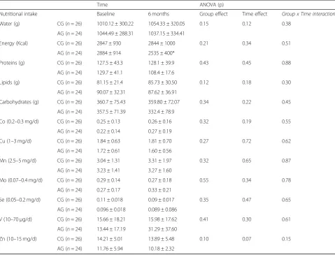
Table 1Nutritional intake of elements in controls and athletes at baseline and after the training program
Time ANOVA (p)
Nutritional intake Baseline 6 months Group effect Time effect Group x Time interaction
Water (g) CG (n= 26) 1010.12 ± 300.22 1054.33 ± 320.05 0.15 0.12 0.38
AG (n= 24) 1044.49 ± 288.31 1037.15 ± 334.41
Energy (Kcal) CG (n= 26) 2847 ± 930 2844 ± 1000 0.21 0.34 0.51
AG (n= 24) 2884 ± 914 2535 ± 400*
Proteins (g) CG (n= 26) 127.5 ± 43.3 128.1 ± 39.9 0.43 0.45 0.88
AG (n= 24) 129.7 ± 41.1 108.4 ± 17.6
Lipids (g) CG (n= 26) 81.15 ± 21.4 85.73 ± 30.50 0.12 0.18 0.30
AG (n= 24) 90.07 ± 32.31 87.62 ± 36.91
Carbohydrates (g) CG (n= 26) 360.7 ± 75.43 359.80 ± 72.07 0.34 0.22 0.45
AG (n= 24) 357.5 ± 71.39 332.4 ± 78.9
Co (0.2–0.3 mg/d) CG (n= 26) 0.25 ± 0.13 0.26 ± 0.16 0.32 0.19 0.55
AG (n= 24) 0.22 ± 0.14 0.27 ± 0.19
Cu (1–3 mg/d) CG (n= 26) 1.84 ± 0.63 1.81 ± 0.70 0.27 0.72 0.62
AG (n= 24) 1.72 ± 0.61 1.60 ± 0.56
Mn (2.5–5 mg/d) CG (n= 26) 3.04 ± 1.31 3.31 ± 1.97 0.32 0.65 0.87
AG (n= 24) 3.23 ± 1.41 3.27 ± 1.60
Mo (0.07–0.4 mg/d) CG (n= 26) 0.29 ± 0.14 0.27 ± 0.18 0.55 0.34 0.78
AG (n= 24) 0.27 ± 0.17 0.33 ± 0.21
Se (0.05–0.2 mg/d) CG (n= 26) 0.11 ± 0.018 0.09 ± 0.017 0.35 0.47 0.65
AG (n= 24) 0.096 ± 0.018 0.089 ± 0.086
V (10–70μg/d) CG (n= 26) 15.66 ± 18.21 15.98 ± 17.62 0.41 0.30 0.61
AG (n= 24) 13.44 ± 17.19 31.29 ± 37.60
Zn (10–15 mg/d) CG (n= 26) 14.21 ± 5.01 13.89 ± 5.48 0.10 0.07 0.15
tests were performed in the morning (between 10 and 12 a.m.) within the recommended parameters [17]. Training intensity and volume were reduced the two previous days applying a regenerative load in order to avoid fatigue in the physical tests.
The exercise test was performed at the beginning and at the end of the experimental period, with the time and conditions being the same for each participant.
Sample collection
At nine o’clock in the morning 5 mL of venous blood were drawn from each participant using a plastic syringe fitted with a stainless-steel needle. The blood samples were collected in a metal-free polypropylene tube (previ-ously washed with diluted nitric acid). Then, the blood samples were centrifuged at 3000 rpm for 15 min at room temperature to separate the serum. Once isolated, the serum was aliquoted into an Eppendorf tube (previ-ously washed with diluted nitric acid) and was conserved at −80 °C until further analysis. Morning midstream urine samples were obtained from all subjects and were collected in polyethylene tubes previously washed with diluted nitric acid and frozen at −80 °C until analysis. Prior to analysis, the samples were thawed and homoge-nized by shaking. This protocol was applied at the begin-ning and at the end of the experimental period.
Experimental design
Urinary creatinine determination
Creatinine concentrations were measured in all urine samples to determine different dilution degrees [18], using Sigma’s Creatinine 555–A kit and a UNICAM 5625 spectrophotometer.
Serum and urinary trace element determination
Sample preparation Co, Cu, Mn, Mo, Se, V and Zn
analyses were performed by inductively coupled plasma mass spectrometry (ICP-MS). To prepare the analysis, the organic matrix was decomposed by heating it for 10 h at 90 °C after the addition of 0.8 mL HNO3and 0.4 mL H2O2 to 2 mL of serum or urine samples. The samples were then dried at 200 °C on a hot plate. Samples recon-stitution was carried out by adding 0.5 mL of nitric acid, 10μL of Indium (In) (10 mg/L) as the internal standard, and ultrapure water to complete 10 mL.
Standard and reference material preparation Reagent
blanks, element standards and certified reference mater-ial (Seronorm, lot 0511545, AS Billingstand, Norway) were prepared identically, and used for accuracy testing. Before the analysis, the commercial control materials were diluted according to the recommendation of the manufacturer.
Sample analysis Digested solutions were assayed with
an ICP-MS Nexion model 300D (PerkinElmer, Inc., Shelton, CT, USA) equipped with a triple quadrupole mass detector and a reaction cell/collision device that al-lows operation in three modes: without reaction gas (STD); by kinetic energy discrimination (KED) with he-lium as the collision gas; and in reaction mode (DRC) with ammonia as the reaction gas. Both collision and re-action gases such as plasmatic argon had a purity of 99.999% and were supplied by Praxair (Madrid, Spain). Two mass flow controllers regulated gas flows. The fre-quency of the generator was free-swinging and worked at 40 Mhz. Three replicates were analyzed per sample. The sample quantifications were performed with indium (In) as the internal standard. The values of the standard materials of each element (10μg/L) used for quality con-trols were in agreement with intra and inter-assay vari-ation coefficients of less than 5%.
Statistical evaluations
Statistical analyses were carried out with IBM SPSS Sta-tistics 22.0 for Windows. The results are expressed as means ± standard deviations. Normality was tested by Shapiro–Wilk test. Two-way ANOVA was used to show differences between study variables. The level of signifi-cance was set atp< 0.05.
Results
Dietary habits
Table 1 shows the results of the nutritional evaluations. None of the participants followed any special diet like e.g., vegetarians and vegans. None of them consumed any mineral supplements either. They reported a similar intake of milk, fish, meat, fruits, and vegetables during the training period. As can be observed in Table 1, no differences were found between groups in any of the nu-tritional variables at baseline, but the caloric content of the diet was lower at the end of the training period in AG group than CG. In this sense, when examining the time effect, no differences were observed.
Anthropometric and ergospirometric characteristics of participants
factors on the dependent variable. As Table2shows, this interaction effect was not significative.
Serum concentrations of metals
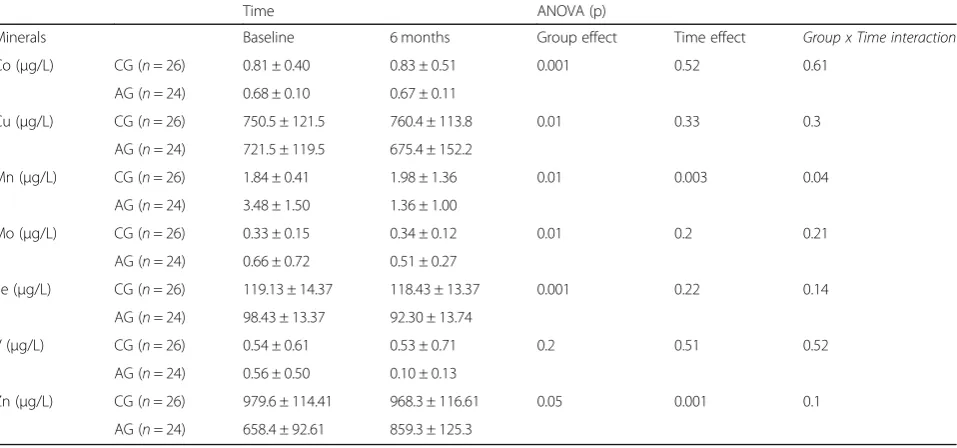
Table 3 shows the serum concentrations of each metal at the start and end of the study in both groups. The ANOVA showed significant differences in Co, Cu, Mn, Mo, Se and V between groups. In addition, there were significant differences in Mn (p = 0.003) an Zn (p = 0.001) across time. We observed a time x group inter-action for serum concentration of Mn (p= 0.04). Thus, a
decrease in this mineral was provoked in AG by training period.
Urinary concentrations of metals
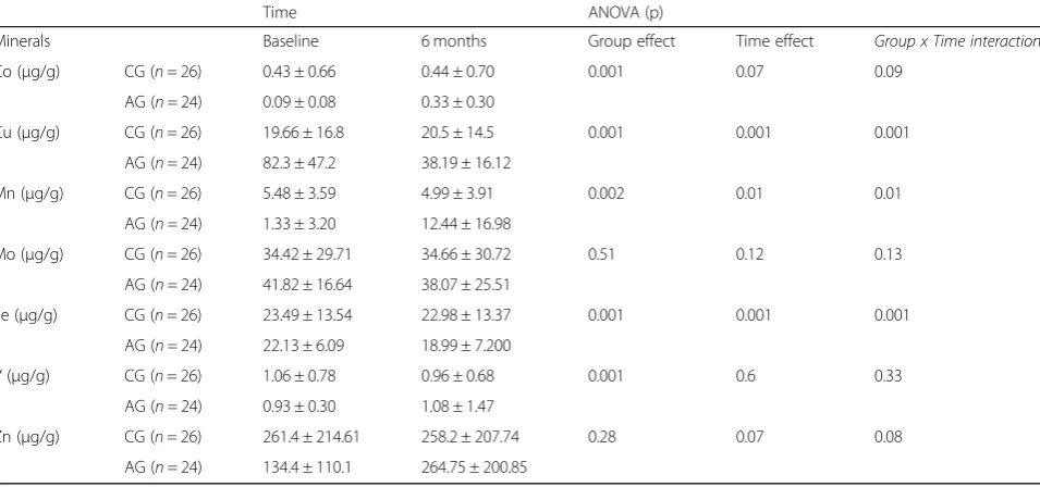
Table 4 shows the urinary concentrations of each metal at the start and at the end of the training period in both study groups. When examining the group effect, there were significant differences in the urinary excretion of all minerals except in the case of Mo. However, after the training period, significant differences were obtained in Cu (p= 0.001), Mn (p= 0.01), and Se (p = 0.001).
Table 2Ergoespirometrics results of controls and athletes at baseline and after the training program
Time ANOVA (p)
Parameters Baseline 6 months Group effect Time effect Group x Time interaction
Total Weight (Kg) CG (n= 26) 76.94 ± 11.07 77.62 ± 12.14 0.001 0.2 0.11
AG (n= 24) 65.55 ± 7.55 64.73 ± 7.83
∑6 Skinfold (mm) CG (n= 26) 62.76 ± 12.23 63.02 ± 13.05 0.001 0.08 0.13 AG (n= 24) 48.74 ± 11.17 46.59 ± 8.89
Rest HR (b/min) CG (n= 26) 65.12 ± 12.42 67.34 ± 11.71 0.001 0.13 0.21
AG (n= 24) 52.12 ± 12.42 49.87 ± 9.27
HR max (b/min) CG (n= 26) 196.33 ± 7.55 197.41 ± 8.01 0.42 0.51 0.72
AG (n= 24) 193.69 ± 7.85 194.30 ± 7.50
VO2max (ml/kg/min) CG (n= 26) 45.72 ± 7.51 46.32 ± 8.16 0.001 0.2 0.31
AG (n= 24) 66.46 ± 10.12 67.94 ± 8.10
VE max (L/min) CG (n= 26) 98.66 ± 11.47 99.26 ± 16.63 0.001 0.42 0.25
AG (n= 24) 137.95 ± 55.34 129.00 ± 26.92 CGControl group,AGAthletes group.HRHearth rate,VO2Oxygen uptake,VEPulmonary ventilation Values are presented as mean ± standard deviation
Table 3Serum concentrations of trace elements in controls and athletes at baseline and after the training program
Time ANOVA (p)
Minerals Baseline 6 months Group effect Time effect Group x Time interaction
Co (μg/L) CG (n= 26) 0.81 ± 0.40 0.83 ± 0.51 0.001 0.52 0.61
AG (n= 24) 0.68 ± 0.10 0.67 ± 0.11
Cu (μg/L) CG (n= 26) 750.5 ± 121.5 760.4 ± 113.8 0.01 0.33 0.3
AG (n= 24) 721.5 ± 119.5 675.4 ± 152.2
Mn (μg/L) CG (n= 26) 1.84 ± 0.41 1.98 ± 1.36 0.01 0.003 0.04
AG (n= 24) 3.48 ± 1.50 1.36 ± 1.00
Mo (μg/L) CG (n= 26) 0.33 ± 0.15 0.34 ± 0.12 0.01 0.2 0.21
AG (n= 24) 0.66 ± 0.72 0.51 ± 0.27
Se (μg/L) CG (n= 26) 119.13 ± 14.37 118.43 ± 13.37 0.001 0.22 0.14
AG (n= 24) 98.43 ± 13.37 92.30 ± 13.74
V (μg/L) CG (n= 26) 0.54 ± 0.61 0.53 ± 0.71 0.2 0.51 0.52
AG (n= 24) 0.56 ± 0.50 0.10 ± 0.13
Zn (μg/L) CG (n= 26) 979.6 ± 114.41 968.3 ± 116.61 0.05 0.001 0.1
Finally, a time x group interaction was observed in urinary excretion of Cu (0.001), Mn (p = 0.01) and Se (p = 0.001), decreasing the urinary excretion of Cu and Se in AG, and increasing in the case of Mn.
Discussion
This study aimed to determine if athletes present differ-ent concdiffer-entrations of essdiffer-ential minerals (Co, Cu, Mn, Mo, Se, V and Zn) with respect to sedentary people and if exercise-induced modifications in the serum and urin-ary concentrations as a result of a period of six months of intense, predominantly aerobic, physical training.
Thus, the discussion of the results related to the min-eral elements studied will be presented, as in the results, analyzing the possible differences between both groups, and then the effects that the 6 months of the study caused in both groups.
All participants lived in the same region and were the same age, this helped to avoid several factors which could have influenced the results. In this respect, control participants did not suffer any anthropometric or ergos-pirometric change, a fact which reinforces the previous statement.
The information used to evaluate the diets and as-certain the specific amounts consumed by the partici-pants is a critical point in this kind of studies. The present survey used different food composition tables [14–16]. In order to ensure reliability, and considering the high variability of amounts of minerals per food reported in the literature, the average amount of min-erals in each food was calculated using the informa-tion in the literature.
When basal results were analyzed, both groups pre-sented a similar intake of nutrients at the start of the study (Table 1), but the caloric intake was lower at the end of the training period in the AG than CG. Table2 shows, as expected, that in high-level athletes (AG), weight, body fat and resting heart rate were significantly lower compared to the CG and, on the contrary, the ergospirometric parameters VO2max and VE max were much higher in the AG athletes regarding the CG, stay-ing similar at the end of the study. These differences are due to the adaptations that aerobic training produces in athletes.
In relation to the elements analyzed, Tables 3 and 4 show that all serum and urinary metal concentrations were within the normal values reported in previous sur-veys [10, 12], developed with a similar technique and expressed in the same units (μg/L).
Serum Co concentration was similar in both groups, but urinary concentration was significantly higher in CG than AG. These results are similar to those found by Muñoz et al. (2019), also in high-level athletes, indicat-ing that they could be due to an adaptive process to maintain normal values in serum and avoid a deficit of the element that could have negative consequences for the maintenance of erythropoiesis [12].
Cu is an essential element in the structure of the im-portant enzyme Cu-Zn-SOD. This enzyme protects the athlete against superoxide anion and is commonly syn-thesized in large amounts among aerobic sportsmen, suggesting a specific exercise-induced metabolic adapta-tion [9]. Our results show higher values in urine of this element in AG than CG at baseline and the end of the
Table 4Urinary concentrations of elements in controls and athletes at baseline and after the training program
Time ANOVA (p)
Minerals Baseline 6 months Group effect Time effect Group x Time interaction
Co (μg/g) CG (n= 26) 0.43 ± 0.66 0.44 ± 0.70 0.001 0.07 0.09
AG (n= 24) 0.09 ± 0.08 0.33 ± 0.30
Cu (μg/g) CG (n= 26) 19.66 ± 16.8 20.5 ± 14.5 0.001 0.001 0.001
AG (n= 24) 82.3 ± 47.2 38.19 ± 16.12
Mn (μg/g) CG (n= 26) 5.48 ± 3.59 4.99 ± 3.91 0.002 0.01 0.01
AG (n= 24) 1.33 ± 3.20 12.44 ± 16.98
Mo (μg/g) CG (n= 26) 34.42 ± 29.71 34.66 ± 30.72 0.51 0.12 0.13
AG (n= 24) 41.82 ± 16.64 38.07 ± 25.51
Se (μg/g) CG (n= 26) 23.49 ± 13.54 22.98 ± 13.37 0.001 0.001 0.001
AG (n= 24) 22.13 ± 6.09 18.99 ± 7.200
V (μg/g) CG (n= 26) 1.06 ± 0.78 0.96 ± 0.68 0.001 0.6 0.33
AG (n= 24) 0.93 ± 0.30 1.08 ± 1.47
Zn (μg/g) CG (n= 26) 261.4 ± 214.61 258.2 ± 207.74 0.28 0.07 0.08
study. However, the main effect was observed between groups. Three previous studies indicated that physical exercise results in large increases in urinary excretion of Cu [12,19,20]. As shown by Muñoz et al. (2019), the in-creased urinary Cu obtained in this survey among AG participants may be related to the biological mobilization of this mineral induced by physical training, as has been described previously [20].
It has been reported that physical exercise increases the activity of Mn-SOD at the myocardial level. So, it has been suggested that the exercise increases the activ-ity of Mn-SOD and that it could be linked to a dimin-ution in the serum concentrations of Mn [21–23].
Furthermore, Mn is an integral part of other important metabolic enzymes such as pyruvate carboxylase, a key enzyme in the process of gluconeogenesis [24]. This en-zyme acts by regulating the whole activity of the Krebs Cycle, using acetyl-CoA as an allosteric activator. Mn is also an integral component of arginase. This enzyme re-quires two molecules of Mn to develop an appropriate function. It takes part in the metabolism of urea, con-verting L-arginine into L-ornithine, and L-ornithine into urea [25,26]. The metabolism of urea is a critical point in endurance exercise, as this chemical compound is an end product in protein metabolism. In this respect, it has been reported that endurance exercise may lead to an increased protein catabolism and affect endurance performance, muscle strength and physical fitness [25].
The high serum Mn concentration found at the start of the study, but not at the end, in AG participants, is similar to other studies and could also be caused by a possible iron deficiency in athletes [10, 12, 13], a fact that would increase Mn absorption, as indicated by Park et al. (2013) [27] or a decrease in urinary elimination in AG. However, it is interesting to highlight that at the end of the study the serum concentration of Mn in the AG is similar to those of the CG, accompanied by a sig-nificant increase in urinary elimination, which would re-veal a possible renal adaptation with training. This response could be produced in order to keep a normal serum concentration of this element.
Mo participates in oxide-reduction processes as an inte-gral part of several enzymes like xanthine dehydrogenase, an enzyme which catalyzes the hypoxanthine transform-ation of xanthine to uric acid which is considered an anti-oxidant [28, 29]. Our results show higher serum concentrations in AG at baseline and final of the training period, with no changes in urinary concentrations.
In a previous study, Maynar et al. (2018) found signifi-cantly elevated values of Mo in all the sports modalities studied with respect to the control group being the low-est in the aerobic athletes. For them, the augmented Mo concentrations would ease the formation of uric acid as well as decrease the damage caused by superoxide
anions generated by xanthine oxidase in ischemia-reperfusion processes, a situation induced by high inten-sity muscular activities [11,30].
Se is an essential element which takes part in several biochemical processes of the antioxidant metabolism. In relation to the effect of exercise on the antioxidant sys-tem, previous studies have concluded that physical train-ing improves the antioxidant response, a fact which has been reported to be reflected in a reduced lipid peroxi-dation among trained athletes throughout the season [30]. Furthermore, Se is an integral component in the catalytic space of the enzyme GPx, so changes in their serum concentrations may influence the activity of this enzyme [31,32], by mean of a reduced bioavailability of this mineral. This enzyme also develops an important role in protecting against oxidative stress and lipid per-oxidation as well, and it is also responsible for the de-toxification of lipid peroxides and hydrogen peroxide (H2O2) [32–34]. In this respect, an increase in the amounts of this enzyme in the erythrocyte has been re-ported as a response to high-level physical training [9], which may affect the metabolism of Se.
In our study, serum concentrations of Se were signifi-cantly lower in AG that CG at baseline and final of the training period, with a similar urinary excretion at the start. However, we found a significant decrease in urin-ary elimination in AG than CG at the end of the study. The same results were obtained by Maynar et al. (2018) and Sánchez et al. (2010) who found lower Se values in an active population in comparison to sedentary people [10, 35]. It could be that Se intake from food was not enough to maintain the constant levels of blood Se dur-ing traindur-ing [36]. The main reason for this affirmation is that Se requirements are increased among athletes [37]. The decrease in urinary elimination would be related to a possible adaptive mechanism to avoid greater losses of Se that would be harmful for the athletes.
V is also closely linked to exercise metabolism, as within its biological properties it includes an insulin-mimetic role [8, 38]. In this respect, Seale et al. (2006) reported that the effects of V on the insulin response are based on a stimulation of insulin sensitization, rein-forced by a stimulation of adiponectin secretion from the adipocytes, as adiponectin is a hormone rich in V [39]. Similar serum and urinary levels of V were found in both groups in basal conditions before and after the study.
prevent oxidative processes by means of an antagonistic role against active metals involved in oxidation-reduction reactions, such as iron and copper [40]. Fur-thermore, Zn also performs an important anti-inflammatory function by reducing cytokine production [41] and it has been reported that high concentrations in serum Zn are associated with a decreased production of lactate and higher blood glucose values during exercise (Khaled et al., 1997), because lactate dehydrogenase is an enzyme that contains Zn [42]. In this respect, adequate concentrations in serum Zn may facilitate the reduction of lactate to pyruvate facilitating the action of LDH ac-tivity in muscle, reducing muscle fatigue [43]. In the present survey, Zn concentrations determined in serum and urine showed significantly lower serum and urinary concentrations in the athletes.
At the beginning of the study, our athletes presented values of Zn similar to those found by Maynar et al. (2018b) in aerobic athletes and that were also signifi-cantly lower than in the respective controls, indicating that the low serum concentrations among athletes, may be due to an exercise-induced body Zn redistribution between body stores, bloodstream and tissues [11]. The urinary concentrations were similar to those presented by Maynar et al. (2018), indicating that this lower elim-ination could correspond to an adaptive mechanism to avoid element losses [13].
Regarding the second section of the discussion an im-portant issue in the research with high-level athletes, be-cause of the high training intensities, is attrition and fatigue, which may affect the results. In this sense, HR (resting and maximal) and VO2max can be valid param-eters to identify fatigue and overtraining [44, 45] . As can be observed in Table 2 no differences were evident among the athletes at the end of the survey, in compari-son to the respective initial values. Furthermore, none of the athletes presented symptoms of overtraining.
The control group did not practice any kind of sport and their nutritional demands were stable during the whole experimental period. This fact served to verify the nutritional analysis, as well as to have a nutritional refer-ence of a population of non-sportsmen. None of the di-ets of any of the participants were manipulated by the researchers.
According to the data from the diets (Table1), CG did not experience any change while the athletes showed a diminution (p< 0.05) in the caloric intake at the end of the experimental period, a fact which was accompanied by an increase in the intake of V.
Regarding the body values of minerals after the six months of the study, no changes were observed among CG either in serum or urine.
When we observe the possible changes occurring in serum and urinary concentrations of minerals, an
increase in urinary Co elimination was reported in ath-letes without modifications in the dietary intake and serum values. It could be due to an increase in the deg-radation of cobalamin, a Co containing vitamin, as a consequence of physical training, a fact which has not been demonstrated yet.
On the other hand, no changes were reported in the ingestion or serum values of Cu after training, so the de-crease found in the urinary elimination among the ath-letes could be explained as a body response to retain this element and ensure adequate amounts which would allow the body to overcome the metabolic demands in-duced by physical training, like enzymatic production. Similarly, no changes in serum concentrations of Cu were found in other studies [46].
The diminution of Mn observed in the serum after the training period of the athletes, could mainly be due to a possible body redistribution of this element to meet cel-lular exercise-induced demands. This diminution in serum was accompanied by an increase in the urinary elimination of Mn. This fact may also be explained by an augmented degradation of proteins rich in Mn as a consequence of exercise. This explanation can be rein-forced by the role of Mn as an antagonist of iron (Fe) [47], a critical element in aerobic metabolism. In this re-spect, the obtained results could be produced as a pre-ventive body response to ensure optimal levels of Fe. Although the real cause of these changes is not entirely clear, the obtained result manifests a real influence of aerobic exercise on the body values of Mn.
The six months of the study did not produce signifi-cant changes of Mo in the serum or urine of the athletes.
In relation to Se, the lower serum concentrations of Se found among athletes after the aerobic training program could be explained by increased cellular metabolic de-mands in order to develop an adequate antioxidant re-sponse induced by the oxidative stress linked to aerobic exercise. Furthermore, considering that in addition to these results, a diminution was observed in the urinary elimination of this element without changes in the daily intake, it seems clear that this framework suggests a pos-sible adaptive response of the body to retain this element in order to prevent major losses and to ensure adequate body concentrations of Se to meet exercise-induced demands.
exercise so it seems most likely that the obtained results may be mainly due to an increased use of this element to exert its insulin mimetic function either to maintain homeostasis or to enhance the metabolism of carbohy-drates or recovery after exercise.
Regarding the Zn results, the significant increase found in the serum among the athletes at the end of the study could be due to an increased disposal from muscle reserves, as it is known that the greater proportion of body Zn is found in skeletal muscle (50–60%) and bone (25–30%) [49].
These increased serum values of Zn could be ex-plained by different hypotheses. The first one could be based on the anti-inflammatory role of Zn, with the in-crease in serum being a possible adaptive mechanism used by the athletes to protect their body against inflam-mation resulting from strenuous physical activity. The second one, could be based on the antioxidant role of this mineral. As aerobic athletes are exposed to in-creased oxidative stress this result could be explained as a response to prevent oxidative damage and to reduce muscle fatigue.
In all cases, it seems clear that physical exercise affects the serum values of this element, so, it could be assumed that a functional Zn redistribution may occur between tissues during exercise in order to meet the demands in-duced by physical training, affecting the serum values. For the abovementioned reports, the obtained results could have a positive impact among the athletes due to a major bioavailability of this mineral, a critical fact for physical performance, and could be explained as an adaptive response to overcome the physical demands of training.
Conclusions
It can be concluded that, except for V, all the mineral studied presented different serum or urinary concentra-tion in athletes with respect to sedentary people in basal conditions, before and after de study. This could be re-lated to mechanisms of adaptation to high intensity aer-obic training.
Six months of aerobic training among well-trained ath-letes can induce important changes in serum and urine concentrations of several essential elements. The main findings in this survey were an increase in the serum concentrations of Zn and a decrease in the concentra-tions of serum Mn, Se and V that can alter the athlete’s physical capacity. The athlete’s body can develop changes in urinary elimination of some elements by re-ducing (Cu and Se) or increasing (Co, Mn, and Zn) ex-cretion rates to maintain the organism in a good state and prevent negative effects.
The results obtained seem to manifest a possible body need of several elements, like Mn, Se and V, a fact which
may indicate specific nutritional supplementation re-quirements, due to reductions in urinary elimination, to maintain concentration as this situation could lead to risks for the performance of the athletes if it is main-tained for long periods. In all cases, further research is required to discover, in more detail, the specific causes of these changes and the possible consequences.
Abbreviations
AG:Athletes group; CG: Control group; Co: Cobalt; Cu: Copper;
DNA: Deoxyribonucleic acid; GPx: Glutathione peroxidase; H2O2: Hydrogen peroxide; ICP-MS: Inductively coupled plasma mass spectrometry; LDH: Lactate dehydrogenase; Mn: Manganese; Mo: Molybdenum;
Se: Selenium; SOD: Superoxide dismutase; V: Vanadium; Zn: Zinc;Σ4: Sum of 4 skinfolds;Σ6: Sum of 6 skinfolds
Acknowledgments
The authors gratefully acknowledge the collaboration of SAIUex.
The research was conducted in the laboratory of Physiology of the School of Sport Sciences (University of Extremadura).
Authors’contributions
MM designed the study; data were collected and analyzed by MM, GB, F-JG and JA; DM, M-CR, and IB undertook data interpretation and manuscript preparation. All authors approved the final version of the paper.
Funding
No funding was received.
Availability of data and materials
All data generated or analyzed during this study are included in this published article.
Ethics approval and consent to participate
This research was carried out under the Helsinki Declaration ethic guidelines, updated at the World Medical Assembly in Seoul in 2008, for research with human subjects. All the participants were informed about the purpose of the study and gave their voluntary signed informed consent.
Consent for publication Not applicable.
Competing interests
The authors declare that they have no competing interests.
Author details
1Sport Sciences Faculty, University of Extremadura, Avenida de la Universidad
s/n 10003, Cáceres, Spain.2Education Faculty, University of Salamanca, /
Henry Collet, 52-70, 37007 Salamanca, Spain.3Education Faculty, University of
Extremadura, Avenida de la Universidad s/n 10003, Cáceres, Spain.
Received: 5 September 2018 Accepted: 5 November 2019
References
1. Lippi G, Franchini M, Guidi GC. Cobalt chloride administration in athletes: a new perspective in blood doping? Br J Sports Med. 2005;39:872–3. 2. Speich M, Pineau A, Ballereau F. Minerals, trace elements and related
biological variables in athletes and during physical activity. Clin Chim Acta. 2001;312:1–11.
3. Ferguson-Miller S, Babcock GT. Heme/copper terminal oxidases. Chem Rev. 1996;96:2889–908.
4. Tapiero H, Tew KD. Trace elements in human physiology and pathology: zinc and metallothioneins. Biomed Pharmacother. 2003;57:399–411. 5. Fridovich I. Superoxide Radical and Superoxide Dismutases. Annu Rev
Biochem. 1995;64:97–112.
7. Marín-García J. Oxidative stress and cell death in cardiovascular disease. In: Post-Genomic Cardiology. Elsevier; 2014. p. 471–98.
8. Rehder D. Is vanadium a more versatile target in the activity of primordial life forms than hitherto anticipated? Org Biomol Chem. 2008;6:957. 9. Mena P, Maynar M, Gutierrez JM, Maynar J, Timon J, Campillo JE. Erythrocyte
free radical scavenger enzymes in bicycle professional racers. Adaptation to training. Int J Sports Med. 1991;12:563–6.
10. Maynar M, Llerena F, Bartolomé I, Alves J, Robles M-C, Grijota F-J, et al. Seric concentrations of copper, chromium, manganesum, nickel and selenium in aerobic, anaerobic and mixed professional sportsmen. J Int Soc Sports Nutr. 2018;15:8.
11. Maynar M, Llerena F, Grijota FJ, Pérez-Quintero M, Bartolomé I, Alves J, et al. Serum concentration of cobalt, molybdenum and zinc in aerobic, anaerobic and aerobic-anaerobic sportsmen. J Int Soc Sports Nutr. 2018;15:1–8. 12. Muñoz D, Maynar M, Barrientos G, Siquier-Coll J, Bartolomé I, Grijota FJ,
et al. Effect of an acute exercise until exhaustion on the serum and urinary concentrations of cobalt, copper, and manganese among well-trained athletes. Biol Trace Elem Res. 2019;189:387–94.
13. Maynar M, Muñoz D, Alves J, Barrientos G, Grijota FJ, Robles MC, et al. Influence of an acute exercise until exhaustion on serum and urinary concentrations of molybdenum, selenium, and zinc in athletes. Biol Trace Elem Res. 2018;186:361–9.
14. Kabata-Pendias A, Mukherjee A. Trace elements from soil to human. Heidelberg: Springer; 2007.
15. Reilly C. The nutritional trace metals. Oxford, UK: Blackwell Publishing Ltd; 2004. 16. Moreiras O. Tablas de composición de alimentos : guía de prácticas. Madrid:
Pirámide; 2016.
17. Niemelä K, Palatsi I, Takkunen J. The oxygen uptake - work-output relationship of runners during graded cycling exercise: sprinters vs. endurance runners. Br J Sports Med. 1980;14:204–9.
18. Shi H, Ma Y, Ma Y. A simple and fast method to determine and quantify urinary creatinine. Anal Chim Acta. 1995;312:79–83.
19. Granell J. Zinc and copper changes in serum and urine after aerobic endurance and muscular strength exercise. J Sports Med Phys Fitness. 2014; 54:232–7.
20. Kikukawa A, Kobayashi A. Changes in urinary zinc and copper with strenuous physical exercise. Aviat Space Environ Med. 2002;73:991–5. 21. Lee Y, Min K, Talbert EE, Kavazis AN, Smuder AJ, Willis WT, et al. Exercise
protects cardiac mitochondria against ischemia-reperfusion injury. Med Sci Sports Exerc. 2012;44:397–405.
22. Bicer M, Gunay M, Baltaci AK, Uney K, Mogulkoc R, Akil M. Effect of zinc supplementation on lipid peroxidation and lactate levels in rats with diabetes induced by streptozotocin and subjected to acute swimming exercise. Bratisl Lek Listy. 2012;113:199–205.
23. De Lisio M, Kaczor JJ, Phan N, Tarnopolsky MA, Boreham DR, Parise G. Exercise training enhances the skeletal muscle response to radiation-induced oxidative stress. Muscle Nerve. 2011;43:58–64.
24. Linder PW, Torrington RG, Seemann UA. Formation constants for the complexes of levulinate and acetate with manganese (II), cobalt (II), nickel (II), copper (II), zinc (II) and hydrogen ions. Talanta. 1983;30:295–8. 25. Gibala MJ. Protein metabolism and endurance exercise. Sport Med. 2007;37:337–40. 26. Morris SM. Regulation of enzymes of the urea cycle and arginine
metabolism. Annu Rev Nutr. 2002;22:87–105.
27. Park S, Sim C-S, Lee H, Kim Y. Blood manganese concentration is elevated in infants with Iron deficiency. Biol Trace Elem Res. 2013;155:184–9.
28. Chan S, Gerson B, Subramaniam S. The role of copper, molybdenum, selenium, and zinc in nutrition and health. Clin Lab Med. 1998;18:673–85. 29. Berger CE, Kröner A, Kluger R, Baron R, Steffan I, Engel A. Effects of
marathon running on the trace minerals chromium, cobalt, nickel, and molybdenum. J Trace Elem Exp Med. 2002;15:201–9.
30. Muñoz D, Barrientos G, Alves J. Grijota FJ. Maynar M. Oxidative stress, lipid peroxidation indexes and antioxidant vitamins in long and middle distance athletes during a sport season. J Sports Med Phys Fitness: Robles MC; 2017. 31. Saïd L, Banni M, Kerkeni A, Saïd K, Messaoudi I. Influence of combined
treatment with zinc and selenium on cadmium induced testicular pathophysiology in rat. Food Chem Toxicol. 2010;48:2759–65.
32. Letavayová L, Vlčková V, Brozmanová J. Selenium: from cancer prevention to DNA damage. Toxicology. 2006;227:1–14.
33. Valko M, Rhodes CJ, Moncol J, Izakovic M, Mazur M. Free radicals, metals and antioxidants in oxidative stress-induced cancer. Chem Biol Interact. 2006;160:1–40.
34. Wasowicz W, Gromadzinska J, Rydzynski K, Tomczak J. Selenium status of low-selenium area residents: polish experience. Toxicol Lett. 2003;137:95– 101.
35. Sánchez C, López-Jurado M, Aranda P, Llopis J. Plasma levels of copper, manganese and selenium in an adult population in southern Spain: influence of age, obesity and lifestyle factors. Sci Total Environ. 2010;408: 1014–20.
36. Pograjc L, Stibilj V, Falnoga I. Impact of intensive physical activity on selenium status. Biol Trace Elem Res. 2012;145:291–9.
37. Margaritis I, Rousseau A-S, Hininger I, Palazzetti S, Arnaud J, Roussel A-M. Increase in selenium requirements with physical activity loads in well-trained athletes is not linear. BioFactors. 2005;23:45–55.
38. Sakurai H. A new concept: the use of vanadium complexes in the treatment of diabetes mellitus. Chem Rec. 2002;2:237–48.
39. Seale AP, de Jesus LA, Park M-C, Kim Y-S. Vanadium and insulin increase adiponectin production in 3T3-L1 adipocytes. Pharmacol Res. 2006;54:30–8. 40. Jomova K, Valko M. Advances in metal-induced oxidative stress and human
disease. Toxicology. 2011;283:65–87.
41. Prasad AS. Impact of the discovery of human zinc deficiency on health. J Am Coll Nutr. 2009;28:257–65.
42. Khaled S, Brun JF, Micallel JP, Bardet L, Cassanas G, Monnier JF, et al. Serum zinc and blood rheology in sportsmen (football players). Clin Hemorheol Microcirc. 1997;17:47–58.
43. Vivoli G, Bergomi M, Rovesti S, Pinotti M, Caselgrandi E. Zinc, copper, and zinc- or copper-dependent enzymes in human hypertension. Biol Trace Elem Res. 1995;49:97–106.
44. Purvis D, Gonsalves S, Deuster PA. Physiological and psychological fatigue in extreme conditions: overtraining and elite athletes. PM&R. 2010;2:442–50. 45. McKenzie DC. Markers of excessive exercise. Can J Appl Physiol. 1999;24:66–73. 46. Pourvaghar MJ, Shahsavar AR. Changes at nano scale level in copper after
an aerobic activity in males. Dig J Nanomater Biostructures. 2009;4:809–12. 47. Rivera-Mancía S, Pérez-Neri I, Ríos C, Tristán-López L, Rivera-Espinosa L,
Montes S. The transition metals copper and iron in neurodegenerative diseases. Chem Biol Interact. 2010;186:184–99.
48. French RJ, Jones PJ. Role of vanadium in nutrition: metabolism, essentiality and dietary considerations. Life Sci. 1993;52:339–46.
49. Combs GF. Geological impacts on nutrition. In: Essentials of medical geology. Dordrecht: Springer Netherlands; 2013. p. 179–94.
Publisher’s Note