R E S E A R C H A R T I C L E
Open Access
Efficacy and safety of interventions to
control myopia progression in children: an
overview of systematic reviews and
meta-analyses
Efthymia Prousali
1,2, Anna-Bettina Haidich
2, Andreas Fontalis
2,3, Nikolaos Ziakas
1, Periklis Brazitikos
1ˆ
and
Asimina Mataftsi
1*Abstract
Background:Myopia is a common visual disorder with increasing prevalence. Halting progression of myopia is
critical, as high myopia can be complicated by a number of vision-compromising conditions.
Methods:Literature search was conducted in the following databases: Medical Literature Analysis and Retrieval
System Online (MEDLINE), Excerpta Medica dataBASE (EMBASE), Cochrane Database of Systematic Reviews (CDSR), Database of Abstracts of Reviews of Effects (DARE) and Centre for Reviews and Dissemination (CRD) Health Technology Assessment (HTA) database. Systematic reviews and meta-analyses investigating the efficacy and safety of multiple myopia interventions vs control conditions, were considered. Methodological quality and quality of evidence of eligible studies were assessed using the ROBIS tool and GRADE rating. The degree of overlapping of index publications in the eligible reviews was calculated with the corrected covered area (CCA).
Results:Forty-four unique primary studies contained in 18 eligible reviews and involving 6400 children were
included in the analysis. CCA was estimated as 6.2% and thus considered moderate. Results demonstrated the superior efficacy of atropine eyedrops; 1% atropine vs placebo (change in refraction: -0.78D, [−1.30 to−0.25] in 1 year), 0.025 to 0.05% atropine vs control (change in refraction: -0.51D, [−0.60 to−0.41] in 1 year), 0.01% atropine vs control (change in refraction: -0.50D, [−0.76 to−0.24] in 1 year). Atropine was followed by orthokeratology (axial elongation: −0.19 mm, [−0.21 to −0.16] in 1 year) and novel multifocal soft contact lenses (change in refraction: -0.15D, [−0.27 to −0.03] in 1 year). As regards adverse events, 1% atropine induced blurred near vision (odds ratio [OR] 9.47, [1.17 to 76.78]) and hypersensitivity reactions (OR 8.91, [1.04 to 76.03]).
Conclusions: Existing evidence has failed to convince doctors to uniformly embrace treatments for myopic
progression control, possibly due to existence of some heterogeneity, reporting of side effects and lack of long-term follow-up. Research geared towards efficient interventions is still necessary.
Keywords: Refractive error, Myopia, Children, Vision, Lenses, Anti-muscarinic agents
© The Author(s). 2019Open AccessThis article is distributed under the terms of the Creative Commons Attribution 4.0
International License (http://creativecommons.org/licenses/by/4.0/), which permits unrestricted use, distribution, and
reproduction in any medium, provided you give appropriate credit to the original author(s) and the source, provide a link to the Creative Commons license, and indicate if changes were made. The Creative Commons Public Domain Dedication waiver (http://creativecommons.org/publicdomain/zero/1.0/) applies to the data made available in this article, unless otherwise stated. * Correspondence:amatafts@auth.gr
ˆDeceased
12nd Department of Ophthalmology, Aristotle University of Thessaloniki,
Thessaloniki, Greece
Background
Myopia is a common condition exhibiting an“epidemic” during the past half-century. East and Southeast Asia ap-pear to have a higher myopia prevalence, compared to Western and European populations, with Singapore, China, Hong Kong and Taiwan representing the regions where the problem is more common [1]. Myopia is in-cluded in the 10 priority eye diseases in VISION 2020 campaign for the prevention of blindness and visual im-pairment, as declared by the World Health Organization [2].
Myopia introduces significant social and psychological impact, as it appears to affect children’s perception of their physical appearance, athletic competence and social acceptance [3]. Myopia also imposes a considerable eco-nomic burden to societies. Annual expenses for myopia treatment are estimated to be greater than for other ocu-lar diseases including age-related macuocu-lar degeneration and primary open-angle glaucoma, as well as for non-ocular chronic pathologies, such as Parkinson’s dis-ease and chronic obstructive pulmonary disdis-ease [4]. A treatment that would halt or at least decelerate myopia’s progression rate is highly desirable, as severe myopia constitutes a substantial risk factor for several ocular conditions which can lead to blindness. These include retinal detachment, primary open angle glaucoma, cata-ract and macular degeneration [1].
Several interventions have been attempted to control myopic progression, some of which showed no effect and others were effective but with limitations [5–8]. Long-term safety and efficacy of interventions to restrict myopia remains unresolved, resulting in the lack of uni-versal consensus in myopia treatment [9–12]. As there appears to be no overview in existing literature, the aim of the present study is to synthesize evidence provided by systematic reviews (SRs) and meta-analyses (MAs) on myopia control.
Methods
Protocol and registration
We used the term ‘overview’ for our synthesis of mul-tiple intervention systematic reviews and meta-analyses, as proposed by the Cochrane Collaboration [13] and reporting followed the PRIO-harms guidelines (Add-itional file 1: Appendix 1) [14]. The protocol of this study is registered in the PROSPERO database (CRD42017068204) [15] and published in Systematic
re-views[15].
Ιnformation sources and search strategy
Purposive literature search was conducted in the Cochrane Database of Systematic Reviews (CDSR), Data-base of Abstracts of Reviews of Effects (DARE) and Centre for Reviews and Dissemination (CRD) Health Technology
Assessment (HTA) Database, using the keyword‘myopia’. A more comprehensive search strategy was applied in MEDLINE and EMBASE, using medical subject headings (MeSH) and text words related to spectacles, contact lenses, anti-muscarinic agents, myopia and children [5,
16] to find any recent primary studies not included in the published systematic reviews. The last search date was March 9, 2018. For all included studies, reference lists were also searched. MEDLINE search strategy is provided (Additional file1: Appendix 2). No language, study type or date restrictions were used.
Eligibility criteria
Participants
Our overview target were children and adolescents, ≤ 18 years of age at baseline, diagnosed with myopia de-fined as spherical equivalent refraction≤ −0.25 dioptres, with or without astigmatism, without any ocular comor-bidities including strabismus and amblyopia. Animals, adult population, patients not suffering from myopia, or patients with myopia and strabismus/amblyopia were ex-cluded. Studies related to surgical interventions for my-opia correction, e.g. refractive surgery were not considered.
Interventions and comparators
We included studies in which any optical or pharmaco-logical intervention for myopia control was compared to single vision spectacles, contact lenses, or placebo. No restriction on duration and dose of treatment, if applic-able, was imposed.
Outcome measures
Our primary outcomes regarded myopia progression and axial elongation as efficacy criteria. Myopia progres-sion was assessed as mean change in refractive error, measured in dioptres. Mean change in axial length, mea-sured in millimetres, was also evaluated. Outcomes reporting change in a 12-month or 24-month period were accepted and described. Reported adverse events (AE) were regarded as safety criteria.
Study design
Unit of analysis of this overview were SRs or meta-analyses of randomized controlled trials (RCTs), pseudo-RCTs, cohort and case-control studies. Network meta-analyses were also reviewed. Only human studies with full text available were analysed. Narrative reviews that do not systematically search the literature and do not critically appraise the quality of included studies were excluded.
meta-analysis and provide effect estimates for myopia control interventions (thereafter referred to as index publications). The total of index publications included in the meta-analysis is provided in Additional file 1: Ap-pendix 3. Levels of evidence as produced by the Oxford Centre for Evidence Based Medicine (OCEBM) were considered [17]. Index publications of low level of evi-dence, i.e. poor quality cohort/case-control studies, case series, case reports or expert opinions were not included. Cohort and case-control studies were considered of low quality if they scored less than 5 points in Newcastle-Ottawa scale. Similarly, index publications without a control group or those comparing two or more different interventions were not included in the statistical analysis.
Study selection and data management
Two independent authors (EP, AF) performed all screen-ing steps. Title and abstract screenscreen-ing were conducted using the Mendeley citation management software. The overview authors screened the titles and abstracts against the eligibility criteria and obtained full reports for all titles that appeared to meet the inclusion criteria or where there was uncertainty. The same two inde-pendent authors (EP, AF) managed data in duplicate from each eligible study, using a data collection form in Microsoft Excel designed to include all the data re-quired. Each SR or MA was initially evaluated to identify whether it matched the eligibility criteria of the over-view. Subsequently, index publications contained in these SRs/MAs were individually reviewed. Data extrac-tion was then performed for inclusion of eligible index publications in the meta-analysis. In cases where risk of bias of index publications was not available by the in-cluded SR or MA, two independent authors (EP, AF) completed the missing assessments. When an index publication was included in more than one SRs or MAs, outcome data were extracted from the most comprehen-sive study. A third author was involved to resolve any discrepancies, using the primary research paper (ABH).
Risk of bias assessment
Two overview authors (EP, AF) independently assessed the methodological quality of each included SR and MA using the Risk Of Bias In Systematic Reviews (ROBIS) tool [18]. The quality of evidence was evaluated by two independent authors (EP, AF) using four domains of the Grading of Recommendations Assessment, Develop-ment, and Evaluation (GRADE) tool: study limitations, imprecision, inconsistency of results, and indirectness and a summary of findings for each outcome of interest was designed using GRADEpro software [19, 20]. In order to minimize the subjectivity of quality assessment
process, a third reviewer was involved to resolve any dis-crepancies (ABH).
The list of included index publications in eligible SRs/ MAs was reviewed in order to identify those contained in two or more reviews. We generated a citation matrix presenting all the SRs/MAs in columns and all included index publications in rows. We estimated the overlap by calculating the corrected covered area (CCA), to assess if specific index publications are overrepresented. The formula for calculating CCA is: CCA = Nrc−−rr where N = sum of the included studies, r = rows (number of unique studies), c = columns (number of reviews). CCA reflected the degree of actual overlap, as it is not influenced by large reviews. Should high or very high overlap be de-tected, which is interpreted as CCA equal to or more than 10%, we planned to retain the review which is (1) the most recent, (2) containing a higher amount of in-formation, and (3) the most rigorous in terms of meth-odology, as assessed by ROBIS tool and GRADE scale [21,22]. In addition, two independent overview authors (EP, AF) examined possible presence of meta-biases, in-cluding publication bias, selective outcome reporting and dual co-authorship. Handling of inconsistency for meta-analyses and other potential sources of bias are also reported (Additional file1: Table S1) [23–26].
Data synthesis and analysis
Index publications included in eligible systematic reviews and meta-analyses were employed as a unit of analysis to perform a meta-analysis using Review Manager software version 5.3. Continuous outcomes were expressed using mean differences with 95% confidence intervals (CIs) and dichotomous outcomes were expressed using odd ratios (ORs) with 95% CIs. Data were synthesized using random-effects models due to the inconsistency across the RCTs and cohort studies. Subgroup analyses according to study design (RCTs and observational studies) were performed and whenever no subgroup differences were obtained the overall effect was reported. We pooled the results referring to one eye only, to each eye separately, or to the average of both eyes, depending on the data ana-lysed by each index publication design (Additional file1: Table S2). Sensitivity analyses excluding studies of lower methodological quality, when each eye was reported sep-arately or those introducing substantial inconsistency were also conducted.
Results
meta-analyses to be included in this overview [5, 6, 9– 12,27–38].
Table1 summarizes the main characteristics of the 18 eligible SRs/MAs. These were published between 2002 and 2017. Five studies are SRs and meta-analyses, four performed systematic review of literature with qualita-tive syntheses of findings, eight performed meta-analyses, and one is a network meta-analysis. Four studies investigated atropine, four analysed orthokeratol-ogy, two focused on outdoors exposure, one examined the efficacy of acupuncture, and two investigated the use of multifocal lenses. The remaining five studies exam-ined multiple interventions for myopia control.
Overlapping
The 18 included SRs and MAs comprised 226 overlap-ping index publications, of which 110 were unique. Two
recently published RCTs not included in the 18 SRs/ MAs were also identified through literature search and added to the index publications. A citation matrix pre-senting all the included SRs/MAs in columns and index publications in rows is provided in Additional file 1: Table S4. Index publications represented in more than one eligible reviews are recognized in the citation matrix. In order to avoid potential double counting of outcomes, we calculated the degree of actual overlap by estimating the CCA:
CCA¼N−r
rc−r¼
226−110 11018−110¼
116
1870¼6:2%
As CCA is estimated at 6.2%, the overlap is in the moderate range reflecting a moderate risk of skewed reporting [21, 22]. Out of the 112 index publications, 44 matched our eligibility criteria and were included in the
Table 1 Characteristics of included studies Review &Type of Study Data bases searc hed and last asses sed No. of primary studies (s ample size) Ethnic ity Age range (average age) in years Tre atmen t Contr ol Overvie w out comes Walline et al. 2011 [ 5 ] SR & meta -analysi s CENTRA L, MED LINE, EMBASE, LILACS, m RCT, Clin icalTrials.gov (10/2 011) 23 (4696 ) Israel , Malay sia, China, USA , Finland, Hong Kon g, Japan , Taiwan, Denm ark 6 – 18 U ndercorrection , mu ltifocal spec tacles, bifo cal sof t contac t le nses, nov el lense s, RG PCLs, anti -mu scarini c me dications Full corr ection spectacles, SVLs, single vis ion contact lense s, placebo Change in RE, change in AL Sherwi n et al. 2012 [ 36 ] SR & meta -analysi s MEDLI NE, EMB ASE, Web of Scienc e, CENTRA L (NA) 23 (80 – 30 09)
Singapore, Australia, Jord
an, China, USA 0.5 – 20 O utdoor expo sure NA Risk for myopi a progression Wen et al. 2015 [ 33 ] SR & meta -analysi s MEDLI NE, EMB ASE, Coc hrane Library, WHO internation al Clinic al Trials Regi stry Pla tform, Clinic alTrials.gov (11/2 014) 8 (769) Chine se, Caucasian , Japan ese 6 – 15 O K Single vision spectacles, contact lense s Change in AL Xiong et al. 2017 [ 12 ] SR & meta -analysi s PubM ed, EMB ASE, Coc hrane Library (12/ 2015) 25 (50 – 50 48)
China, Taiwan, Singapore, Australia,
UK , USA, Turkey 6 – 18 a O utdoor expo sure NA Risk of myopi a progression Gong et al. 2017 [ 9 ] SR & meta -analysi s PubM ed, EMB ASE, Coc hrane Central Re gister of Contr olled Trials (04/2 016) 19 (3137 ) Taiwan, U SA, Singapore, China, Ho ng Kon g 5 – 17 Atr opine Atropine , cont rol cond itions
Myopia progression, adverse
Table 1 Characteristics of included studies (Continued) Review &Type of Study Data bases searc hed and last asses sed No. of primary studies (s ample size) Ethnic ity Age range (average age) in years Tre atmen t Contr ol Overvie w out comes Li et al. 2017 [ 11 ] Met a-analysi s MEDLI NE, EMB ASE, Coc hrane Library, Chine se Clinical Trial Regi stry, WHO intern ational Clinic al Trials Regi stry Pla tform, Clinic alTrials.gov (05/2 016) 8 (587) USA, China, Hong Kon g, New Zealand, Japan , Spain 6 – 18 SC Ls with conc entric ring bifoc al and pe ripheral add mu ltifocal de signs single vis ion SCLs or spectacles Change in RE, change in AL Cui et al. 2017 [ 35 ] Met a-analysi s MEDLI NE, Coc hrane, EMB ASE, Goog le Scholar (09/201 5) 5 (673)
USA, Singapore, East
Asi a 6 – 16 RG PCLs SCLs, spec tacles, OK Change in RE, change in AL Huang et al. 2016 [ 34 ] Netw ork meta -analysi s MEDLI NE, EMB ASE, Coc hrane Central Re gister of Contr olled Trials, WHO intern ational Clinic al Trials Regi stry Pla tform, Clinic alTrials.gov (08/2 014) 30 (5387 ) Israel , Malay sia, Hong Kon g, USA, Denm ark, Finland, Japan , China, New
England, Singapore, Taiwan
< 18 Atr opine , pirenzepine, ti
molol, cyclopen
meta-analysis. These consisted of 28 RCTs and 16 obser-vational studies, and reported data on a total of 6400 patients.
Assessment of methodological quality
Qualitative, domain-based rating of methodological quality of eligible studies with ROBIS tool is provided in Additional file 1: Table S5 [18]. The overall risk of bias was ‘low’ in 14 reviews [5, 6, 9–11, 27–35],‘unclear’ in three [12,36,37] and‘high’in one review [38].
With regard to ‘Study eligibility criteria’ domain, two studies [6, 38] were judged with ‘high concern’ due to imprecisely defined eligibility criteria, publication status and/or language limitations. One study [38] was judged with‘high’and one [29] with‘unclear concern’in‘ Identi-fication and selection of studies’domain because of lim-ited details regarding search strategy and unclear study selection process. Domain 3 assessed the methodology used for data collection and study appraisal, in which two studies [6, 28] were judged with ‘unclear’ and one [38] with ‘high concern’, due to lack of information on included studies’ characteristics for appropriate inter-pretation of findings, or because of inappropriate or no risk of bias assessment of index publications. Fifteen eli-gible reviews reported quality assessment of the included
index publications. Four [29–32] used the Jadad scale and four [10,11,33,37] combined Jadad with Newcastle Ottawa scale for observational studies, four [5, 27, 34,
35] used the Cochrane Collaboration Risk of Bias tool, one [9] combined the Cochrane tool with Newcastle Ottawa scale and two [12, 36] used other tools. Three reviews [6, 28, 38] did not report formal assessment of included index publications. In ‘Synthesis and findings’ domain, three studies [36–38] were judged with ‘high’ and one [12] with‘unclear concern’, due to inappropriate quantitative or qualitative synthesis.
None of the eligible studies reported a GRADE assess-ment. Two authors (EP, AF) independently rated the quality of evidence for our outcomes using the GRADE scale (Tables 2, 3, 4, 5 and 6), [19–21]. Quality was assessed as ‘high’in one outcome,‘moderate’in 41,‘low’ in 12 and ‘very low’ in 23 outcomes examining efficacy or safety (Additional file 1: Appendix 6). Low/very low quality is due to a number of index publications being at risk of bias from elements involving imprecision, incon-sistency and limitations including lack of blinding or al-location concealment and loss to follow-up.
Quality of the 44 index publications included in the meta-analysis was adequate. More than 50% of the RCTs were at low risk of bias for random sequence generation
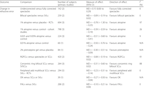
Table 2Primary outcomes from baseline (1 year) - Change in refractive error
Outcome Comparison Number of subjects
(primary studies)
Measure of effect (95% CI)
Direction of effect I2 (%)
Change in refractive error
Undercorrected versus fully corrected spectacles
142 (2) MD = 0.15 (0.00 to
0.29)
Favours fully corrected spectacles
0
Bifocal spectacles versus SVLs 259 (2) MD =−0.09 (−0.19 to 0.02)
Favours bifocal spectacles 0
1% atropine versus placebo - RCTs 604 (3) MD =−0.78 (−1.30 to
−0.25)
Favours atropine 97
1% atropine versus control - cohort studies
798 (3) MD =−0.39 (−0.59 to
−0.19)
Favours atropine 26
0.025 and 0.05% atropine versus control
224 (3) MD =−0.51 (−0.60 to
−0.41)
Favours atropine 9
0.01% atropine versus control 60 (1) MD =−0.50 (−0.76 to
−0.24)
Favours atropine N/A
2% pirenzepine gel versus placebo 84 (1) MD =−0.30 (−0.51 to
−0.09)
Favours pirenzepine N/A
RGPCLs versus spectacles or SCLs 420 (2) MD =−0.08 (−0.19 to 0.02)
Favours RGPCLs 91
Concentric ring bifocal SCLs versus SVSCLs
264 (3) MD =−0.31 (−0.60 to 0.02)
Favours concentric ring bifocal SCLs
88
Peripheral add multifocal SCLs versus SVLs - RCTs
294 (5) MD =−0.23 (−0.31 to
−0.14)
Favours peripheral add multifocal SCLs
0
ΟΚversus SCLs or SVLs 39 (1) MD =−0.27 (−0.50 to
−0.04)
Favours OK N/A
PALs versus SVLs 206 (2) MD =−0.10 (−0.21 to
0.00)
Favours PALs 0
and allocation concealment and more than 80% of RCTs were at low risk of bias for selective outcome reporting. Nonetheless, only 20% of RCTs achieved appropriate blinding of participants and outcome assessors, while al-most 70% were at high risk for incomplete outcome data. Three RCTs were assessed with Jadad scale and scored 4 or above, while one scored 2. More than 90% of included cohort studies were awarded with 8 or more stars in Newcastle-Ottawa Quality Assessment scale. Risk of bias assessments of index publications as
presented in the eligible SRs/MAs are shown in Add-itional file1: Tables S6, S7 and S8.
Synthesis of results
The included SRs and MAs provided outcome data re-lating to the following comparisons: Undercorrected vs fully-corrected spectacles, bifocal spectacles vs single vi-sion lens spectacles (SVLs), atropine vs placebo, pirenze-pine gel vs placebo, rigid gas permeable contact lenses (RGPCLs) vs spectacles or soft contact lenses (SCLs),
Table 3Primary outcomes from baseline (1 year)–Change in axial length
Outcome Comparison Number of subjects
(primary studies)
Measure of effect (95% CI)
Direction of effect I2 (%)
Change in axial length
Undercorrected versus fully corrected spectacles
94 (1) MD = 0.05 (−0.01 to
0.11)
Favours full correction N/A
RGPCLs versus spectacles or SCLs 415 (2) MD =−0.02 (−0.05 to 0.10)
Favours spectacles/SCLs 0
2% pirenzepine gel versus placebo 264 (2) MD =−0.10 (−0.18 to
−0.01)
Favours pirenzepine 0
Concentric ring bifocal SCLs versus SVSCLs
264 (3) MD =−0.12 (−0.19 to
−0.06)
Favours concentric ring bifocal SCLs
66
1% atropine versus control 586 (3) MD =−0.36 (−0.41 to
−0.30)
Favours atropine 46
Peripheral add multifocal SCLs versus SVLs - RCTs
294 (5) MD =−0.10 (−0.14 to
−0.05)
Favours peripheral add multifocal SCLs
37
ΟΚversus SCLs or SVLs 524 (8) MD =−0.19 (−0.21 to
−0.16)
Favours OK 0
PALs versus SVLs 211 (2) MD =−0.08 (−0.14 to
0.02)
Favours PALs 65
CIconfidence interval,MDMean Difference,N/Anot applicable,OKOrthokeratology,PALsprogressive addition lenses,RGPCLsrigid gas permeable contact lenses,
SCLssoft contact lenses,SVLssingle vision lenses,SVSCLssingle vision soft contact lenses
Table 4Primary outcomes from baseline (2 years)–Change in refractive error
Outcome Comparison Number of subjects
(primary studies)
Measure of effect (95% CI)
Direction of effect I2
(%)
Change in refractive error
Undercorrected versus fully corrected spectacles
142 (2) MD = 0.17 (0.12 to
0.23)
Favours fully corrected spectacles
0
Bifocal spectacles versus single vision lens spectacles
351 (3) MD =−0.19 (−0.59 to 0.21)
Favours bifocal spectacles 85
1% atropine versus placebo 400 (1) MD =−0.92 (−1.08 to−0.76)
Favours atropine N/A
2% pirenzepine gel versus placebo 74 (1) MD =−0.41 (−0.70 to−0.12)
Favours pirenzepine N/A
RGPCLs versus spectacles or SCLs 398 (2) MD =−0.16 (−0.33 to−0.00)
Favours RGPCLs 92
Concentric ring bifocal SCLs versus SVSCLs
128 (1) MD =−0.20 (−0.38 to−0.02)
Favours concentric ring bifocal SCLs
N/A
Peripheral add multifocal SCLs versus SVLs
99 (2) MD =−0.50 (−0.65
to−0.35)
Favours peripheral add multifocal SCLs
0
ΟΚversus SCLs or SVLs 39 (1) MD =−0.66 (−1.01 to
−0.31)
Favours OK N/A
PALs versus SVLs 940 (4) MD =−0.15 (−0.40
to 0.11)
Favours PALs 89
concentric ring bifocal SCLs vs single vision soft contact lenses (SVSCLs), peripheral add multifocal SCLs vs SCLs or SVLs, OK vs SCLs or SVLs, progressive addition lenses (PALs) vs SVLs. The outcomes assessed included change in refractive error and change in axial length from baseline to 1 year and from baseline to 2 years. These outcomes were identified a priori as being of interest for this overview [15]. Safety of myopia inter-ventions was assessed by quantitative analysis of the number and type of reported adverse events.
Effects of interventions
Undercorrected vs fully-corrected spectacles
Two RCTS encompassing 142 children investigated the effect of undercorrection in myopic progression. The overall pooled analysis revealed that the undercorrected group showed greater change in refractive error (RE) in 1 year (MD 0.15, 95% CI 0.00 to 0.29), and in 2 years from baseline (MD 0.20, 95% CI 0.01 to 0.39) and the evidence quality of this outcome was considered moder-ate (Tables2,3,4and 5& Additional file1: Appendices 4 and 6).
Bifocal spectacles vs single vision lens spectacles
Two RCTs (259 children) examined the effect of bifocal spectacles in myopia control and showed no change in RE in 1 year from baseline (MD -0.09, 95% CI -0.19 to 0.02; GRADE evidence: moderate; Table 2 & Additional file 1: Appendices 4 and 6). Three RCTs (351 children) reported no change in RE using bifocal spectacles in 2 years from baseline (MD -0.19, 95% CI -0.59 to 0.21;
GRADE evidence: low; Table 3 & Additional file 1: Ap-pendices 4 and 6). These 3 trials appeared to be incon-sistent (I2= 85%). Sensitivity analysis excluding
Parsinnenet al. demonstrated no difference in the effect
of bifocal spectacles.
1% atropine vs placebo
Three RCTs (604 children) and three cohort studies (798 children) provided outcomes on the effect of 1% at-ropine eyedrops in refraction change in 1 year, (Table 2). Subgroup analysis of the three trials reported a change of−0.78D, favouring atropine (95% CI,−1.30 to−0.25) with moderate quality of evidence (Additional file1: Ap-pendix 6). Due to high inconsistency (I2= 97%), sensitiv-ity analysis excluding Yi et al. revealed a change of − 0.54D, also favouring atropine (95% CI,−0.76 to−0.33), with moderate inconsistency among the two studies (I2 = 54%). Treatment effect reported by cohort studies showed an increase in refraction for the subgroup re-ceiving placebo. Mean change in RE over 1 year was − 0.39D, favouring the use of atropine (95% CI, −0.59 to
−0.19).
Two RCTs (540 children) and one cohort study (46 children) compared mean axial length (AL) change be-tween 1% atropine eyedrops and placebo in 1 year (Table 4). Two trials revealed that atropine administra-tion decreased AL change by−0.35 mm (95% CI,−0.38 to−0.31). Treatment effect provided by the cohort study also favoured atropine, which showed AL change of − 0.61 mm (95% CI, −0.88 to −0.34). The overall treat-ment effect (586 children) showed that 1% atropine
Table 5Primary outcomes from baseline (2 years)–Change in axial length
Outcome Comparison Number of subjects
(primary studies)
Measure of effect (95% CI)
Direction of effect I2 (%)
Change in axial length
Undercorrected versus fully corrected spectacles
94 (1) MD = 0.06 (−0.04 to
0.16)
Favours full correction N/A
Bifocal spectacles versus single vision lens spectacles
89 (1) MD =−0.20 (−0.31 to
−0.09)
Favours bifocal spectacles N/A
1% atropine versus placebo 400 (1) MD =−0.36 (−0.43 to
−0.29)
Favours atropine N/A
2% pirenzepine gel versus placebo 74 (1) MD =−0.12 (−0.29 to 0.05)
Favours pirenzepine N/A
RGPCLs versus spectacles or SCLs 394 (2) MD = 0.03 (−0.05 to 0.12)
Favours spectacles or SCLs 0
Concentric ring bifocal SCLs versus SVSCLs
128 (1) MD =−0.12 (−0.20 to
−0.04)
Favours concentric ring bifocal SCLs
N/A
Peripheral add multifocal SCLs versus SVLs
99 (2) MD =−0.13 (−0.20 to
−0.06)
Favours peripheral add multifocal SCLs
0
ΟΚversus SCLs or SVLs 663 (11) MD =−0.27 (−0.31 to
−0.23)
Favours OK 0
PALs versus SVLs 791 (3) MD =−0.10 (−0.20 to
0.00)
Favours PALs 78
Table 6Primary outcomes from baseline–Adverse Events
Outcome Comparison Number of subjects
(primary studies)
Measure of effect (95% CI)
Direction of effect I2 (%)
Allergic or hypersensitivity reactions or discomfort
1% atropine versus control 446 (2) OR = 8.91 (1.04, 76.03)
Favours control 0
Blurred near vision 1% atropine versus control 540 (2) OR = 9.47 (1.17, 76.78)
Favours control 0
Contact lens-related discomfort/Unwilling-ness to wear contact lenses
Concentric ring bifocal SCLs versus SVSCLs
261 (2) OR = 0.95 (0.49, 1.81)
Favours concentric ring bifocal SCLs
0
Mild corneal erosion ΟΚversus SCLs or SVLs 151 (2) 0R = 4.56 (0.49,
42.25)
Favours SCLs/SVLs 0
Papillae/Follicles 2% pirenzepine gel versus control
323 (3) OR = 3.21 (0.95, 10.88)
Favours control 74
Medication residue on eyelids or eye 2% pirenzepine gel versus control
323 (3) OR = 0.77 (0.38, 1.59)
Favours pirenzepine 33
Abnormality of accommodation 2% pirenzepine gel versus control
323 (3) OR = 16.92 (6.27, 45.64)
Favours control 0
Itching, eye 2% pirenzepine gel versus
control
323 (3) OR = 1.01 (0.54, 1.90)
No difference 0
Visual acuity decreased (subjectively) 2% pirenzepine gel versus control
323 (3) OR = 3.89 (0.93, 16.27)
Favours control 33
Injection 2% pirenzepine gel versus
control
323 (3) OR = 0.92 (0.22, 3.73)
Favours pirenzepine 74
Fluorescein staining 2% pirenzepine gel versus control
323 (3) OR = 0.57 (0.23, 1.44)
Favours pirenzepine 45
Burn/Sting, eye, on instillation 2% pirenzepine gel versus control
323 (3) OR = 1.84 (0.76, 4.46)
Favours control 0
Eye/Vision, blurred 2% pirenzepine gel versus control
323 (3) OR = 1.17 (0.52, 2.63)
Favours control 0
Erythema, eyelids 2% pirenzepine gel versus control
110 (2) OR = 0.69 (0.01, 41.23)
Favours pirenzepine 76
Eyelid abnormality 2% pirenzepine gel versus control
110 (2) OR = 1.73 (0.27, 11.12)
Favours control 0
Photophobia 2% pirenzepine gel versus
control
110 (2) OR = 1.57 (0.35, 6.96)
Favours control 0
Eye pain 2% pirenzepine gel versus
control
110 (2) OR = 2.07 (0.33, 12.98)
Favours control 0
Cough, increased 2% pirenzepine gel versus
control
323 (3) OR = 1.06 (0.59, 1.92)
No difference 0
Infection, respiratory 2% pirenzepine gel versus control
297 (2) OR = 1.32 (0.69, 2.51)
Favours control 0
Rhinitis/Sinusitis 2% pirenzepine gel versus control
323 (3) OR = 1.08 (0.42, 2.76)
No difference 28
Fever 2% pirenzepine gel versus
control
297 (2) OR = 1.07 (0.51, 2.24)
No difference 0
Abdominal pain 2% pirenzepine gel versus
control
323 (3) OR = 2.42 (0.88, 6.62)
Favours control 0
Headache 2% pirenzepine gel versus
control
323 (3) OR = 1.30 (0.66, 2.56)
Favours control 0
Flu syndrome 2% pirenzepine gel versus
control
297 (2) OR = 0.54 (0.26, 1.13)
Favours pirenzepine 0
Pharyngitis 2% pirenzepine gel versus
control
323 (3) OR = 1.07 (0.48, 2.37)
No difference 0
Rash/Allergic reaction 2% pirenzepine gel versus control
323 (3) OR = 1.77 (0.51, 6.12)
Favours control 22
eyedrops can reduce AL change in 1 year (MD -0.36, 95% CI -0.41 to −0.30), with moderate inconsistency among studies (I2= 46%) and moderate evidence quality (Additional file1: Appendix 6).
Two adverse events, including blurred near vision and allergic/hypersensitivity reactions or discomfort, were separately reported by two index publications. Two RCTs (540 children) showed that 1% atropine solution may induce blurred near vision (OR 9.47, 95% CI 1.17 to 76.78; Table 6& Additional file1: Appendices 5 and 6). One RCT and one cohort study (446 children) revealed an effect of 1% atropine for hypersensitivity reactions (OR 8.91, 95% CI 1.04 to 76.03), while no inconsistency exists between these two studies (I2= 0%). One RCT (400 children) provided data on myopic progression and axial elongation for 2 years. Atropine appeared to reduce RE change (MD -0.92, 95% CI -1.08 to −0.76) and favour AL change (MD -0.36, 95% CI -0.43 to −0.29) compared to placebo (Tables4and5& Additional file1: Appendices 4 and 6).
0.025 To 0.05% atropine vs control
Three cohort studies (224 children) examined this com-parison. An effect on refraction change in 1 year was re-ported (MD -0.51, 95% CI -0.60 to −0.41), favouring atropine, while low inconsistency exists among these studies (I2= 9%; Table2 & Additional file 1: Appendices 4 and 6).
0.01% atropine vs control
One cohort study (60 children) reported favourable ef-fect of 0.01% atropine on RE change in 1 year (MD -0.50, 95% CI -0.76 to −0.24, GRADE evidence quality: very low, Table2 & Additional file 1: Appendices 4 and 6).
2% Pirenzepine gel vs placebo
Two RCTs (264 children) examined the effect of piren-zepine in myopic progression. Findings showed that pir-enzepine has a favourable effect on AL change, reducing it by −0.10 mm in 1 year (95% CI, −0.18 to −0.01; GRADE evidence: moderate; Table4& Additional file1: Appendix 6). Nonetheless, a number of reactions have been reported for this agent. Pirenzepine is more likely to induce abnormality of accommodation (OR 16.92, 95% CI 6.27 to 45.64) and subjectively reduce visual
acuity (OR 3.89, 95% CI 0.93 to 16.27), while other ad-verse reactions had a smaller measure of effect (Add-itional file1: Appendix 5). A full list of AE is provided in Table6.
RGPCLs vs spectacles or SCLs
Two RCTs (420 children) failed to identify any effect of RGPCLs on myopic progression (Tables2and 4& Add-itional file 1: Appendices 4 and 6). Although findings favour RGPCLs in reduction of refractive change, sub-stantial inconsistency exists for both 1-year (p= 0.0008,
I2
= 91%), and 2-year outcomes (p= 0.0005, I2= 92%). Mean AL change did not differ between the two groups according to 1- and 2- year findings (Tables 3 and 5 & Additional file1: Appendices 4 and 6).
Concentric ring bifocal SCLs vs SVSCLs
Three RCTs (264 children) showed an effect of concen-tric ring bifocal SCLs on myopia control, with low qual-ity of evidence (Tables2,3,4 and5& Additional file1: Appendices 4 and 6). These trials reported a change of
−0.31D in 1 year, favouring concentric ring bifocal lenses (95% CI,−0.60 to−0.02). Due to high inconsistency (p
= 0.0003, I2= 88%), sensitivity analysis was performed. Exclusion of Aller et al. revealed a change of −0.15D, favouring concentric ring bifocal lenses (95% CI, −0.27 to −0.03), with no existing inconsistency between stud-ies (p= 0.35,I2= 0%).The three trials (264 children) also compared mean AL change between concentric ring bi-focal lenses and control in 1 year. Treatment with this type of lenses decreased AL change by −0.12 mm (95% CI, −0.19 to −0.06). Two trials (261 children) reported contact lens-related discomfort or unwillingness to wear contact lenses (OR 0.95, 95% CI 0.49 to 1.81, Table6 & Additional file1: Appendices 5 and 6).
Peripheral add multifocal SCLs vs SCLs or SVLs
Two RCTs (105 children) and three cohort studies (189 children) examined this comparison. Subgroup analysis of two RCTs showed no change in refraction in 1 year (MD -0.13D, 95% CI -0.28 to 0.02), but revealed an ef-fect in AL change in 1 year (MD -0.11, 95% CI -0.17 to
−0.05), favouring peripheral add multifocal lenses. Sub-group analysis of cohort studies revealed a treatment ef-fect of multifocal lenses in refraction and AL change in 1 year, (MD -0.27D, 95% CI -0.38 to −0.17) and (MD−
Table 6Primary outcomes from baseline–Adverse Events(Continued)
Outcome Comparison Number of subjects
(primary studies)
Measure of effect (95% CI)
Direction of effect I2 (%)
control 1.42)
Accidental injury 2% pirenzepine gel versus control
110 (2) OR = 2.32 (0.74, 7.22)
Favours control 0
0.08 mm, 95% CI -0.16 to−0.01), respectively. The over-all treatment effect (294 children) showed that periph-eral add multifocal lenses can slow refractive change in 1 year (MD -0.23D, 95% CI -0.31 to −0.14), with no existing inconsistency among studies (I2= 0%) and very low evidence quality (Table 2 & Additional file 1: Ap-pendices 4 and 6). Two cohort studies (99 children) pro-vided outcomes of the effect of peripheral add multifocal lenses in 2 years with very low evidence quality (Table3
& Additional file 1: Appendices 4 and 6). Findings re-vealed that multifocal lenses can slow myopic progres-sion, by reducing RE change (MD -0.50D, 95% CI -0.65 to−0.36) and by restricting axial elongation (MD−0.13 mm, 95% CI -0.20 to−0.06).
OK vs SCLs or SVLs
Three RCTs (115 children) and 8 cohort studies (548 children) investigated the use of orthokeratology for my-opia control. Subgroup analysis of two RCTs (113 chil-dren) showed a change of−0.19 mm in axial elongation in 1 year, favouring OK (95% CI,−0.25 to−0.13). Simi-larly, subgroup analysis of six cohort studies (411 chil-dren) revealed favourable effect of OK in AL change in 1 year, which was reduced by−0.18 mm (95% CI,−0.22 to −0.15). The overall treatment effect (524 children) with moderate evidence quality showed that OK can re-duce AL change in 1 year by −0.19 mm compared to control (95% CI,−0.21 to−0.16), with no inconsistency among studies (I2= 0%; Table4& Additional file 1: Ap-pendices 4 and 6). Three RCTs (108 children) investi-gated AL change in 2 years. Subgroup analysis of the clinical trials showed that mean AL change was −0.27 mm, favouring OK (95% CI, −0.36 to −0.18). Chan et al. reported on each eye separately and due to unit of analysis issues, sensitivity analysis excluding Chan et al. revealed mean AL change of −0.28 mm, favouring OK (95% CI,−0.38 to−0.19). Eight cohort studies (555 chil-dren) reported on the same outcome for 2 years of OK treatment. Subgroup analysis revealed that OK induced AL change of −0.27 mm (95% CI, −0.31 to −0.22). Total effect of RCTs and cohort studies (663 children) revealed that OK restricts axial elongation (MD −0.27 mm, 95% CI -0.31 to −0.23), with no inconsistency among studies (I2= 0%). Mild corneal erosion was re-ported by two cohort studies (151 children) as an ad-verse event (OR 4.56, 95% CI 0.49 to 42.25; Table6).
PALs vs SVLs
Six trials (1151 children) provided moderate quality evi-dence on the effect of PALs in progression of myopia. The overall pooled analysis of two RCTs (206 children) showed that children treated with PALs achieved greater reduction in RE change in 1 year, (Table 2). Two RCTs (211 children) investigated the effect of PALs on AL
change in 1 year, which was restricted by −0.06 mm, favouring PALs (95% CI, −0.12 to −0.00; Table 4). Two-year results on refraction change were reported by four RCTs (940 children). PALs appeared to reduce RE change by−0.26D (95% CI,−0.39 to−0.12), with mod-erate inconsistency among studies (I2= 59%). Three RCTs (791 children) estimated AL change in 2 years. PALs induced a change of −0.10 mm (95% CI -0.20 to 0.00) but with considerable inconsistency among studies (I2= 78%). Sensitivity analysis excluding Leung et al. demonstrated no difference in this case.
Discussion
This overview represents a comprehensive and thorough review of high level evidence from systematic reviews and meta-analyses on the efficacy and safety of optical and pharmaceutical modalities for restriction of myopic progression in children. Through this study, care was taken to identify and include all relevant methodologic-ally robust primary studies and utilize them to perform an extensive meta-analysis, in order to fully depict current knowledge for retarding juvenile myopia. Owing to the reasonably limited number of published RCTs in this field so far, we incorporated high quality cohort studies in our analysis.
Existing high-level evidence suggests that atropine eye-drops appear to be more effective for myopia control compared to spectacles or CLs (Additional file 1: Table S9). Our findings are also in line with the consensus published by the World Society of Paediatric Ophthal-mology and Strabismus (WSPOS), which reported that atropine is the most beneficial intervention for myopia progression control [40]. In addition, modern orthokera-tology also demonstrates efficacy in retarding myopia development compared to other types of lenses [10,41], though its use is considerably limited by the associated high risk for microbial keratitis [42, 43]. Multifocal CLs designed with novel technology appear as an emerging treatment which has also proved to be effective, and has a low reported risk for infectious keratitis [11]. Finally, there is increasing evidence that outdoor exposure in children has a protective effect on myopia development and should be readily encouraged [12].
Despite the apparent beneficial effect of atropine, it has not been widely adopted for myopia treatment [28,
scarce, subgroup of myopic individuals who do not re-spond to this treatment. Notably lacking is an evidence-based and widely accepted management plan that would define indications for treatment, timing of initiation and discontinuation, taking into account age, severity of myopia, rate of myopia progression, family history of myopia, race etc. [28]. Wu et al. proposed a treatment strategy for myopia control with the use of 0.01% atropine solution. Authors advocated initial treat-ment with atropine for 2 years and in case of rapid pro-gress, combination of atropine with time outdoors, stepwise increase in concentration or implementation of alternative therapy, such as orthokeratology. Decision on continuation of treatment after 2 years relied on the my-opia progression rate. However, uncertainty still remains regarding poor responders, as well as treatment duration and whether a wash-out period is deemed necessary [45].
When it comes to optimal atropine dose choice, find-ings from our meta-analysis are concordant with recent evidence from a network meta-analysis in 2016 [34] and another meta-analysis in 2017 [9] which showed no dose dependence and no difference in the efficacy of atropine across different doses in the range of 0.01–1%. Nonethe-less, latest findings from Phase 1 of the LAMP study un-veiled a concentration-dependent pattern of decelerating myopic progression among low dosages (0.01–0.05%) of atropine. These 1-year findings demonstrated that 0.01% atropine was effective in reducing refractive change, but not in restricting axial elongation [46]. Concordant sults after 1 year of follow-up had been previously re-ported by ATOM 2 study [47]. LAMP proposed the use of 0.05% atropine as an optimal dose for obtaining clin-ically important outcomes, with a minimum risk for ad-verse reactions including photophobia, reduction in accommodative amplitude and pupillary dilation [46,
48]. Notwithstanding, five-year results from ATOM2 study supported binocular daily application of 0.01% at-ropine as the safest and most effective concentration for restricting myopia, as it appears that a plateau effect oc-curs following prolonged use of atropine with regard to clinically meaningful results [47]. Furthermore, higher doses of atropine have been associated with increased risk for adverse events, such as photophobia, poor near visual acuity, allergy and rebound effect [9, 47]. An in-verse dose-related rebound effect upon treatment dis-continuation has also been described [28]. Pirenzepine, which acts only to M1 anti-muscarinic receptors that are less concentrated in ciliary body and iris, is believed to have a lower impact on dilatation of the pupil or ac-commodation compared to atropine. Despite the en-couraging findings shown by two RCTs, research on this agent has been abandoned, due to related costs and regulatory purposes [49, 50]. Further research in this
area is warranted to investigate long-term efficacy of lower atropine concentrations, long-term adverse reac-tions, as well as the rebound phenomenon [48].
Modern orthokeratology has been described as a major effective alternative to atropine for myopia treatment. Orthokeratology lenses are worn overnight and provide the advantage of clear vision during the day without the need for optical correction. Findings from a recent RCT showed that stopping OK use after 2 years of treatment results in greater axial growth compared to individuals who continued treatment, but similar to those who wore spectacles during this 2-year period. Interestingly, axial elongation was retarded after resuming the lenses for a 6-month period. However, more evidence on the effect of OK is needed [51], especially with regards to its safety whereby major concerns have been raised [10,32]. A re-cent systematic review reported on the infectious kera-titis clinical profile following OK lens use. The study included 173 eyes of 166 patients with this complication and suggested that in spite of early treatment, most in-fections caused formation of corneal scars and nearly 10% of the cases required surgical treatment [43]. Ro-bust evidence on the overall incidence of keratitis was not available. Another systematic review on the safety of OK reported corneal staining as the most prominent side effect, along with lens binding and reduced tear film stability in long-term use. Orthokeratology side effects have resulted in this treatment presenting higher drop-out rates compared to other myopia interventions. Pa-tient training on proper fitting of the lenses and advice on timely attendance in case signs of ocular infection ap-pear, is crucial [33,39,42,43].
multifocal lenses should aim to provide higher retinal image quality [11].
Increased outdoor exposure is yet another myopia-controlling intervention for which the mechan-ism of action has not been clarified. Index publications assessing outdoor exposure are not statistically analysed in this overview, due to serious limitations of the studies assessing this intervention: a) outcome measures and study design vary largely between these studies and add-itionally outcomes are distinct from ours, b) a number of them have broad age range of participants involving adults, c) observational studies present several types of biases such as recall bias and loss to follow-up, finally d) synthesizing evidence from RCTs and observational studies, mainly cross-sectional ones, would probably provide imprecise estimates. Lastly, current evidence on the effect of outdoor exposure reflects controversy. A systematic review and meta-analysis analysing up-to-date evidence showed that outdoor exposure ap-pears to provide protection from myopia onset in non-myopes, but does not result in restriction of myopia progression in already myopic individuals [12]. In con-trast, a recent RCT reports a beneficial effect of outdoor exposure in both nonmyopic and myopic individuals [55]. Additional evidence on this area is expected from clinical trials underway (NCT02980445, NCT03552016).
Optical undercorrection has been another debatable issue, as studies have produced contradictory results over the years. Our meta-analysis showed that full cor-rection reduces progression of myopia compared to undercorrection over a 2-year period of treatment [56,
57]. A retrospective cohort study by Vasudevan et al. also supports this finding [58]. Nonetheless, a recent co-hort study on 121 Chinese children proposed that ab-stinence from correction is effective in slowing myopic progression and axial elongation compared to full cor-rection [59], which is in line with former findings from animal studies [60, 61]. Undercorrection on animal models imposes myopic defocus which was considered to slow myopic progression. This intervention proved ef-fective in animals, possibly because it was implemented at a very early stage of development, in contrast to the majority of human studies [58].
To our knowledge, this is the first overview of system-atic reviews and meta-analyses on interventions for my-opia control. Through this study, we identified and synthesized all available high level evidence, estimated the actual overlap of index publications that composed eligible reviews, and reported on efficacy and safety of myopia in-terventions. Certain limitations stand out in this overview. A number of treatments, such as atropine and OK, were represented by a larger number of reviews compared to other therapies, including bifocal or multifocal lenses. In one large SR [5], dual co-authorship was identified, as two
of the authors were principal investigators in two included trials and both of them were involved in quality assess-ment of the included index publications. A protocol was not available for the majority of eligible reviews, and one protocol amendment was reported [5]. A large proportion of the eligible index publications contained in the system-atic reviews were at high risk of bias for selective outcome reporting. Publication bias was suspected in eleven re-views, due to language restrictions and exclusion of un-published material or conference abstracts. Included index publications were largely unable to achieve appropriate blinding and allocation concealment, mainly due to the nature of the investigated interventions (eyedrops, spectacles, contact lenses). Follow-up periods varied sig-nificantly among the trials, and losses to follow-up were also noted, mainly depending on the type of treatment and related adverse events. The majority of index publica-tions were conducted in Asian ethnicities, which could compromise the external validity of their findings. Due to small sample sizes analysed, treatment effects are likely to be overestimated. Index publications either reported on one affected eye, or each eye separately, or provided the measure of effect as the average of both eyes [62]. Finally, only 9 index publications reported on adverse events.
Conclusions
Our data suggest that atropine followed by orthokeratol-ogy and novel multifocal soft contact lenses demonstrate efficacy in controlling myopic progression. Future re-search should be geared towards effective interventions and their potential combinations. More evidence on low-dose atropine is needed and several parameters re-main to be defined, such as the appropriate onset and duration of treatment, as well as the period needed for tapering off the medication without causing a rebound effect. ATOM3 study (NCT03140358) is underway and is expected to provide some answers to outstanding is-sues. It remains unclear if atropine or orthokeratology could lead to a permanent long-term effect on myopia control. Possible rebound effect upon treatment cessa-tion should also be assessed for OK and multifocal lenses. In addition, more research in non-Asian ethnici-ties is needed. Methodologically rigorous trials with long-term follow-up and large sample sizes constitute the optimal study design for further investigating myopia interventions. Finally, systematic collection of evidence on safety issues is essential, as these treatments gradually enter routine practice all over the world.
Additional file
Additional file 1:“Efficacy and safety of interventions to control myopia progression in children: An overview of systematic reviews and
strategy, forest plots, citation matrix, methodological quality assessment and a summary of the findings of each included study. (DOCX 662 kb)
Abbreviations
AE:Adverse events; AL: Axial length; CCA: Corrected Covered Area;
CLs: Contact lenses; CRD: Centre for Reviews and Dissemination; DARE: Database of Abstracts of Reviews of Effects; FDA: Food and Drug Administration; HTA: Health Technology Assessment; MAs: Meta-analyses; MeSH: Medical subject headings; OK: Orthokeratology; PALs: Progressive addition lenses; PRIO: Preferred Reporting Items for Overviews; PROSPERO: International prospective register of systematic reviews; RCTs: Randomized Controlled Trials; RGPSCLs: Rigid gas permeable soft contact lenses; ROBIS: Risk of Bias in Systematic Reviews; SCLs: Soft contact lenses; SRs: Systematic reviews; SVLs: Single vision lens spectacles; SVSCLs: Single vision soft contact lenses
Acknowledgements
This project was partly presented at the 43rd Annual Meeting of the
European Paediatric Ophthalmological Society, 31 August–2 September
2017, Oxford, United Kingdom.
Funding
«This research is co-financed by Greece and the European Union (European Social Fund- ESF) through the Operational Programme «Human Resources Development, Education and Lifelong Learning» in the context of the project
“Strengthening Human Resources Research Potential via Doctorate Research”
(MIS-5000432), implemented by the State Scholarships Foundation (ΙΚΥ)».
The funders had no role in study design, data collection and analysis, inter-pretation of data, or writing the manuscript.
Availability of data and materials
All data generated or analysed during this study are included in this published article [and its supplementary information files].
Authors’contributions
All authors have made substantive intellectual contributions to this study. All authors (EP, ABH, AF, NZ, PB and AM) contributed to the conceptualisation of the manuscript, overview of literature and interpretation of data. EP, AM, ABH contributed to the design of this work. EP prepared the draft manuscript of this overview. EP and AF contributed to data acquisition and extraction. EP and ABH performed the statistical analysis. All authors reviewed and approved the final version of the manuscript.
Ethics approval and consent to participate
Not applicable.
Consent for publication
Not applicable.
Competing interests
The authors declare that they have no competing interests.
Publisher’s Note
Springer Nature remains neutral with regard to jurisdictional claims in published maps and institutional affiliations.
Author details
12nd Department of Ophthalmology, Aristotle University of Thessaloniki,
Thessaloniki, Greece.2Department of Hygiene, Social-Preventive Medicine and Medical Statistics, Aristotle University of Thessaloniki, Thessaloniki, Greece.3Sheffield Teaching Hospitals NHS Foundation Trust, Northern General Hospital, Herries Rd, Sheffield, UK.
Received: 5 February 2019 Accepted: 22 April 2019
References
1. Wu P-C, Huang H-M, Yu H-J, Fang P-C, Chen C-T. Epidemiology of Myopia.
Asia-Pacific J Ophthalmol (Philadelphia, Pa). 2016;5(6):386–93.
2. Bhatnagar K. Childhood blindness: a priority eye disease. Med J Dr DY Patil
Univ. 2016;9(4):455.
3. Walline JJ, Jones LA, Sinnott L, Chitkara M, Coffey B, Jackson JM, et al.
Randomized trial of the effect of contact lens wear on self-perception in
children. Optom Vis Sci. 2009;86(3):222–32 Lippincott Williams and Wilkins
(530 Walnut Street, P O Box 327, Philadelphia PA 19106-3621, United States).
4. Zheng Y-F, Pan C-W, Chay J, Wong TY, Finkelstein E, Saw S-M. The
economic cost of myopia in adults aged over 40 years in Singapore.
Investig Ophthalmol Vis Sci. 2013;54(12):7532–7 Association for Research in
Vision and Ophthalmology Inc. (12300 Twinbrook Parkway, Suite 250, Rockville MD 20852-1606, United States).
5. Walline JJ, Lindsley K, Vedula SS, Cotter SA, Mutti DO, Twelker JD.
Interventions to slow progression of myopia in children. Cochrane Database
Syst Rev. 2011;(12):CD004916.https://www.ncbi.nlm.nih.gov/pubmed/
22161388.
6. Saw S-M, Shih-Yen EC, Koh A, Tan D, et al. Interventions to retard myopia
progression in children: An evidence-based update. Ophthalmology. 2002;
109(3):415–21 United States: Elsevier Inc. (360 Park Avenue South, New York
NY 10010, United States).
7. Schwartz JT. Results of a monozygotic cotwin control study on a treatment
for myopia. Prog Clin Biol Res. 1981;69:Pt C:249-58.
8. Jensen H. Myopia progression in young school children. A prospective
study of myopia progression and the effect of a trial with bifocal lenses and
beta blocker eye drops. Acta Ophthalmol Suppl (Oxf ). 1991;(200):1–79.
Denmark.https://www.ncbi.nlm.nih.gov/pubmed/?term=1663308.
9. Gong Q, Janowski M, Luo M, Wei H, Chen B, Yang G, et al. Efficacy and
adverse effects of atropine in childhood myopia: a meta-analysis. JAMA
Ophthalmol. 2017;135(6):624–30.
10. Li S-M, Kang M-T, Wu S-S, Liu L-R, Li H, Chen Z, et al. Efficacy, safety and
acceptability of orthokeratology on slowing axial elongation in myopic
children by meta-analysis. Curr Eye Res. 2016;41(5):600–8 Taylor and Francis
Ltd (E-mail:healthcare.enquiries@informa.com).
11. Li S-M, Kang M-T, Wu S-S, Meng B, Sun Y-Y, Wei S-F, et al. Studies using
concentric ring bifocal and peripheral add multifocal contact lenses to slow myopia progression in school-aged children: a meta-analysis. Ophthalmic
Physiol Opt. 2017;37(1):51–9.
12. Xiong S, Sankaridurg P, Naduvilath T, Zang J, Zou H, Zhu J, et al. Time spent
in outdoor activities in relation to myopia prevention and control: a
meta-analysis and systematic review. Acta Ophthalmol. 2017:1–16.https://doi.org/
10.1111/aos.13403.
13. Becker LA, Oxman AD. Chapter 22: overviews of reviews. In: Higgins JPT,
Green S, editors. Cochrane handbook for systematic reviews of interventions (version 5.1.0). The Cochrane Collaboration; 2011. In: Cochrane Handbook
for Systematic Reviews of Interventions. Chichester: Wiley, Ltd; 2011. p. 607–
31. Available from:http://doi.wiley.com/10.1002/9780470712184.ch22.
14. Bougioukas KI, Liakos A, Tsapas A, Ntzani E, Haidich A-B. Preferred reporting
items for overviews of systematic reviews including harms checklist: a pilot tool to be used for balanced reporting of benefits and harms. J Clin
Epidemiol. 2018;93:9–24.
15. Prousali E, Mataftsi A, Ziakas N, Fontalis A, Brazitikos P, Haidich A-B.
Interventions to control myopia progression in children: protocol for an overview of systematic reviews and meta-analyses. Syst Rev. 2017;6(1):188
Available from:http://www.ncbi.nlm.nih.gov/pubmed/28893307.
16. Boluyt N, Tjosvold L, Lefebvre C, Klassen TP, Offringa M. Usefulness of
systematic review search strategies in finding child health systematic
reviews in MEDLINE. Arch Pediatr Adolesc Med. 2008;162(2):111–6.
17. Howick J, Chalmers I, Glasziou P, Greenhalgh T, Heneghan C, Liberati A, et al.
The Oxford 2011 levels of evidence. Oxford Centre evidence-based medicine,
vol. 1; 2011. Group. Available from:http://www.cebm.net/index.aspx?o=1025
18. Whiting P, SavovićJ, Higgins JPT, Caldwell DM, Reeves BC, Shea B, et al.
ROBIS: a new tool to assess risk of bias in systematic reviews was
developed. J Clin Epidemiol. 2016;69:225–34.
19. Balshem H, Helfand M, Schünemann HJ, Oxman AD, Kunz R, Brozek J, et al.
GRADE guidelines: 3. Rating the quality of evidence. J Clin Epidemiol. 2011;
64(4):401–6.
20. Guyatt GH, Oxman AD, Kunz R, Vist GE, Falck-Ytter Y, Schünemann HJ.
GRADE: what is“quality of evidence”and why is it important to clinicians?
Chinese J Evidence-Based Med. 2009;9(2):133–7.
21. Ballard M, Montgomery P. Risk of bias in overviews of reviews: a scoping
review of methodological guidance and four-item checklist. Res Synth
22. Pieper D, Antoine SL, Mathes T, Neugebauer EAM, Eikermann M. Systematic review finds overlapping reviews were not mentioned in every other
overview. J Clin Epidemiol. 2014;67(4):368–75 Elsevier Inc.
23. Thornton A, Lee P. Publication bias in meta-analysis: its causes and
consequences. J Clin Epidemiol. 2000;53(2):207–16.
24. Aromataris E, Fernandez R, Godfrey C, Holly C, Tungpunkom P.
Methodology for JBI umbrella reviews. Joanna Briggs Inst Rev Man. 2014:5–
34.https://ro.uow.edu.au/cgi/viewcontent.cgi?referer=https://www.google. com/&httpsredir=1&article=4367&context=smhpapers.
25. Higgins J, Green S. Cochrane handbook for systematic. Cochrane handbook
for systematic reviews of interventions; 2008.
26. Büchter RB, Pieper D. Most overviews of Cochrane reviews neglected
potential biases from dual authorship. J Clin Epidemiol. 2016;77:91–4.
27. Wei ML, Liu JP, Li N, Liu M. Acupuncture for slowing the progression of
myopia in children and adolescents. Cochrane Database Syst Rev. 2011;9(9): CD007842.
28. Shih KC, Chan TC-Y, Ng AL-K, Lai JS-M, Li WW-T, Cheng AC-K, et al. Use of
atropine for prevention of childhood myopia progression in clinical practice.
Eye Contact Lens. 2016;42(1):16–23 Lippincott Williams and Wilkins (E-mail:
kathiestclai@aptaorg).
29. Song Y, Wang H, Wang B, Qi H, Rong Z, Chen H, et al. Atropine in
ameliorating the progression of myopia in children with mild to moderate myopia: a meta-analysis of controlled clinical trials. J Ocul Pharmacol Ther.
2011;27(4):361–8 United States: Mary Ann Liebert Inc. (140 Huguenot Street,
New Rochelle NY 10801-5215, United States).
30. Li S-M, Ji Y-Z, Wu S-S, Zhan S-Y, Wang B, Liu L-R, et al. Multifocal versus
single vision lenses intervention to slow progression of myopia in
school-age children: a meta-analysis. Surv Ophthalmol. 2011;56(5):451–60.
31. Sun Y, Xu F, Zhang T, Liu M, Wang D, Chen Y, et al. Orthokeratology to
control myopia progression: a meta-analysis. Al-Ghoul KJ editor. PLoS One. 2015;10(4):e0124535.
32. Si J-K, Tang K, Bi H-S, Guo D-D, Guo J-G, Wang X-R. Orthokeratology for
myopia control: a meta-analysis. Optom Vis Sci. 2015;92(3):252–7.
33. Wen D, Huang J, Chen H, Bao F, Savini G, Calossi A, et al. Efficacy and
acceptability of orthokeratology for slowing myopic progression in children: a systematic review and meta-analysis. J Ophthalmol. 2015;2015:360806.
34. Huang J, Wen D, Wang Q, McAlinden C, Flitcroft I, Chen H, et al. Efficacy
comparison of 16 interventions for myopia control in children: a network
meta-analysis. Ophthalmology. 2016;123(4):697–708.
35. Cui Y, Li L, Wu Q, Zhao J, Chu H, Yu G, et al. Myopia correction in children: a
meta-analysis. Clin Invest Med. 2017;40(3):E117.
36. Sherwin JC, Reacher MH, Keogh RH, Khawaja AP, Mackey DA, Foster PJ. The
association between time spent outdoors and myopia in children and adolescents: a systematic review and meta-analysis. Ophthalmology. 2012;
119(10):2141–51.
37. Li S-M, Wu S-S, Kang M-T, Liu Y, Jia S-M, Li S-Y, et al. Atropine slows myopia
progression more in Asian than white children by meta-analysis. Optom Vis
Sci. 2014;91(3):342–50.
38. Chassine T, Villain M, Hamel CP, Daien V. How can we prevent myopia
progression? Eur J Ophthalmol. 2015;25(4):280–5.
39. Li X, Friedman IB, Medow NB, Zhang C. Update on orthokeratology in
managing progressive myopia in children: efficacy, mechanisms, and
concerns. J Pediatr Ophthalmol Strabismus. 2017;54(3):142–8.
40. States U, Westernu, States U, Factors G. World Society of Paediatric
Ophthalmology and Strabismus. Myopia consensus statement.http://www.
wspos.org/wspos-myopia-consensus-statement/.
41. Swarbrick HA, Alharbi A, Watt K, Lum E, Kang P. Myopia control during
orthokeratology lens wear in children using a novel study design.
Ophthalmology. 2015;122(3):620–30 Elsevier Inc. (E-mail:usjcs@elsevier.com).
42. Liu YM, Xie P. The safety of orthokeratology--a systematic review. Eye
Contact Lens. 2016;42(1):35–42.
43. Kam KW, Yung W, Li GKH, Chen LJ, Young AL. Infectious keratitis and
orthokeratology lens use: a systematic review. Infection. 2017;45(6):727–35.
44. Shih YF, Hsiao CK, Chen CJ, Chang CW, Hung PT, Lin LL. An intervention
trial on efficacy of atropine and multi-focal glasses in controlling myopic
progression. Acta Ophthalmol Scand. 2001;79(3):233–6 Denmark: Blackwell
publishing ltd (9600 Garsington road, Oxford OX4 2XG, United Kingdom).
45. Wu P-C, Chuang M-N, Choi J, Chen H, Wu G, Ohno-Matsui K, et al. Update
in myopia and treatment strategy of atropine use in myopia control. Eye.
2019;33(1):3–13.
46. Yam JC, Jiang Y, Tang SM, Law AKP, Chan JJ, Wong E, et al.
Low-concentration atropine for myopia progression (LAMP) study: a randomized, double-blinded, placebo-controlled trial of 0.05, 0.025, and 0.01% atropine
eye drops in myopia control. Ophthalmology. 2018;126(1):113–24 American
Academy of Ophthalmology.
47. Chia A, Lu Q-S, Tan D. Five-year clinical trial on atropine for the treatment of
myopia 2: myopia control with atropine 0.01% Eyedrops. Ophthalmology. 2016;
123(2):391–9.
48. Sankaridurg P, Tran HDM. The lowdown on low-concentration atropine for
myopia progression. Ophthalmology. 2019;126(1):125–6 American Academy of
Ophthalmology.
49. Siatkowski RM, S a C, Crockett RS, Miller JM, Novack GD, Zadnik K. Two-year
multicenter, randomized, double-masked, placebo-controlled, parallel safety and efficacy study of 2% pirenzepine ophthalmic gel in children with
myopia. J AAPOS. 2008;12(4):332–9.
50. Tan DTH, Lam DS, Chua WH, Shu-Ping DF, Crockett RS, Group APS, et al.
One-year multicenter, double-masked, placebo-controlled, parallel safety and efficacy study of 2% pirenzepine ophthalmic gel in children with
myopia. Ophthalmology. 2005;112(1):84–91 Elsevier Inc. (360 Park Avenue
South, New York NY 10010, United States).
51. Cho P, Cheung SW. Discontinuation of orthokeratology on eyeball
elongation (DOEE). Cont Lens Anterior Eye. 2017;40(2):82–7.
52. Alvarez TL, Kim EH, Granger-Donetti B. Adaptation to Progressive Additive
Lenses: Potential Factors to Consider. Sci Rep. 2017;7(1):2529 Available from: http://www.nature.com/articles/s41598-017-02851-5.
53. Kang P. Optical and pharmacological strategies of myopia control. Clin Exp
Optom. 2018;101(3):321–32.
54. Lam CSY, Tang WC, Tse DY-Y, Tang YY, To CH. Defocus incorporated soft contact
(DISC) lens slows myopia progression in Hong Kong Chinese schoolchildren: a
2-year randomised clinical trial. Br J Ophthalmol. 2014;98(1):40–5.
55. Wu P-C, Chen C-T, Lin K-K, Sun C-C, Kuo C-N, Huang H-M, et al. Myopia
prevention and outdoor light Intensity in a School-Based Cluster
Randomized Trial. Ophthalmology. 2018;125(8):1239–50.
56. Adler D, Millodot M. The possible effect of undercorrection on myopic
progression in children. Clin Exp Optom. 2006;89(5):315–21 (Adler, Millodot)
Department of Optometry, Hadassah College of technology, Jerusalem, Israel.
57. Chung K, Mohidin N, O’Leary DJ. Undercorrection of myopia enhances
rather than inhibits myopia progression. Vis Res. 2002;42(22):2555–9.
58. Vasudevan B, Esposito C, Peterson C, Coronado C, Ciuffreda KJ.
Under-correction of human myopia--is it myopigenic?: a retrospective analysis of
clinical refraction data. J Optom. 2014;7(3):147–52.
59. Sun YY, Li S-M, Li SY, Kang M-T, Liu LR, Meng B, et al. Effect of uncorrection
versus full correction on myopia progression in 12-year-old children.
Graefe’s Arch Clin Exp Ophthalmol. 2017;255(1):189–95.
60. Smith EL, Hung L-F. The role of optical defocus in regulating refractive
development in infant monkeys. Vision Res. 1999;39(8):1415–35 Elsevier ltd
(Langford lane, Kidlington, Oxford OX5 1GB, United Kingdom).
61. Schaeffel F, Troilo D, Wallman J, Howland HC. Developing eyes that lack
accommodation grow to compensate for imposed defocus. Vis Neurosci.
1990;4(2):177–83.
62. Bunce C, Patel KV, Xing W, Freemantle N, Doré CJ. Ophthalmic statistics
note 1: unit of analysis. Br J Ophthalmol. 2014;98(3):408–12 Available from: