C E L L B I O L O G Y
Monoubiquitination of p120-catenin is essential
for TGF
-induced epithelial-mesenchymal transition
and tumor metastasis
Qingang Wu1*, Gao Li1*, Chengwen Wen1*, Taoling Zeng1,2*, Yuxi Fan1, Chunyan Liu1,
Guo-Feng Fu1, Changchuan Xie1, Qi Lin1, Liping Xie1, Lei Huang1, Pengpeng Pu3, Zhong Ouyang3,
Hong-Lin Chan4, Tong-Jin Zhao1, Xiao Lei Chen1†, Guo Fu1†, Hong-Rui Wang1,2†
Disassembly of intercellular junctions is a hallmark of epithelial-mesenchymal transition (EMT). However, how the junctions disassemble remains largely unknown. Here, we report that E3 ubiquitin ligase Smurf1 targets p120- catenin, a core component of adherens junction (AJ) complex, for monoubiquitination during transforming growth factor (TGF)–induced EMT, thereby leading to AJ dissociation. Upon TGF treatment, activated extracellular signal–regulated kinase 1/2 (ERK1/2) phosphorylates T900 of p120-catenin to promote its interaction with Smurf1 and subsequent monoubiquitination. Inhibition of T900 phosphorylation or ubiquitination of p120-catenin abrogates TGF-induced AJ dissociation and consequent tight junction (TJ) dissociation and cytoskeleton re-arrangement, hence markedly blocking lung metastasis of murine breast cancer. Moreover, the T900 phosphoryl-ation level of p120-catenin is positively correlated with malignancy of human breast cancer. Hence, our study reveals the underlying mechanism by which TGF induces dissociation of AJs during EMT and provides a potential strategy to block tumor metastasis.
INTRODUCTION
Epithelial cells display apical-basal polarity and are tightly held together by cell-cell junctions, in particular via tight junctions (TJs) and ad-herens junctions (AJs) (1, 2). TJs are located at the apex of lateral plasma membrane and primarily function as a barrier to prevent diffusion of solutes through intercellular space (3). They are formed by homophilic interaction between extracellular domains of trans-membrane proteins including claudin and occludin (4). The intra-cellular domains of these transmembrane proteins bind to cytoplasmic adaptor proteins such as zonula occludens proteins (ZO-1, ZO-2, and ZO-3), which in turn interact with F-actin, thereby linking TJs to actin cytoskeleton (3). AJs are necessary for initiating and main-taining cell-cell adhesion. They are positioned immediately below TJs and composed of transmembrane classical cadherins such as E-cadherin and cytoplasmic catenin proteins including p120-catenin, -catenin, and -catenin (5, 6). The extracellular cadherin repeat domains of E-cadherin form Ca2+-dependent trans-interactions
be-tween neighboring cells. The intracellular tail of E-cadherin interacts with p120-catenin and -catenin, forming the cadherin-catenin core complex together with -catenin, which concurrently interacts with -catenin and F-actin (6). Dissociation of p120-catenin from the cadherin-catenin core complex results in endocytosis of cadherins, leading to disruption of AJs. Thus, binding of p120-catenin to the cadherin cytoplasmic tail is critical for the assembly and stabilization of AJs (7).
Epithelial-mesenchymal transition (EMT) is the conversion of epithelial cells to mesenchymal cells, a fundamental and complicated multistep process that occurs not only in embryonic development but also in wound healing, fibrosis, and cancer progression (8). During EMT, epithelial cells undergo morphological and molecular changes to acquire mesenchymal phenotype, including loss of the intercellular junctions and apical-basal polarity, reorganization of the actin cyto-skeleton, reprogramming of gene expression, and dissociation from surrounding cells, thereby becoming able to migrate away from the original tissue (9). Transforming growth factor (TGF) family proteins are potent inducers of EMT and have a predominant role in regulating this transdifferentiation process in nearly all scenarios in which EMT occurs, including tumor metastasis (9–11). Conse-quently, TGF signaling is the most well-characterized one among the known pathways that can induce EMT (11).
TGF signaling regulates EMT through both Smad-dependent and Smad-independent pathways (9, 11). In response to TGF signal, type I and type II TGF receptors (TRI and TRII) form a hetero-tetrameric complex, in which TRI is phosphorylated and activated by TRII. The receptor-regulated Smads (R-Smads) can then get phosphorylated by activated TRI and subsequently translocated into nucleus to regulate gene expression in a complex with co-Smad (Smad4) (12). TGF/Smad pathway induces the expression of EMT transcription factors including Snail1/2, ZEB1/2, and Twist (9, 11, 13) and suppresses the expression of epithelial marker proteins E-cadherin, claudin, and occludin (14, 15). In addition to the Smad-dependent pathway, TGF signaling also controls EMT through Smad-independent pathways such as ERK/MAPK (extracellular signal–regulated kinase/ mitogen-activated protein kinase), PI3K/AKT (phosphatidylinositol 3-kinase/AKT), and Rho-like GTPase (guanosine triphosphate) path-ways (16–19). In the early stage of EMT, the partitioning-defective protein 6 (Par6), a regulator of epithelial cell polarity assembly, is phosphorylated by TRII. Phosphorylated Par6 recruits the E3 ubiquitin ligase Smad ubiquitination regulatory factor 1 (Smurf1) to the TJ
1State Key Laboratory of Cellular Stress Biology, Innovation Center for Cell Biology,
School of Life Sciences, Xiamen University, Fujian 361102, China. 2Cancer Research
Center of Xiamen University, Xiamen, Fujian 361102, China. 3Department of Breast
Surgery, First Affiliated Hospital of Xiamen University, Xiamen, Fujian 361003, China.
4Institute of Bioinformatics and Structural Biology, Department of Medical Sciences,
National Tsing Hua University, Hsinchu 30013, Taiwan. *These authors contributed equally to this work.
†Corresponding author. Email: wanghr@xmu.edu.cn (H.-R.W.); guofu@xmu.edu.cn (G.F.); cxl2015@xmu.edu.cn (X.L.C.)
Copyright © 2020 The Authors, some rights reserved; exclusive licensee American Association for the Advancement of Science. No claim to original U.S. Government Works. Distributed under a Creative Commons Attribution NonCommercial License 4.0 (CC BY-NC).
on September 17, 2020
http://advances.sciencemag.org/
region, where Smurf1 mediates localized RhoA for degradation, leading to dissolution of cortical actin (20). However, how AJs and TJs are dissolved during EMT still remains largely unknown. It is not clear whether dissociation of cellular junctions is due to the collapse of cortical actin. Also, whether dissociation of TJs and dissociation of AJs are concomitant events or sequential events is still elusive.
In this study, we demonstrate that in response to TGF treatment, activated ERK1/2 phosphorylates p120-catenin, thereby promoting binding of p120-catenin to Smurf1 and subsequent monoubiquiti-nation. The monoubiquitination of p120-catenin is required for its dissociation from AJ complex, which is critical for AJ dissociation. Inhibition of AJ dissociation also impeded TJ dissociation and cyto-skeleton rearrangement, indicating that epithelial cells go through a stepwise procedure to dissolve the cell-cell junctions during EMT. Therefore, our study identifies an underlying molecular mechanism for AJ dissociation in TGF-induced EMT, providing a new insight to fully understand the regulation of epithelial cell plasticity during EMT.
RESULTS
Smurf1 mediates monoubiquitination of p120-catenin
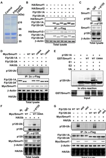
To identify new interacting proteins for Smurf1, we carried out affinity purification of Flag-tagged Smurf1-C699A, a catalytically inactive form of Smurf1 that may be used to trap the ligase substrates, followed by mass spectrometry analysis to determine the identities of Smurf1 binding proteins. Among the detected proteins, p120-catenin was identified (Fig. 1A). Because p120-catenin 1A and 3A are two predom-inantly expressed alternative splicing isoforms (3A lacks N-terminal 101 amino acids compared with 1A) in mesenchymal and epithelial cells (21–24), we therefore used p120-catenin 1A and 3A to examine the interaction between p120-catenin and Smurf1 by coimmunopre-cipitation assay. We found that both p120-catenin 1A and 3A specifi-cally bound to Smurf1 but not Smurf2, a closely related family member of Smurf1 (Fig. 1B). We further confirmed that p120-catenin interacts with Smurf1 endogenously (Fig. 1C). Moreover, we performed in vitro glutathione S-transferase (GST) pull-down assay using bacterially produced GST-tagged p120-catenin and His-tagged Smurf1 and found that p120-catenin directly interacts with Smurf1 (fig. S1A).
Because Smurf1 is an E3 ubiquitin ligase, we asked whether p120-catenin is a substrate for Smurf1. Overexpression of Smurf1 did not affect steady-state levels of either endogenous or exogenous p120-catenin (fig. S1, B and C) but induced a monoubiquitin mod-ification on both p120-catenin 1A and 3A isoforms (Fig. 1D). We also performed in vitro ubiquitination assay using bacterially pro-duced Smurf1 and p120-catenin and ascertained that Smurf1 could directly catalyze monoubiquitination of p120-catenin 1A and 3A (Fig. 1E and fig. S1D). Furthermore, we also confirmed that endogenous p120-catenin was able to be monoubiquitinated by Smurf1 (Fig. 1F). Thus, our results indicated that Smurf1 targets p120-catenin for a monoubiquitin, rather than a polyubiquitin, modification.
To identify the ubiquitination site(s) on p120-catenin, we first examined to which domain ubiquitin is conjugated. As shown in fig. S1E, p120-catenin and p120-catenin truncates containing the armadillo domain were able to be monoubiquitinated by Smurf1, whereas p120-catenin truncates without the armadillo domain were resistant to Smurf1-mediated ubiquitination, suggesting that the ubiquitination occurred in the armadillo domain. Therefore, we carried out in vitro ubiquitination reaction of p120-catenin in the presence of Smurf1 to obtain ubiquitinated p120-catenin for mass
spectrometry analysis. Ubiquitin conjugation was detected on four lysine residues (Lys355, Lys421, Lys422, and Lys517) in the armadillo
do-main of human p120-catenin 1A (Lys254, Lys320, Lys321, and Lys416 for 3A) (fig. S1F). Mutations of all the four lysine residues to arginines (4KR) completely abolished Smurf1-mediated ubiquitination of p120-catenin (Fig. 1G).
Smurf1-mediated monoubiquitination of p120-catenin is necessary for TGF-induced AJ and TJ dissociation
A previous study showed that overexpression of Smurf1 leads to dissociation of TJs between epithelial cells and transformation of epithelial cells to mesenchymal phenotype (20). We sought to in-vestigate whether Smurf1-mediated ubiquitination of p120-catenin has any role in regulating cell-cell junctions. As predicted, Smurf1-C699A showed well colocalization with endogenous p120-catenin and E-cadherin, whereas overexpression of wild-type Smurf1, but not Smurf2, resulted in dissociation of cell-cell junctions (fig. S2, A and B), confirming that catalytic activity of Smurf1 is responsible for cell-cell junction dissociation.
We next examined the role of p120-catenin ubiquitination in Smurf1-mediated junction dissociation using the ubiquitination- resistant p120-catenin 4KR mutants. Knockdown of p120-catenin totally disrupted AJs and drove Madin-Darby canine kidney (MDCK) cells from epithelial to mesenchymal morphology (fig. S2, C and D). Reintroduction of both wild-type and 4KR p120-catenin (1A or 3A) restored the AJs and epithelial phenotype of MDCK cells (fig. S2, E to G). Coexpression of Smurf1 could only break down cell-cell junctions in cells reintroduced with wild-type p120-catenin, but not the 4KR mutants (fig. S3, A and B), indicating that the ubiquitination of p120-catenin is required for Smurf1-mediated junction dissociation.
Because Smurf1 is essential for TJ dissociation during TGF- mediated EMT (20), we investigated whether Smurf1 also has a role in regulating AJ dissociation in this process. Knockdown of Smurf1, but not Smurf2, blocked TGF-induced dissociation of AJs (fig. S4, A and B), indicating that Smurf1 is also required for AJ dissociation. As p120-catenin is an important component of cadherin-catenin complex for maintaining the integrity of AJs, we therefore hypothe-sized that Smurf1-mediated monoubiquitination of p120-catenin might be required for dissociation of AJs in the process of TG-F-induced EMT. To verify this, we first examined whether TGF signaling could up-regulate p120-catenin monoubiquitination. TGF treatment notably enhanced monoubiquitination of p120-catenin (Fig. 2A), and knockdown of Smurf1 abolished TGF-induced p120- catenin monoubiquitination (Fig. 2B), indicating that TGF signaling induces p120-catenin monoubiquitination through Smurf1. Meanwhile, TGF only promoted ubiquitination of wild-type p120-catenin but not 4KR mutants (Fig. 2C), confirming that TGF-promoted monoubi-quitination of p120-catenin occurs at the Smurf1-targeted ubiquiti-nation site(s). In line with this, TGF treatment recruited Smurf1 to the AJ region and notably enhanced interaction between endogenous Smurf1 and p120-catenin (fig. S4, C and D).
We next examined whether the ubiquitination of p120-catenin is required for TGF-induced EMT. TGF treatment caused AJ disso-ciation in p120-catenin knockdown cells with reintroduction of wild- type human p120-catenin 1A or 3A; however, reintroduction of p120-catenin 4KR mutants significantly blocked TGF-induced AJ dissociation (Fig. 2, D and E). The 4KR mutants blocked not only TGF-induced dissociation of AJs but also the dissociation of TJs and reorganization of actin cytoskeleton (Fig. 2, F and G), indicating
on September 17, 2020
http://advances.sciencemag.org/
Fig. 1. Smurf1 monoubiquitinates p120-catenin. (A) Smurf1 interacts with p120-catenin. MDCK cells transfected with Flag-tagged catalytically inactive mutant
Smurf1-C699A (F/Smurf1-Smurf1-C699A) were subjected to anti-Flag immunoprecipitation (IP), followed by SDS–polyacrylamide gel electrophoresis and Coomassie brilliant blue staining. The arrow-indicated band was p120-catenin–analyzed by mass spectrometry. (B) Smurf1, but not Smurf2, interacts with p120-catenin. HEK293T cells transfected with indicated combinations of hemagglutinin (HA)–tagged Smurf1 (HA/Smurf1) or Smurf2 (HA/Smurf2) and Flag-tagged p120-catenin 1A (F/p120-1A) or p120-catenin 3A (F/p120-3A) were subjected to anti-Flag IP followed by immunoblotting assay to detect associated Smurfs. (C) Smurf1 and p120-catenin interact endogenously. Cell lysates from MDCK were subjected to anti–p120-catenin IP followed by immunoblotting to detect associated Smurf1. (D) Smurf1 mediates monoubiquitination of p120-catenin. HEK293T cells transfected with indicated combinations of HA-tagged ubiquitin (HA/Ub), Myc-tagged wild-type (WT) or C699A mutant Smurf1 (Myc/Smurf1), and Flag-tagged p120-catenin 1A or 3A (F/p120-1A or F/p120-3A) were subjected to anti-Flag IP, eluted by boiling in 1% SDS, and then reprecipitated with anti-Flag antibody (2× anti-Flag IP). Ubiquitin- conjugated p120-catenin (p120-Ub) was detected by immunoblotting with anti-HA antibody. (E) Smurf1 directly ubiquitinates p120-catenin 1A in vitro. GST-tagged Smurf1 (GST/Smurf1) (WT or C699A) and p120-catenin 1A (p120-1A) purified from bacteria were subjected to an in vitro ubiquitination assay. p120-1A and ubiquitinated p120-1A were detected with anti–p120-catenin antibody. Asterisk (*) indicates the monoubiquitinated p120-1A. (F) Smurf1 targets endogenous p120-catenin for monoubiquitination. MDCK cells transduced with lentivirus encoding Myc/Smurf1 (WT or C699A) and HA/Ub were subjected to anti–p120-catenin IP followed by immunoblotting with anti-HA antibody to detect conjugation of Ub to p120-catenin. (G) The 4KR mutation blocks Smurf1-mediated ubiquitination of p120-catenin. HEK293T cells transfected with in-dicated combinations of HA/Ub, Myc/Smurf1 (WT or C699A), and F/p120-1A (WT or 4KR) or F/p120-3A (WT or 4KR) were subjected to ubiquitination assay as in (D).
on September 17, 2020
http://advances.sciencemag.org/
Fig. 2. TGF promotes monoubiquitination of p120-catenin through Smurf1 to induce junction dissociation. (A) TGF treatment promotes monoubiquitination of endogenous p120-catenin. MDCK cells transduced with lentivirus encoding HA/Ub were treated for 8 hours with or without 200 pM TGF before being subjected to anti–p120-catenin IP followed by immunoblotting with anti-HA antibody to detect conjugation of Ub to p120-catenin. (B) TGF-promoted monoubiquitination of endogenous p120-catenin is through Smurf1. MDCK cells transduced with lentivirus encoding HA/Ub and control shRNA (sh-Con) or shRNA against Smurf1 (sh-Smurf1-1 or 2) were treated with TGF and subjected to ubiquitination assay. (C) 4KR mutation blocks TGF-promoted monoubiquitination of p120-catenin. MDCK cells transduced with lentivirus encoding HA/Ub and 1A or 3A F/p120 (WT or 4KR) were treated for 8 hours with or without 200 pM TGF and then subjected to ubiquitination assay. (D and E) Ubiquitination of p120-catenin is required for TGF-induced AJ dissociation. p120-KD MDCK cells transduced with lentivirus encoding WT or 4KR F/p120 (1A or 3A) were treated for 24 hours with or without 200 pM TGF and then subjected to immunofluorescence assay to examine AJs. Scale bars, 10 m (D). The percentages of cells with AJs were quantified and represented as mean ± SD in (E). (F and G) Ubiquitination of p120-catenin is required for TGF-induced TJ dissociation. The same cells as in (D) were subjected to immunofluorescence assay to examine TJs and actin cytoskeleton. Scale bars, 10 m (F). The percentages of cells with TJs were quantified and represented as mean ± SD in (G).
on September 17, 2020
http://advances.sciencemag.org/
that AJ dissociation is a prerequisite step for TJ dissociation during TGF-induced EMT. In agreement with this, reintroduction of the 4KR mutants notably blocked TGF-induced down-regulation of RhoA (fig. S4E), suggesting that Smurf1 has to target p120-catenin for ubiquitination to lead to AJ dissociation before it can target RhoA for degradation to reorganize actin cytoskeleton.
ERK1/2 phosphorylates p120-catenin to promote its binding to Smurf1
During our testing of the interaction between Smurf1 and p120- catenin, we observed that GST-Smurf1 showed a higher affinity to p120-catenin in the lysate of cells treated with TGF than that of cells without TGF treatment in GST pull-down assays (Fig. 3A), suggesting that TGF-enhanced binding of Smurf1 to p120-catenin is due not only to recruitment of Smurf1 to the AJ region but also to involvement of other factor(s). Because protein kinases such as ERK, JNK (c-Jun N-terminal kinase), p38, and PI3K/AKT are also engaged in TGF-induced EMT (18, 19), we therefore examined whether these kinases are responsible for TGF-enhanced interaction between Smurf1 and p120-catenin. The ERK inhibitor U0126 notably blocked TGF-enhanced Smurf1 and p120-catenin interaction, whereas the p38 inhibitor SB203580, the JNK inhibitor SP600125, and the AKT inhibitor wortmannin did not (Fig. 3B and fig. S5A), suggesting that ERK kinase activity is important for this interaction.
We further examined the role of ERK1 and ERK2 in regulating the interaction between Smurf1 and p120-catenin by knocking down ERK1 and/or ERK2. As shown in Fig. 3C, knocking down either ERK1 or ERK2 notably attenuated the endogenous interaction between Smurf1 and p120-catenin, and knocking down both ERK1 and ERK2 simultaneously nearly totally blocked this interaction, suggesting that both ERK1 and ERK2 are involved in controlling binding of Smurf1 to p120-catenin. Accordingly, both ERK1 and ERK2 could interact with p120-catenin 1A and 3A, respectively (Fig. 3D). Furthermore, we used kinase-dead mutant ERK1-K71R or ERK2-K52R to trap endogenous p120-catenin and found that interaction between ERK1/2 and endogenous p120-catenin was notably enhanced by TGF treatment (Fig. 3E).
We next carried out in vitro phosphorylation assay using consti-tutively active forms of ERK1/2 (ERK1-R84S and ERK2-R67S) and catalytically inactive mutants (ERK1-K71R and ERK2-K52R) as negative controls. We observed that ERK1/2 could phosphorylate both p120-catenin 1A and 3A on their threonine but not serine residue(s) by using phospho-Thr– and phospho-Ser–specific anti-bodies (Fig. 3F and fig. S5B). We identified that T308, T310, and T900 of human p120-catenin 1A (T207, T209, and T799 of human p120-catenin 3A, correspondingly, and hereafter referred to as same sites as in 1A) were phosphorylated by ERK1/2 using matrix-assisted laser desorption/ionization time-of-flight mass spectrometry (MALDI-TOF-MS), which are all conserved in mouse, dog, and human (fig. S5C). The phosphorylation of T310 had been previously reported (25), and there is commercial phosphospecific antibody for T310; however, the phosphorylation of T308 and T900 has not been reported before. We therefore generated phosphospecific antibodies for T308 and T900 of human p120-catenin 1A. We could detect the phosphorylation of T308, T310, and T900 using these phosphorylation site–specific anti-bodies, and mutations of all three threonine residues to alanines (3TA) totally abolished the ERK1/2-mediated phosphorylation of p120-catenin in the in vitro phosphorylation assay (Fig. 3G and fig. S5D), confirming that the ERK1/2-mediated p120-catenine
Fig. 3. ERK phosphorylates p120-catenin, and its activity is required for TGF- enhanced Smurf1 and p120-catenin interaction. (A) TGF treatment enhances interaction between Smurf1 and p120-catenin in vitro. Cell lysates from MDCK cells treated for 2 hours with or without 200 pM TGF were subjected to GST pull-down assay with GST/Smurf1 to detect associated p120-catenin. (B) ERK activity is required for TGF-enhanced interaction between Smurf1 and p120-catenin 1A. MDCK cells transduced with lentivirus encoding F/p120-1A were pretreated for 2 hours with indi-cated kinase inhibitors (20 M SB203580, 20 M SP600125, 0.25 M U0126, and 0.5 M wortmannin) before being treated for another 2 hours with or without 200 pM TGF. The cells were then subjected to anti-Flag IP followed by immunoblotting assay to detect associated Smurf1. (C) Both ERK1 and ERK2 are required for TGF-enhanced Smurf1 and p120-catenin interaction. MDCK cells transduced with lentivirus encoding sh-Con or shRNA against ERK1 or ERK2 (sh-ERK1 or sh-ERK2) were treated for 2 hours with or without 200 pM TGF and then subjected to anti–p120-catenin IP followed by immunoblotting assay to detect associated Smurf1. (D) Both ERK1 and ERK2 interact with p120-catenin 1A or 3A. HEK293T cells transfected with F/p120 (1A or 3A) and HA-tagged ERK1 or ERK2 (HA/ERK1 or HA/ERK2) were subjected to anti-Flag IP followed by immunoblotting assay to examine associated ERK1/2. (E) TGF treatment enhances interaction between ERK and endogenous p120-catenin. MDCK cells transduced with lentivirus encoding Flag-tagged kinase-dead mutant ERK1-K71R or ERK2-K52R (F/ERK1-K71R or F/ERK2-K52R) were treated for 1.5 hours with or without 200 pM TGF and then subjected to anti-Flag IP followed by immunoblotting assay to detect associated endogenous p120-catenin. (F) ERK2 phosphorylates p120-catenin at its threonine residues. In vitro kinase assay was carried out by incubating bacterially ex-pressed and purified p120-catenin (1A or 3A) with constitutive active mutant ERK2-R67S or kinase-dead mutant ERK2-K52R. Phosphorylated p120-catenin was detected by immunoblotting using phosphothreonine or phosphoserine antibodies. (G) 3TA mutation blocks ERK2-mediated phosphorylation of p120-catenin. Bacterially ex-pressed and purified WT or 3TA mutant p120-catenin (1A or 3A) was subjected to in vitro kinase assay with ERK2-R67S. Phosphorylated p120-catenin was detected by im-munoblotting using phosphothreonine or phosphorylation site–specific antibodies.
on September 17, 2020
http://advances.sciencemag.org/
phosphorylation in vitro is on these sites. Moreover, the interaction of Smurf1 with wild-type but not 3TA p120-catenin was markedly enhanced by the ERK-mediated phosphorylation in vitro (fig. S5, E and F), indicating that the phosphorylation of p120-catenin is required for its sufficient binding to Smurf1.
Phosphorylation of p120-catenin is required for TGF-induced monoubiquitination of p120-catenin and junction dissociation
Next, we examined whether p120-catenin phosphorylation plays a role in regulating p120-catenin monoubiquitination and AJ dissocia-tion in response to TGF treatment. Treatment with the ERK inhibitor U0126 markedly attenuated TGF-induced monoubiquitination of p120-catenin (Fig. 4A), which is in good agreement with the fact that ERK activity is required for the Smurf1 and p120-catenin interaction as shown above. In addition, U0126 significantly blocked TGF- induced AJ dissociation (fig. S6, A and B), indicating that ERK activity is necessary for TGF-mediated p120-catenin monoubiq-uitination and AJ dissociation.
We could only detect an enhanced ERK-mediated phosphorylation of p120-catenin after TGF treatment using phospho-T900 anti-body (Fig. 4B), suggesting that T900 is the major phosphorylation site in response to TGF treatment in cells. We confirmed the effect of TGF on inducing phosphorylation of T900, and the specificity of the phospho-T900 antibody by reintroducing human wild-type or T900A p120-catenin into MDCK cells with stable knockdown of endogenous p120-catenin (Fig. 4C and fig. S6C). The T900A muta-tion markedly attenuated TGF-promoted binding of endogenous Smurf1 to p120-catenin (Fig. 4D) and, accordingly, notably blocked TGF-induced monoubiquitination of p120-catenin (Fig. 4E). In line with this, reintroducing p120-catenin T900A mutant remarkably blocked TGF-induced AJ and TJ dissociation, and RhoA degrada-tion (Fig. 4F and fig. S6, D to H), indicating that the phospho ryladegrada-tion of T900 of p120-catenin is required for TGF-mediated p120-catenin monoubiquitination and junction dissociation. In addition, the 4KR mutations did not affect the TGF-induced phosphorylation of T900 (fig. S6I), further confirming that the T900 phosphorylation of p120-catenin is upstream of its monoubiquitination.
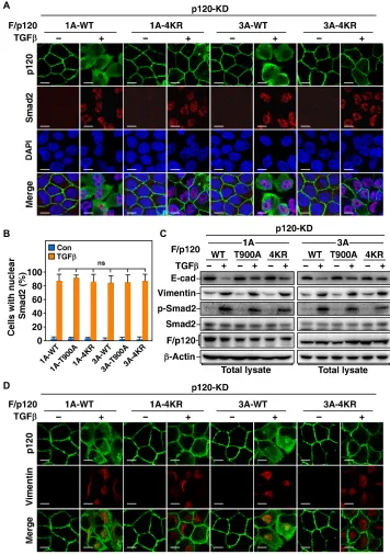
Smad activation is independent of the phosphorylation and monoubiquitination of p120-catenin
A previous study showed that Smad signaling can still be activated upon TGF treatment even though TJ dissociation is blocked (20), and we therefore investigated whether phosphorylation or monoubiquiti-nation of p120-catenin is required for activation of the Smad pathway. As predicted, although TGF-induced AJ dissociation was markedly blocked by impeding phosphorylation or monoubiquitination of p120-catenin, blockade of either phosphorylation or monoubiquiti-nation of p120-catenin had no significant effect on TGF-induced nuclear accumulation of Smad2 (Fig. 5, A and B, and fig. S7A), in-dicating that activation of Smad signaling by TGF is independent of the phosphorylation and monoubiquitination of p120-catenin and AJ dissociation. In line with this, blockade of phosphorylation or monoubiquitination of p120-catenin notably inhibited down- regulation of E-cadherin induced by TGF treatment; however, the levels of phosphorylation of Smad2 or expression of the mesenchy-mal marker protein vimentin were not affected (Fig. 5C). Accord-ingly, the cells with p120-catenin 4KR or T900A mutants showed similar expression of vimentin as that in the cells with wild-type
p120-catenin, even though the cells with the mutants still main-tained AJs after TGF treatment (Fig. 5D and fig. S7B).
Phosphorylation and monoubiquitination of p120-catenin are required for breast cancer metastasis
To determine the role of phosphorylation and monoubiquitination of p120-catenin in vivo, we reintroduced wild-type, T900A, or 4KR p120-catenin 1A and 3A to mouse breast cancer 4T1 p120-catenin knockout cells (fig. S8A) and then injected the cells into the mammary fat pad of female BALB/c mice to examine the primary tumor growth and lung metastasis. The phosphorylation and ubiquitination-resistant mutants of p120-catenin had no significant effect on primary tumor growth and AJ formation in the primary tumors (Fig. 6, A and B, and fig. S8B); however, they markedly attenuated the lung metastasis of the breast cancer cells (Fig. 6, C to E, and fig. S8C), indicating that the T900 phosphorylation and ubiquitination of p120-catenin cer-tainly play a pivotal role in breast cancer tumor metastasis. In line with the result obtained in cultured cells, phosphorylation of T900 was able to be detected in primary tumors that originated from 4T1 cells with wild-type and 4KR p120-catenin, but not from 4T1 cells with T900A p120-catenin (Fig. 6F), confirming that phosphorylation of p120-catenin at T900 happens in vivo.
To evaluate the correlation between T900 phosphorylation of p120-catenin and breast tumor invasiveness, we analyzed specimens from patients with breast cancer. For this purpose, we categorized the tumor samples into an invasive group and a noninvasive group, which were reported with or without lymph node metastasis on the basis of the pathological diagnosis, respectively. We found that the levels of T900 phosphorylation of p120-catenin are markedly higher in tumors with lymph node metastasis than in tumors without lymph node metastasis (Fig. 6, G and H, and fig. S8D), indicating a positive correlation between T900 phosphorylation of p120-catenin and malignancy of human breast cancer. Hence, our study revealed an underlying mechanism of AJ dissociation that plays a key role in TGF-induced EMT and tumor metastasis.
DISCUSSION
EMT is a complex process that involves changes in cell morphology and gene expression. Disassembly of cell-cell junctions including both TJs and AJs is essential for epithelial cells undergoing EMT to become individual mesenchymal cells. A previous study showed that TGF- induced EMT requires a Smurf1-mediated degradation of RhoA to dis-assemble cortical actin filaments (20). However, it is still vague as to how the junctions are dissolved. Our study here demonstrates that instead of simply by disassembling cortical actin filaments to break down the junctions, epithelial cells go through a stepwise procedure to dissolve the cell-cell junctions during EMT. The cells have to disrupt AJs by monoubiquitinating p120-catenin in the cadherin-catenin core com-plex before dismantling TJs and cortical actin filaments. The ubiq-uitination site is located in the armadillo domain of p120-catenin, which is responsible for the binding of p120-catenin to E-cadherin (26). It has been shown that the inter action between p120-catenin and E-cadherin through the armadillo domain of p120-catenin and juxtamembrane do-main of E-cadherin is critical for the stability of cell-cell adhesion com-plexes. Disruption of this interaction could lead to down-regulation of E-cadherin at the cell surface and destabilization of cadherin-mediated AJs (26). Ubiquitination of p120-catenin in the armadillo domain may interrupt its binding to E-cadherin and therefore causes dissociation
on September 17, 2020
http://advances.sciencemag.org/
Fig. 4. Phosphorylation of p120-catenin is necessary for TGF-promoted monoubiquitination of p120-catenin and junction dissociation. (A) ERK activity is
required for TGF-promoted monoubiquitination of p120-catenin. MDCK cells transduced with lentivirus encoding HA/Ub were pretreated for 2 hours with or without 0.25 M U0126 before being treated for another 8 hours with 200 pM TGF. The cells were then subjected to anti–p120-catenin IP followed by immunoblotting assay using anti-HA antibody to detect ubiquitin conjugation of endogenous p120-catenin. (B) TGF induces phosphorylation of T900 of p120-catenin 1A and 3A, respectively. MDCK cells transduced with lentivirus encoding F/p120-1A or F/p120-3A were pretreated for 2 hours with or without 0.25 M U0126 before being treated for another 2 hours with or without 200 pM TGF and then subjected to anti-FLAG IP followed by immunoblotting assay using phospho-specific antibody to detect the phosphoryl-ation of p120-catenin. (C) TGF-induced phosphorylation of p120-catenin 1A at T900. p120-KD MDCK cells transduced with lentivirus encoding WT or T900A mutant F/p120-1A were treated for 2 hours with or without 200 pM TGF and then subjected to immunoblotting assay. (D) TGF-mediated phosphorylation of p120-catenin is critical for the interaction between Smurf1 and p120-catenin. MDCK cells transduced with lentivirus encoding F/p120-1A or F/p120-3A (WT or T900A) were treated for 2 hours with or without 200 pM TGF and then subjected to anti-Flag IP followed by immunoblotting assay to detect associated Smurf1. (E) TGF-mediated phosphoryl-ation of p120-catenin is required for monoubiquitinphosphoryl-ation of p120-catenin. MDCK cells transduced with indicated combinphosphoryl-ations of lentivirus encoding HA/Ub, F/p120-1A or F/p120-3A (WT or T900A), were treated for 8 hours with or without 200 pM TGF and then subjected to ubiquitination assay with 2× anti-Flag IP. (F) Phosphorylation of p120-catenin is required for TGF-induced AJ dissociation. p120-KD MDCK cells transduced with lentivirus encoding F/p120-1A or F/p120-3A (WT or T900A) were treated for 24 hours with or without 200 pM TGF and then subjected to immunofluorescence assay to examine AJs. Scale bars, 10 m.
on September 17, 2020
http://advances.sciencemag.org/
of AJs. Hence, our study reveals a pivotal role of Smurf1-mediated monoubiquitination of p120-catenin in regulating the stability of AJs.
Because p120-catenin was originally identified as a substrate of Src kinase (27, 28), phosphorylation of p120-catenin has been extensively
studied (29). However, although many phosphorylation sites includ-ing both tyrosine and serine/threonine residues have been identified and proposed to exert important roles in development and tumor progress, little is known about how exactly the phosphoryl ation affects
Fig. 5. Phosphorylation and ubiquitination of p120-catenin are dispensable for TGF-induced nuclear translocation of Smad2. (A) Ubiquitination of p120-catenin
is not required for TGF-induced nuclear translocation of Smad2. p120-KD MDCK cells with expression of F/p120-1A or F/p120-3A (WT or 4KR) were treated for 24 hours with or without 200 pM TGF and then subjected to immunofluorescence assay to examine nuclear localization of Smad2. Scale bars, 10 m. (B) Phosphorylation and ubiquitination are not required for nuclear translocation of Smad2 in response to TGF treatment. p120-KD MDCK cells with expression of F/p120-1A or F/p120-3A (WT, 4KR, or T900A) were treated as in (A), and the percentages of cells with nuclear accumulation of Smad2 were quantified and presented as mean ± SD of three independent experiments. (C) Blockade of p120-catenin phosphorylation or ubiquitination protects E-cadherin from TGF-promoted turnover but does not affect Smad2 phosphoryl-ation or vimentin expression. p120-KD MDCK cells with expression of F/p120-1A or F/p120-3A (WT, T900A, or 4KR) were treated for 24 hours with or without 200 pM TGF and then subjected to immunoblotting assay. (D) Blockade of p120-catenin ubiquitination does not affect TGF-induced Smad2 phosphorylation or vimentin expression. The same cells as in (A) were subjected to immunofluorescence assay to examine vimentin expression.
on September 17, 2020
http://advances.sciencemag.org/
Fig. 6. Phosphorylation and ubiquitination of p120-catenin are essential for breast cancer metastasis. (A) Phosphorylation- and ubiquitination-resistant mutations of
p120-catenin do not affect primary tumor growth. p120-catenin knockout (p120-KO) 4T1 cells transduced with lentivirus encoding F/p120-1A and F/p120-3A (WT, T900A, or 4KR) (1 × 10⁶) were orthotopically injected into the mammary fat pad of BALB/c mice to form primary breast tumor. The mice were sacrificed 25 days later, the primary tumors
were weighted, and the data were presented as mean ± SD of eight mice per group. (B) The p120-catenin mutations have no influence on AJ formation in primary tumors. Primary tumors in (A) were subjected to histological assay to examine AJs. Scale bars, 50 m. (C to E) Impeded phosphorylation or ubiquitination of p120-catenin significant-ly blocks lung metastasis of mouse breast cancer cells. Representative images of lung metastasis nodules (C) and hematoxylin and eosin (H&E)–stained lung sections (D) were obtained from mice in (A). The numbers of lung metastasis nodules were counted and presented as mean ± SD of eight mice per group (E). (F) The 4KR mutation does not block the T900 phosphorylation of p120-catenin in primary tumors. Lysates of primary tumors in (A) were subjected to immunoblotting assay. (G) Comparison of T900 phos-phorylation in invasive and noninvasive human breast cancer primary tumors. The phosphos-phorylation levels of T900 were determined by immunoblotting the extracts from three representative invasive tumor tissues and three noninvasive tumor tissues. (H) Phosphorylation of p120-catenin at T900 in primary tumors is positively correlated with lymph node invasion in human breast cancer specimens. A total of 27 samples of human breast cancer were classified into two groups based on the status of lymph node invasion, and the relative levels of T900 phosphorylation of p120-catenin were examined by immunoblotting assay (photo credit: Qingang Wu, Xiamen University).
on September 17, 2020
http://advances.sciencemag.org/
the interaction between p120-catenin and E-cadherin (29). The phos-phorylation of T900 in p120-catenin identified in this study presents a direct linkage between phosphorylation and mono ubiquitination of p120-catenin, revealing a mechanism of how phos phorylation regu-lates the binding of p120-catenin to E-cadherin. We observed that the levels of T900 phosphorylation and mono ubiquitination of p120- catenin increase and then decrease after TGF treatment, suggesting a dynamic regulation of the phosphoryl ation and monoubiquitination of p120-catenin during EMT through a yet unknown mechanism.
In good agreement with a previous study that blockade of TGF signaling–induced disassembly of cortical actin by abolishing phos-phorylation of Par6, a key component of polarity complex, does not affect Smad activation and downstream gene expression (20), hin-drance of AJ dissociation by abrogating TGF-triggered phosphoryl-ation or ubiquitinphosphoryl-ation of p120-catenin does not block Smad activphosphoryl-ation and downstream gene expression either, indicating that TGF- mediated AJ dissociation through phosphorylation and ubiquitination of p120-catenin is in the same signaling cascade with TGF-mediated cortical actin disassembly via phosphorylation of Par6 and ubiquitina-tion of RhoA. Unlike TGF-induced polyubiquitination of RhoA that results in a degradation, the ubiquitination pattern of p120-catenin induced by TGF signaling is monoubiquitination, which affects the interaction between p120-catenin and E-cadherin but does not cause p120-catenin degradation. It has been reported that p120-catenin is shifted from cell-cell junctions to cytoplasm during EMT (30); however, the underlying mechanism as to how p120-catenin is translo-cated into cytosol remains unclear. Further exploration of how the monoubiquitination affects the localization switch of p120- catenin might provide more insight to understand this molecular event.
It is well known that EMT has a fundamental role in many physi-ological and pathphysi-ological processes such as embryonic development, tissue repair, fibrosis, and tumor metastasis (8, 11). In addition to TGF signaling, many other signaling pathways including bone morphogenetic protein (BMP), Wnt, Notch, Hedgehog, epidermal growth factor (EGF), fibroblast growth factor (FGF), platelet-derived growth factor (PDGF), and integrin are also involved in the regula-tion of EMT to respond to various extracellular signals (31–36). The signals have to be integrated through cross-talk among these path-ways to properly control EMT. In line with our result, a previous report indicated that ERK1/2 activity is necessary for TGF-induced EMT (18). Meanwhile, ERK1/2 pathway has been shown to be involved in most of the signaling pathways that induce EMT (37). Therefore, it would be of great interest to investigate whether ERK/Smurf1 or potential other E3 ubiquitin ligase(s)–mediated ubiquitination of p120-catenin is a general requisite of the EMT process in different biological contexts. Abolishing either the ERK-mediated phosphoryl-ation or Smurf1-mediated ubiquitinphosphoryl-ation of p120-catenin markedly attenuated metastasis of breast cancer in mice, and consistently, the T900 phosphorylation levels of p120-catenin in the specimens from breast cancer patients present a significant positive correlation with cancer malignancy, suggesting that the mechanism we identified in this study plays a key role in tumor metastasis. Hence, our study points out a potential strategy to target tumor metastasis.
MATERIALS AND METHODS
DNA constructs
The complementary DNAs (cDNAs) of human p120-catenin, ERK1, and ERK2 were gifts from J. Han. Mutations of p120-catenin and
ERK1/2 were generated by polymerase chain reaction (PCR)–based site-directed mutagenesis. Cloning for protein expression in mam-malian cells was carried out using a pCMV6 vector for transfection and pBOBI and pCDH-EF1-MCS-IRES-puro vectors for lentivirus infection. pGEX-4T-1 and pRroEX were used for bacterial expression of proteins. Human Smurf1 (wild-type and C699A) and Smurf2 had been previously reported (38). Yellow fluorescent protein (YFP)– tagged Smurf1 had been previously described (39). The lentiviral- based vector pLL3.7 was used for short hairpin RNA (shRNA) expression. The sequences used in MDCK cells for expression of p120-catenin shRNA-1 and shRNA-2 are 5′-GCACTTGTGC-TATCGCAAT-3′ and 5′-GCACGAACGGGGAAGTTTAGC-3′, respectively; 5′-GCATTCTGGCTGAGATGCTCT-3′ for ERK1 shRNA; 5′-GCGCTTCAGACATGAGAAC-3′ for ERK2 shRNA; 5′-TATTCTACGGACAACATTT-3′ and 5′-GATAGGCACTG-GAGGCTCTGT-3′ for Smurf1 shRNA-1 and shRNA-2, respectively; and 5′-GCTGGATTTCTTGGTTGTGTT-3′ and 5′-GTGTGGA-TACTTGAGAATGAT-3′ for Smurf2 shRNA-1 and shRNA-2, respectively. The scramble sequence 5′-TTCTCCGAACGTGG-CACGA-3′ was used for a control shRNA.
Antibodies and chemical reagents
Rabbit anti-ERK1/2 (1:2000, #9102s), anti–phospho-ERK1/2 (1:2000, #4370T), anti-AKT (pan) (C67E7) (1:2000, #4691s), anti– phospho-AKT (Ser473) (193H12) (1:2000, #4058), anti–c-Jun (60A8) (1:2000, #9165), anti–phospho–c-Jun (Ser73) (1:2000, #9164), anti–
MAPKAPK-2 (1:2000, #3042), anti–phospho-MAPKAPK-2 (Thr334) (27B7) (1:2000, #3007), anti–phosphothreonine (1:1000,
9381), anti-Smad2 (86F7) (1:2000, #3122s), and anti–phospho- Smad2 (S465/467) (1:2000, #3108s) were purchased from Cell Signal-ing Technology. Mouse anti- Myc (1:2000, sc-40), anti-GST (B-14) (1:2000, sc-138), and anti- actin (C4) (1:2000, sc-47778) were pur-chased from Santa Cruz Biotechnology (Santa Cruz, CA, USA). Mouse anti-Smurf1 (1:2000, ab57573) and goat anti-rabbit immu-noglobulin G (IgG) H&L [horseradish peroxidase (HRP)] (1:5000, ab27236), anti-mouse IgG H&L (HRP) (1:5000, ab27241), and anti- rat IgG H&L (HRP) (1:5000, ab97057) were purchased from Abcam. Mouse anti-p120 (1:2000, 610134), anti–phospho-human p120 catenin (T310) (1:2000, 558203), and anti-vimentin (1:2000, 550513) were purchased from BD. Alexa Fluor 488 donkey anti-mouse (1:500, A21202), Alexa Fluor 555 donkey anti-mouse (1:500, A31570), Alexa Fluor 488 donkey anti-rat (1:500, A21208), Alexa Fluor 647 donkey anti-rat (1:500, A21247), Alexa Fluor 488 donkey anti- rabbit (1:500, A21206), Alexa Fluor 555 donkey anti-rabbit (1:500, A31572), and Alexa Fluor TM 555 phalloidin (1:200, A34055) were purchased from Thermo Fisher Scientific. Mouse anti-Flag (M2) (1:2000, F1804) and rat anti– E-cadherin (1:2000, U3254) were pur-chased from Sigma-Aldrich. Rat anti–ZO-1 (1:2000, MABT11) and mouse anti-phosphoserine (1:1000, #05-1000) were purchased from Merck Millipore. Rabbit anti- Flag (1:200, #20543-1-AP) was purchased from Proteintech. Rat anti- hemagglutinin (HA) (1:2000, #11867423001) was purchased from Roche (Mannheim, Germany). Inhibitors for MEK (MAPK kinase) U0126-EtOH (HY-12031), P38 MAPK SB203580 (HY-10256), JNK SP600125 (HY-12041), pro-tease inhibitor cocktail (HY-K0010), phosphatase inhibitor cocktail І (HY-K0021), and phosphatase inhibitor cocktail ІI (HY-K0022) were purchased from MedChem Express. Inhibitor for AKT wort-mannin (#9951) was purchased from Cell Signaling Technology. 4′,6-Diamidino-2-phenylindole (DAPI; D1306) was purchased
on September 17, 2020
http://advances.sciencemag.org/
from Thermo Fisher Scientific. N-ethylmaleimide (A600450) was purchased from Sangon Biotech.
Cell culture and TGF treatment
Human embryonic kidney (HEK) 293T and mouse breast cancer 4T1 were purchased from the American Type Culture Collection. MDCK (NBL-2) cells were obtained from the Cell Bank of the Chinese Academy of Sciences (Shanghai). HEK293T and MDCK cells were cultured in high-glucose Dulbecco’s modified Eagle’s medium (DMEM), and 4T1 was cultured in RPMI 1640, all supple-mented with 10% (v/v) fetal bovine serum (FBS) (Thermo Fisher Scientific) and streptomycin and penicillin (100 U/ml; Millipore) at 37°C in a humidified 5% CO2 incubator. The cell lines were
routinely tested and found negative for mycoplasma. For TGF treatment, cells were washed with phosphate-buffered saline (PBS), cultured in DMEM with 0.05% FBS for 8 hours, and then treated with TGF for a determined time.
Transfection and lentivirus infection
Plasmid transient transfection was performed using the polyethyleni-mine (PEI) method. Plasmids and PEI mixture with a ratio of 3:1 (w/w) were directly added to cell culture, and it was replaced with fresh medium 12 hours after transfection to avoid notable toxicity. Recom-binant lentivirus for infection was generated using the ViraPower Lentiviral Expression System (Invitrogen).
Generation of p120-catenin knockout cell line
Mouse 4T1 cells were used to generate p120-catenin knockout cell line using the pX330 CRISPR-Cas9 vector (40), and the gene-specific region of the guide RNA (gRNA) sequences was designed by the CRISPR design tool from the Zhang laboratory (http://crispr.mit.edu/). The two gRNA sequences for mouse p120-catenin are 5′-AGTTACAG-TAGCTTCCGGAG-3′ and 5′-GTTTGGATGCGTAGTTGCCA-3′. Single clones were picked up, and the efficiency of p120-catenin knockout was assessed by Western blot.
Immunoprecipitation and immunoblotting assays
Immunoprecipitation and immunoblotting assays were performed as previously described (41). Briefly, cells were lysed on ice with lysis buf-fer TNTE 0.5% [50 mM tris-HCl (pH 7.5), 150 mM NaCl, 1 mM EDTA, and 0.5% Triton X-100, containing pepstatin A (10 g/ml), leupeptin (10 g/ml), and 1 mM phenylmethylsulfonyl fluoride (PMSF)] and then subjected to immunoprecipitation or immuno-blotting assays with appropriate antibodies.
GST pull-down assay
For GST pull-down assay, bacterially expressed GST–p120-catenin and GST-Smurf1 were purified using glutathione Sepharose beads in TNTE 0.5% buffer. Bacterially expressed His-Smurf1 was puri-fied using nickel beads in 0.5% TNT [50 mM tris-HCl (pH 7.5), 150 mM NaCl, 0.5% Triton X-100, containing pepstatin A (10 mg/ml), leupeptin (10 mg/ml), and 1 mM PMSF]. Purified His-Smurf1 was incubated with beads bound with GST (control) or GST–p120- catenin in TNTE 0.5% buffer at 4°C for 3 hours before washing five times with the same buffer and then subjected to immunoblot-ting assay. The lysates of cells treated with or without TGF were incubated with GST-Smurf1 in TNTE 0.5% buffer at 4°C for 3 hours to detect the interaction between GST-Smurf1 and endogenous p120-catenin.
Ubiquitination assay
Ubiquitination assay in cells or in vitro had been previously de-scribed (41). For ubiquitination assay of Flag-tagged p120-catenin in cells, cell lysates were subjected to anti-Flag immunoprecipi-tation, eluted by boiling 5 min in 1% SDS, diluted 10 times in lysis buffer TNTE 0.5%, and then reimmunoprecipitated with anti-Flag (2× immunoprecipitation). The ubiquitin-conjugated proteins were detected by immunoblotting. For ubiquitination assay of endogenous p120-catenin, cell lysates were subjected to anti–p120-catenin immunoprecipitation followed by immuno-blotting. For in vitro ubiquitination assay of p120-catenin, free p120-catenin proteins were obtained by the tobacco etch virus (TEV) protease cleavage of purified GST-p120.
In vitro kinase assay
For in vitro kinase assay, bacterially expressed wild-type or mutant ERK1/2 and p120-1A/3A proteins were incubated in kinase reac-tion buffer [20 mM tris-HCl (pH 7.5), 10 mM MgCl2, 1 mM
dithio-threitol, 25 M adenosine triphosphate (ATP)] at 37°C for 1 hour before being subjected to immunoblotting assay.
Immunofluorescence assay
Cells grown on glass coverslips were washed three times with PBS, fixed with 4% paraformaldehyde, and permeabilized with 0.25% Triton X-100. The cells were then stained using appro-priate primary and proper fluorescently conjugated secondary antibodies. Alexa Fluor 555–conjugated phalloidin was used for F-actin staining. Images were obtained using a Zeiss LSM 780 con-focal microscope with ZEN 2010 software (Carl Zeiss GmbH, Jena, Germany).
Histological assays
For immunohistochemistry analysis of the lung metastasis, the lungs were fixed in 4% (v/v) paraformaldehyde for 24 hours, em-bedded in paraffin, and sectioned at 5 m. The slides were dried at 60°C for at least 1 hour, deparaffinized with 100% xylene and a graduated series of ethanol from 100 to 50% ethanol, and then stained with hematoxylin and eosin (H&E) followed by washing with H2O. The primary tumor tissues of mice were removed and
subsequently embedded in tissue-freezing medium in cassettes for facilitation of tissue sectioning. The slides were incubated with ap-propriate primary antibodies and fluorescently conjugated secondary antibodies and then subjected to imaging using a Zeiss LSM 780 confocal microscope with ZEN 2010 software (Carl Zeiss GmbH, Jena, Germany).
In vivo metastasis assays
Female BALB/c mice (6 weeks old) were purchased from and housed in the Laboratory Animal Center of Xiamen University (China) in a facility with 12-hour light/12-hour dark cycles under pathogen-free conditions. Mouse experiments were performed in accordance with protocols approved by the Institutional Animal Care and Use Committee of Xiamen University. For the lung metas-tasis experiments, wild-type or p120-catenin knockout 4T1 cells with or without reintroduction of wild-type, T900A, or 4KR p120- catenin (1 × 10⁶) were harvested in PBS (40 l) and injected into the
mammary fat pad of the mice. The mice were sacrificed 25 days after injection, the number of lung metastasis colonies was counted, and the primary tumors were weighted.
on September 17, 2020
http://advances.sciencemag.org/
Patient samples
Primary human breast cancer tissue samples and corresponding adjacent normal tissues were obtained in accordance with research ethics board approval from Xiamen University and the First Affiliated Hospital. All samples taken after surgery were stocked in liquid nitro-gen for further immunoblotting assay. Informed consent was ob-tained from all patients.
Statistical analysis
One-way analysis of variance (ANOVA) with least significant dif-ference post hoc test was used to compare values among different experimental groups using the GraphPad Prism program version 6.01.
P < 0.05 was considered a statistically significant change. *P < 0.05;
**P < 0.01; ***P < 0.001; NS, not significant. All the values were
presented as mean ± SD of at least triplicate experiments.
SUPPLEMENTARY MATERIALS
Supplementary material for this article is available at http://advances.sciencemag.org/cgi/ content/full/6/4/eaay9819/DC1
Fig. S1. Smurf1 targets p120-catenin for monoubiquitination. Fig. S2. 4KR mutation does not affect assembly of cell-cell junctions.
Fig. S3. Overexpression of Smurf1 disrupts cell junctions by ubiquitinating p120-catenin. Fig. S4. Smurf1-mediated monoubiquitination of p120-catenin is required for TGF-induced junction dissociation.
Fig. S5. p120-catenin is phosphorylated by ERK.
Fig. S6. Phosphorylation of p120-catenin is required for p120-catenin monoubiquitination and junction dissociation induced by TGF treatment.
Fig. S7. TGF-induced nuclear translocation of Smad2 is not dependent on phosphorylation of p120-catenin.
Fig. S8. Both phosphorylation and ubiquitination of p120-catenin are required for breast cancer metastasis but not tumorigenesis.
View/request a protocol for this paper from Bio-protocol.
REFERENCES AND NOTES
1. M. A. Garcia, W. J. Nelson, N. Chavez, Cell-cell junctions organize structural and signaling networks. Cold Spring Harb. Perspect. Biol. 10, a029181 (2018).
2. T. Yano, H. Kanoh, A. Tamura, S. Tsukita, Apical cytoskeletons and junctional complexes as a combined system in epithelial cell sheets. Ann. N. Y. Acad. Sci. 1405, 32–43 (2017).
3. C. Zihni, C. Mills, K. Matter, M. S. Balda, Tight junctions: From simple barriers to multifunctional molecular gates. Nat. Rev. Mol. Cell Biol. 17, 564–580 (2016). 4. J. Piontek, L. Winkler, H. Wolburg, S. L. Muller, N. Zuleger, C. Piehl, B. Wiesner, G. Krause,
I. E. Blasig, Formation of tight junction: Determinants of homophilic interaction between classic claudins. FASEB J. 22, 146–158 (2008).
5. P. Coopman, A. Djiane, Adherens junction and E-cadherin complex regulation by epithelial polarity. Cell. Mol. Life Sci. 73, 3535–3553 (2016).
6. M. Takeichi, Dynamic contacts: Rearranging adherens junctions to drive epithelial remodelling. Nat. Rev. Mol. Cell Biol. 15, 397–410 (2014).
7. A. Kourtidis, S. P. Ngok, P. Z. Anastasiadis, p120 catenin: An essential regulator of cadherin stability, adhesion-induced signaling, and cancer progression. Prog. Mol. Biol. Transl. Sci.
116, 409–432 (2013).
8. M. A. Nieto, R. Y. Huang, R. A. Jackson, J. P. Thiery, EMT: 2016. Cell 166, 21–45 (2016). 9. S. Lamouille, J. Xu, R. Derynck, Molecular mechanisms of epithelial-mesenchymal
transition. Nat. Rev. Mol. Cell Biol. 15, 178–196 (2014).
10. R. Derynck, B. P. Muthusamy, K. Y. Saeteurn, Signaling pathway cooperation in TGF--induced epithelial-mesenchymal transition. Curr. Opin. Cell Biol. 31, 56–66 (2014).
11. D. M. Gonzalez, D. Medici, Signaling mechanisms of the epithelial-mesenchymal transition.
Sci. Signal. 7, re8 (2014).
12. C. J. David, J. Massague, Contextual determinants of TGF action in development, immunity and cancer. Nat. Rev. Mol. Cell Biol. 19, 419–435 (2018).
13. H. Peinado, D. Olmeda, A. Cano, Snail, Zeb and bHLH factors in tumour progression: An alliance against the epithelial phenotype? Nat. Rev. Cancer 7, 415–428 (2007). 14. H. Peinado, M. Quintanilla, A. Cano, Transforming growth factor -1 induces snail
transcription factor in epithelial cell lines: Mechanisms for epithelial mesenchymal transitions. J. Biol. Chem. 278, 21113–21123 (2003).
15. T. Vincent, E. P. Neve, J. R. Johnson, A. Kukalev, F. Rojo, J. Albanell, K. Pietras, I. Virtanen, L. Philipson, P. L. Leopold, R. G. Crystal, A. G. de Herreros, A. Moustakas, R. F. Pettersson,
J. Fuxe, A SNAIL1-SMAD3/4 transcriptional repressor complex promotes TGF- mediated epithelial-mesenchymal transition. Nat. Cell Biol. 11, 943–950 (2009).
16. R. Derynck, Y. E. Zhang, Smad-dependent and Smad-independent pathways in TGF- family signalling. Nature 425, 577–584 (2003).
17. A. Moustakas, C.-H. Heldin, Non-Smad TGF- signals. J. Cell Sci. 118, 3573–3584 (2005). 18. L. Xie, B. K. Law, A. M. Chytil, K. A. Brown, M. E. Aakre, H. L. Moses, Activation of the Erk
pathway is required for TGF-1-induced EMT in vitro. Neoplasia 6, 603–610 (2004). 19. J. Xu, S. Lamouille, R. Derynck, TGF--induced epithelial to mesenchymal transition.
Cell Res. 19, 156–172 (2009).
20. B. Ozdamar, R. Bose, M. Barrios-Rodiles, H. R. Wang, Y. Zhang, J. L. Wrana, Regulation of the polarity protein Par6 by TGF receptors controls epithelial cell plasticity.
Science 307, 1603–1609 (2005).
21. S. Aho, L. Levänsuo, O. Montonen, C. Kari, U. Rodeck, J. Uitto, Specific sequences in p120ctn determine subcellular distribution of its multiple isoforms involved in cellular adhesion of normal and malignant epithelial cells. J. Cell Sci. 115, 1391–1402 (2002). 22. A. Keirsebilck, S. Bonne, K. Staes, J. van Hengel, F. Nollet, A. Reynolds, F. van Roy,
Molecular cloning of the human p120ctn catenin gene (CTNND1): Expression of multiple alternatively spliced isoforms. Genomics 50, 129–146 (1998).
23. Y. Y. Mo, A. B. Reynolds, Identification of murine p120 isoforms and heterogeneous expression of p120cas isoforms in human tumor cell lines. Cancer Res. 56, 2633–2640 (1996). 24. M. Yanagisawa, D. Huveldt, P. Kreinest, C. M. Lohse, J. C. Cheville, A. S. Parker,
J. A. Copland, P. Z. Anastasiadis, A p120 catenin isoform switch affects Rho activity, induces tumor cell invasion, and predicts metastatic disease. J. Biol. Chem. 283, 18344–18354 (2008).
25. X. Xia, J. Brooks, R. Campos-Gonzalez, A. B. Reynolds, Serine and threonine phospho-specific antibodies to p120-catenin. Hybrid Hybridomics 23, 343–351 (2004). 26. N. Ishiyama, S. H. Lee, S. Liu, G. Y. Li, M. J. Smith, L. F. Reichardt, M. Ikura, Dynamic
and static interactions between p120 catenin and E-cadherin regulate the stability of cell-cell adhesion. Cell 141, 117–128 (2010).
27. D. F. Aghib, P. D. McCrea, The E-cadherin complex contains the src substrate p120.
Exp. Cell Res. 218, 359–369 (1995).
28. A. B. Reynolds, D. J. Roesel, S. B. Kanner, J. T. Parsons, Transformation-specific tyrosine phosphorylation of a novel cellular protein in chicken cells expressing oncogenic variants of the avian cellular src gene. Mol. Cell. Biol. 9, 629–638 (1989).
29. J. Y. Hong, I. H. Oh, P. D. McCrea, Phosphorylation and isoform use in p120-catenin during development and tumorigenesis. Biochim. Biophys. Acta 1863, 102–114 (2016). 30. D. I. Bellovin, R. C. Bates, A. Muzikansky, D. L. Rimm, A. M. Mercurio, Altered localization
of p120 catenin during epithelial to mesenchymal transition of colon carcinoma is prognostic for aggressive disease. Cancer Res. 65, 10938–10945 (2005). 31. A. E. Al Moustafa, A. Achkhar, A. Yasmeen, EGF-receptor signaling and
epithelial-mesenchymal transition in human carcinomas. Front. Biosci. 4, 671–684 (2012). 32. E. M. De Francesco, M. Maggiolini, A. M. Musti, Crosstalk between Notch, HIF-1alpha
and GPER in breast cancer EMT. Int. J. Mol. Sci. 19, E2011 (2018).
33. M. Katoh, H. Nakagama, FGF receptors: Cancer biology and therapeutics. Med. Res. Rev.
34, 280–300 (2014).
34. F. A. Mamuya, M. K. Duncan, aV integrins and TGF--induced EMT: A circle of regulation.
J. Cell. Mol. Med. 16, 445–455 (2012).
35. N. McCormack, S. O'Dea, Regulation of epithelial to mesenchymal transition by bone morphogenetic proteins. Cell. Signal. 25, 2856–2862 (2013).
36. J. Taipale, P. A. Beachy, The Hedgehog and Wnt signalling pathways in cancer.
Nature 411, 349–354 (2001).
37. M. Olea-Flores, M. D. Zuniga-Eulogio, M. A. Mendoza-Catalan, H. A. Rodriguez-Ruiz, E. Castaneda-Saucedo, C. Ortuno-Pineda, T. Padilla-Benavides, N. Navarro-Tito, Extracellular-signal regulated kinase: A central molecule driving epithelial-mesenchymal transition in cancer. Int. J. Mol. Sci. 20, E2885 (2019).
38. M. Wang, L. Guo, Q. Wu, T. Zeng, Q. Lin, Y. Qiao, Q. Wang, M. Liu, X. Zhang, L. Ren, S. Zhang, Y. Pei, Z. Yin, F. Ding, H. R. Wang, ATR/Chk1/Smurf1 pathway determines cell fate after DNA damage by controlling RhoB abundance. Nat. Commun. 5, 4901 (2014). 39. H. R. Wang, Y. Zhang, B. Ozdamar, A. A. Ogunjimi, E. Alexandrova, G. H. Thomsen,
J. L. Wrana, Regulation of cell polarity and protrusion formation by targeting RhoA for degradation. Science 302, 1775–1779 (2003).
40. L. Cong, F. A. Ran, D. Cox, S. Lin, R. Barretto, N. Habib, P. D. Hsu, X. Wu, W. Jiang, L. A. Marraffini, F. Zhang, Multiplex genome engineering using CRISPR/Cas systems.
Science 339, 819–823 (2013).
41. H. R. Wang, A. A. Ogunjimi, Y. Zhang, B. Ozdamar, R. Bose, J. L. Wrana, Degradation of RhoA by Smurf1 ubiquitin ligase. Methods Enzymol. 406, 437–447 (2006).
Acknowledgments
Funding: This work was supported by the National Natural Science Foundation of China
(U1605222, 31970742), the National Key Research and Development Project of China (2016YFC1302400), and the Open Research Fund of the State Key Laboratory of Cellular Stress
on September 17, 2020
http://advances.sciencemag.org/
Biology, Xiamen University (SKLCSB2019KF009). Author contributions: Q.W., G.L., C.W., T.Z., Y.F., C.L., and G.-F.F. conducted the experiments and analyzed the data. Q.L. performed molecular biology experiments. L.H. carried out immunohistochemistry assay, and C.X. performed mass spectrometry. L.X. and H.-L.C. analyzed the data. T.-J.Z. generated knockout cells. P.P. and Z.O. provided study materials. G.F., X.L.C., and H.-R.W. designed the experiments and wrote the manuscript. Competing interests: The authors declare that they have no competing interests. Data and materials availability: All data needed to evaluate the conclusions in the paper are present in the paper and/or the Supplementary Materials. Additional data related to this paper may be requested from the authors.
Submitted 2 August 2019 Accepted 20 November 2019 Published 22 January 2020 10.1126/sciadv.aay9819
Citation: Q. Wu, G. Li, C. Wen, T. Zeng, Y. Fan, C. Liu, G.-F. Fu, C. Xie, Q. Lin, L. Xie, L. Huang, P. Pu, Z. Ouyang, H.-L. Chan, T.-J. Zhao, X. L. Chen, G. Fu, H.-R. Wang, Monoubiquitination of p120-catenin is essential for TGF-induced epithelial-mesenchymal transition and tumor metastasis. Sci. Adv. 6, eaay9819 (2020).
on September 17, 2020
http://advances.sciencemag.org/
Xie, Lei Huang, Pengpeng Pu, Zhong Ouyang, Hong-Lin Chan, Tong-Jin Zhao, Xiao Lei Chen, Guo Fu and Hong-Rui Wang Qingang Wu, Gao Li, Chengwen Wen, Taoling Zeng, Yuxi Fan, Chunyan Liu, Guo-Feng Fu, Changchuan Xie, Qi Lin, Liping
DOI: 10.1126/sciadv.aay9819 (4), eaay9819.
6
Sci Adv
ARTICLE TOOLS http://advances.sciencemag.org/content/6/4/eaay9819
MATERIALS
SUPPLEMENTARY http://advances.sciencemag.org/content/suppl/2020/01/17/6.4.eaay9819.DC1
REFERENCES
http://advances.sciencemag.org/content/6/4/eaay9819#BIBL
This article cites 41 articles, 12 of which you can access for free
PERMISSIONS http://www.sciencemag.org/help/reprints-and-permissions
Terms of Service
Use of this article is subject to the
is a registered trademark of AAAS.
Science Advances
York Avenue NW, Washington, DC 20005. The title
(ISSN 2375-2548) is published by the American Association for the Advancement of Science, 1200 New
Science Advances
License 4.0 (CC BY-NC).
Science. No claim to original U.S. Government Works. Distributed under a Creative Commons Attribution NonCommercial Copyright © 2020 The Authors, some rights reserved; exclusive licensee American Association for the Advancement of
on September 17, 2020
http://advances.sciencemag.org/