only to restoring lost functionality, but also in-cludes reminding patients and their families that pharmacological and invasive treatment must be complemented by lifestyle changes. Today, cardi-ac rehabilitation is one of the basic objectives in-herent in the secondary prevention of cardiovas-cular disease [1].
The strictly controlled physical training which is part of cardiac rehabilitation produces some changes in the functioning of the cardiovascular The last decade of the 20th century was
a peri-od of fascination with the undeniable achievements in cardiology, cardiac surgery, intensive care and the progress of pharmacotherapy. Although treat-ments such as coronary artery bypass grafting or percutaneous coronary interventions were perfec-tion, medical science failed to eliminate or at least reduce the underlying cause of cardiovascular dis-ease: Atherosclerosis. In its complex action and assumptions, cardiac rehabilitation is not limited
Ireneusz Jurczak
1, A–E, Ksenia Jurczak
2, B–D, Robert Irzmański
3, A, E, FThe Effects of Controlled Physical Training
on Peripheral Circulation Following Interventional
Treatment of Coronary Artery Disease*
1 Institute of Health Sciences, University of Social Sciences, Łódź, Poland
2 Clinical Division of Post Traumatic Rehabilitation, University Hospital of the Military Medical Academy, Łódź,
Poland
3 Clinic of Internal Diseases and Cardiac Rehabilitation, Medical University of Lodz, Poland
A – research concept and design; B – collection and/or assembly of data; C – data analysis and interpretation;
D – writing the article; E – critical revision of the article; F – final approval of article
Abstract
Background. Controlled physical training induces specific changes in the peripheral circulatory system and can lead to positive changes in the vascular perfusion of the lower extremities.
Objectives. The aim of the study was to evaluate changes in peripheral circulation in the calf in patients with acute coronary disease (ACD) undergoing controlled physical training. Impedance plethysmography was used to moni-tor peripheral circulation during the training.
Material and Methods. A total of 90 patients were divided into three study groups. Group 1 (n = 30) participated in a two-week cardiac rehabilitation program consisting of interval training on a cycle ergometer and exercise to improve the participants’ general physical condition. Group 2 (n = 30) went through the same cardiac rehabilita-tion program for four weeks. The control group (n = 30) was assigned breathing exercises, active free exercises of the peripheral joints and different muscle groups, and relaxation exercises. All the patients underwent impedance plethysmography tests before and after the training sessions.
Results. In Group 1, the systolic slope (PSlope) increased by 2%, pulse wave amplitude (PAmpl) increased by 4.2%, crest time (CT) increased by 1.5% and propagation time (PT) decreased by 1.2% (p > 0.05). In Group 2, the PSlope and PAmpl increased by 19% and 17% respectively, while the CT and PT decreased by 8% and 6.5% respectively (p < 0.05). In the control group, only the CT decreased, by 5% (p < 0.05).
Conclusions. The study confirmed that cardiac rehabilitation improves blood flow in lower limb vessels in patients with ACD. The results depend on the duration and the type of physical training. Impedance plethysmography allows for precise and repeatable monitoring of local blood flow (Adv Clin Exp Med 2016, 25, 5, 851–859).
Key words: controlled physical training, peripheral circulation, impedance plethysmography.
ORIGINAL PAPERS
Adv Clin Exp Med 2016, 25, 5, 851–859
DOI: 10.17219/acem/36358 © Copyright by Wroclaw Medical University ISSN 1899–5276
system, which in turn have hemodynamic, ana-tomical and metabolic consequences in the central and peripheral circulation. This obliges the coro-nary rehabilitation staff to monitor and evaluate not only changes in the coronary arteries, but al-so in the peripheral circulation. This enables the rehabilitation program to be fully effective and to achieve the aims of decreasing rehospitalization, improving patients’ psychophysical condition and allowing patients more fully independent func-tioning [2].
Every physical workout is associated with changes in the cardiovascular system. Function-al adaptive changes include decreases in the heart rate and increases in cardiac output, left ventricu-lar stroke volume, the arteriovenous oxidation dif-ference and arterial tension. This occurs simulta-neously with pump muscle activation responsible for maintaining peripheral venous return and dia-stolic filling of the heart [3]. The central adaptive mechanisms are closely connected with the pe-ripheral vascular and skeletal system. This coop-eration is required to provide proper blood flow at rest as well as during exercise.The most important function of both the central and peripheral sys-tems is to provide proper distribution of oxygen-ated blood to all body organs, especially to working muscles. The aim of this mechanism is to redistrib-ute the blood flow through different vascular areas, especially to supply the right amount of oxygen to working muscles during exercise. The activation of the sympathetic nervous system leads to increases in the rate of metabolism and the local accumula-tion of metabolic products (lactate, adenosine, hy-drogen ions, potassium ions, CO2), an increase in muscle temperature, hypoxia and osmotic pres-sure. During physical exercise coronary perfusion is almost five times faster. However, the most spec-tacular changes can be observed in skeletal mus-cles, which, during physical exercise, are provid-ed with 80–85% of the left ventricular output [4]. In recent years, a number of hypotheses have aris-en involving the pathogaris-enesis of cardiovascular disease with peripheral pathomechanisms, such as neurohormonal activation, impaired anabolic-catabolic balance, impaired autonomic balance, muscle dysfunction or the activation of pro-in-flammatory cytokines. These hypotheses are as-sociated with patients’ reduced exercise tolerance and the progression of vascular disease [5]. Chron-ic functional disorders of the heart and its structur-al changes translate into a cardiovascular disorder adaptation in the form of lower peripheral blood redistribution. The blood flow through the skeletal muscles and the amount of oxygen provided both during exercise and at rest becomes limited. This is connected with a decrease in capillary density in
the muscles, peripheral arterial vasoconstriction, increased activity ergoreceptors, impaired endo-thelial vasodilation function and reduced relax-ation of peripheral vessels. An important role is played by changes in the quality, type and archi-tecture of muscle fibers [6].
It is understood that regular exercise training can result in the development of collateral circu-lation, and post-exercise changes within it can be caused by the development of new collateral vessels or the expansion of existing ones. Improved endo-thelial vasodilator function is one of the reasons for the increase in blood flow. Some authors sug-gest that chronic increased shear forces can stimu-late the release of growth factors such as vascular endothelial growth factor (VEGF) and basic fibro-blast growth factor (bFGF). The result is stimula-tion of the vascular adaptastimula-tion processes, and a re-sulting increase in collateral blood vessels [7]. As an effect of physical activity, which plays a role in flow-dependent vasodilatation, secretion of nitric oxide (NO) is observed to increase. This is due to increases in the activity and expression of endothe-lial NO synthase and the increased bioavailability of L-arginine; as well as to increases in the activity and expression of superoxide dismutase, catalase and peroxidase, and antioxidants (vitamins E and C), which inhibit the degradation of NO. As blood flow increases, shear force intensifies. Nitric oxide mediates the expansion of arteries, reducing shear force to its initial level; decreasing the amount of NO in the vessel walls leads to an increase in leuko-cyte adhesion and aggregation of platelets, initiat-ing the process of atherosclerosis [8]. It is believed that long-term physical training has the effect of increasing the synthesis of NO and prostacyclin. Improvement of vascular endothelial vasodilator function, which has been observed in response to exercise training in patients with cardiovascular disease (CVD) and confirmed by experiments in animals, leads to increased blood flow as well as in-creased release of NO. This leads to a reduction in vascular resistance and improves patients’ periph-eral blood flow [9, 10].
Monitoring cardiovascular activity is a neces-sary element of supervising the patient during car-diac rehabilitation. An important element of this supervision is to evaluate the efficiency of the pe-ripheral circulation, especially in patients who have undergone coronary artery bypass grafting (CABG). Despite the availability of tests designed to evaluate vascular perfusion (Doppler ultrasound, thermodi-lution) their use is a significant nuisance for both the subject and the examiner, and the equipment and required training are quite costly.
reactance. The result of these two is impedance. Each tissue is different in terms of impedance dur-ing the flow of alternatdur-ing current, which differ-entiates its functional state [11]. Thanks to these relationships it is possible to use bioimpendance, or, to be more precise, a method of measuring the volume and pace of local blood flow, i.e. imped-ance plethysmography, in the diagnostics of the peripheral circulatory system. Changes in the vol-ume and the pace of blood flow can be detected by measuring changes in the electrical resistance in particular body parts. The blood demonstrates better conduction properties than fixed body parts, which are mostly composed of membranous struc-tures and therefore have insulating properties. The arterial pulse wave is very small but regular, so it is easily detected and measured [12]. Regis-tration of the plethysmographic curve is a useful method of analyzing blood flow [13]. Impedance plethysmography is a comfortable method for the patient. The authors therefore believe that it could be used to evaluate peripheral circulation in pa-tients who are undergoing cardiac rehabilitation.
The aim of the study was to evaluate chang-es in peripheral circulation in the calf induced by controlled physical exercise performed by patients with acute coronary disease (ACD). Impedance plethysmography was used to monitor the periph-eral circulation during different types of physical training.
Material and Methods
Ninety patients, aged 45–78 (mean: 61 ± 7.1) were included in the study. Among them were 67 men and 27 women who suffered from ACD. All the patients were referred for cardiac rehabili-tation at the Clinic of Internal Diseases and Cardi-ac Rehabilitation of the Medical University of Lodz no more than one month after the occurrence of acute coronary disease.
The selection criteria included a diagnosis of ACD (myocardial infarction, coronary artery by-pass grafting surgery, percutaneous intervention), a lack of absolute and relative contraindications to physical exercise, and a lack of comorbidities (symptomatic atherosclerosis of lower extremities, diagnosed advanced diabetes, neurological inci-dents). The patients were qualified for the training program on the basis of the results of an exer-cise test performed on a Cardiovit CS-200 tread-mill (Schiller AG, Baar, Switzerland), following the Bruce protocol; during the test the patients’ blood pressure was monitored and they underwent elec-trocardiogram examination. Achieving an exercise workload of ≥ 5 MET was the criterion for
inclu-sion in the study. This result qualified the patients for at least phase II cardiac rehabilitationaccording to the guidelines of the Polish Cardiac Society [14]. The occurrence of at least one of the following car-diac rehabilitation risk stratification factors was grounds for exclusion from the study groups: ejec-tion fracejec-tion (EF) < 40%, horizontal depression of the ST segment on exertion, complex ventricular arrhythmia at rest and on exertion, physical effi-ciency < 5 MET, a pathological reaction to phys-ical training in terms of systolic arterial pressure or heart rate. All the patients included in the study were undergoing pharmacological treatment in compliance with the guidelines of the Polish Car-diac Society, and had not changed their treatment in the previous three weeks.
Training Sessions
After the initial tests, the participants were randomly assigned to three groups of 30 patients. Group 1 consisted of 18 men and 12 women with an average age of 61.18 ± 8.53 years. Group 2 consisted of 22 men and 8 women; average age 59.69 ± 9.24 years. Group 3 (the control group) consisted of 20 men and 10 women with a mean age of 75.56 ± 7.50 years.
Group 1 participated in a two-week cardiac rehabilitation program, and Group 2 participated in a four-week program. Both rehabilitation pro-grams consisted of sessions of controlled inter-val training on Ergoline cycle ergometers (Ergo-line GmbH, Bitz, Germany). The training sessions were held five times a week before noon. Each in-terval training session lasted 32 min and was con-ducted according to a pre-established schedule. It started with a warm-up with about a 10-Watt load, followed by five 4-min intervals during which the load was gradually increased. The load in each interval increased up to the middle of the session; next, it decreased to the initial level; and there were five 2-min periods of rest with a 10-Watt load. The peak load (and maximum heart rate) dur-ing the initial traindur-ing sessions was equal to 50% of the load (and heart rate) during the electro-graphic exercise test. In addition to interval exer-cises, breathing exerexer-cises, resistance training ex-ercises for the small muscle groups of the upper and lower limbs and relaxation exercises were al-so introduced. These exercises were initiated after the interval training and lasted 20 min. During the entire training session the exertion did not exceed category 11–13 on the 6–20 Borg scale.
character-istics. The control group performed only breath-ing exercises, general fitness exercises and relax-ation exercises. Each exercise session lasted 30 min and did not exceed 8–9 points on the Borg scale. The average stay in the group was 18 ± 32 days.
Before and after each exercise session, the heart rate and blood pressure of all the patients in all three groups were checked.
Evaluation of Blood Flow
in the Lower Extremities
Before and after the two- and four-week train-ing programs,all the patients underwent imped-ance plethysmography tests to evaluate peripheral circulation in the lower extremities.
Six months after the completion of the training programs the impedance plethysmography tests were repeated to evaluate the long-term effects.
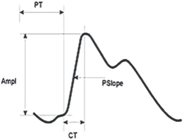
Measurements of the blood flow were made in accordance with the instruction manual of the Niccomo cardiac output monitor (Medis Med-izinische Messtechnik GmbH, Ilmenau, Ger many). During the test the authors evaluated the following plethysmographic parameters (Fig. 1):
Four electrodes were placed on the patient’s low-er limb. The yellow electrodes in the calf area are the so-called measuring electrodes; the black electrodes on the dorsum of the foot and the distal anterior ar-ea of the thigh are the so-called voltage electrodes. The measurements were taken with the patient at rest in a lying position, after a 10-min rest. During the ex-amination the patient was lying flat while electrodes placed on his lower limb measured the blood flow pa-rameters within the examined area (Fig. 2). The final values were gathered after 8 min.
The data analysis was performed using PQStat software (v. 1.4.2.324, PQStat Software, Poznań,
Poland). In the first stage of the analysis descrip-tive statistics characterizing the study group were generated. For the interval variables, average val-ues with 25–75% confidence intervals were cal-culated, as well as the standard deviation, median and upper and lower limits of quartiles. The results of the analyzed parameters were compared using the Kruskal-Wallis test, a post-hoc Dunn test and Wilcoxon’s test. The Friedman test for dependent variables was used to analyze the data obtained at the three measurements (before, immediately after and six months after the completion of the train-ing program). Differences were considered statis-tically significant when the p-value was < 0.05, and were assumed to be highly significant at p < 0.01.
The study was approved by the Ethical Review Board of the Medical University of Lodz (number RNN/226/11/KB).
Results
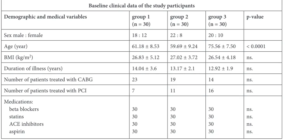
The baseline clinical data of the study partici-pantsare presented in Table 1. Other than the ag-es of patients, there was no statistically significant difference between the three groups in terms of baseline clinical variables and treatment.
In all three groups the authors observed chang-es in the four plethysmographic parameters stud-ied (PSlope, PAmpl, CT and PT) in comparison to their baseline values. In Group 1 PSlope increased by 2%, PAmpl by 4.2% and CT by 1.5%, while PT decreased by 1.2%. The change in the mean val-ues of the parameters was not statistically signifi-cant (Table 2).
In Group 2, after four weeks of rehabilitation, PSlope and PAmpl parameters increased by 19% and 17% respectively, while CT and PT decreased by 8% and 6.5% respectively. The mean values of the flow parameters appeared to be highly statisti-cally significant (Table 3).
At the end of cardiac rehabilitation (after an average of 18 days),in the control group there was
Fig. 1. Diagram of a plethysmographic curve [15] PT [ms] – propagation time; PAmpl [‰] – pulse wave amplitude; CT [ms] – crest time; PSlope [‰/s] – sys-tolic slope.
Fig. 2.
Table 1. Baseline clinical data of the studied patients
Baseline clinical data of the study participants
Demographic and medical variables group 1
(n = 30) group 2(n = 30) group 3(n = 30) p-value
Sex male : female 18 : 12 22 : 8 20 : 10
Age (year) 61.18 ± 8.53 59.69 ± 9.24 75.56 ± 7.50 < 0.0001 BMI (kg/m2) 26.83 ± 5.12 27.02 ± 3.72 26.54 ± 4.18 ns.
Duration of illness (years) 14.04 ± 3.6 13.17 ± 2.1 12.92 ± 1.9 ns. Number of patients treated with CABG 23 19 14 ns. Number of patients treated with PCI 7 11 16 ns. Medications:
beta blockers statins ACE inhibitors aspirin
30 30 30 30
30 30 30 30
30 30 30 30
ns. ns. ns. ns.
Values are expressed as mean ± SD; ns. = not significant.
Table 2. Values of the studied parameters in Group 1
Plethysmo-graphic parameters
Period Descriptive statistics Wilcoxon test
average sd min lower
quartile median upper quartile max
PAmpl [‰] before 0.56 0.12 0.35 0.44 0.58 0.65 0.76 0.4140 after 0.58 0.16 0.35 0.45 0.57 0.76 0.83
PSlope [‰/s] before 5.79 1.36 3.30 4.50 5.85 6.70 7.80 0.3986 after 5.82 1.38 3.70 4.80 5.95 6.70 8.30
CT [ms] before 170.32 16.46 143.00 162.00 166.50 175.00 200.00 0.9032 after 172.36 21.65 139.00 157.00 167.50 185.00 222.00
PT [ms] before 269.82 39.01 200.00 239.00 271.00 297.00 328.00 0.5781 after 268.55 36.95 199.00 233.00 271.00 291.00 334.00
Table 3. Values of the studied parameters in Group 2
Plethysmo-graphic parameters
Period Descriptive statistics Wilcoxon test
average sd min lower
quartile median upper quartile max
PAmpl [‰] before 0.54 0.17 0.22 0.44 0.54 0.66 0.86 0.0003 after 0.63 0.14 0.41 0.52 0.63 0.74 0.93
PSlope [‰/s] before 5.59 1.65 2.60 5.00 5.80 6.60 9.30 0.0002 after 6.56 1.56 4.00 5.70 6.50 7.50 10.90
CT [ms] before 168.65 15.27 144.00 156.00 167.50 178.00 201.00 0.0011 after 159.12 14.07 139.00 147.00 157.50 173.00 191.00
a 3% increase in PSlope, a 2.5% increase in PAmpl, and a 1% decrease in PT, but these values were not statistically significant. The only statistically signif-icant change in the control group parameters was the CT, which decreased by 5% (Table 4).
Highly significant differences were found be-tween Groups 1 and 2 at the end of the training pro-gram in terms of PSlope and PAmpl (p = 0.0003), as well as CT (p = 0.0233). The difference in the values of the propagation time (PT) were not sta-tistically significant (p = 0.2481).
Discussion
Among the participants in the present study, men constituted a distinct majority (70%). This re-flects epidemiological data, which shows that twice as many men as women suffer and die from coro-nary artery disease (CAD) [16]. This is confirmed by the data obtained in the Reversing Atherosclero-sis with Aggressive Lipid Lowering (REVERSAL), the Comparsion of Amlodipine Versus Enarapril to Limit Occurences of Thrombosis (CAMELOT) and ACAT Intravascular Atherosclerosis Treat-ment Evaluation (ACTIVATE) studies, conduct-ed among 978 people (251 female and 727 male) Table 4. Values of the studied parameters in the control group
Plethysmo-graphic parameters
Period Descriptive statistics Wilcoxon
test
average sd min lower
quartile median upper quartile max
PAmpl [‰] before 0.39 0.14 0.16 0.31 0.42 0.48 0.59 0.2343 after 0.40 0.15 0.20 0.33 0.40 0.48 0.67
PSlope [‰/s] before 4.18 1.35 2.00 3.30 4.50 4.85 6.30 0.5067 after 4.31 1.38 2.30 3.70 4.45 4.85 6.90
CT [ms] before 165.25 6.97 154.00 158.00 167.50 171.00 173.00 0.0013 after 157.00 9.62 148.00 150.00 153.00 161.00 174.00
PT [ms] before 269.94 13.25 244.00 263.00 274.50 276.00 286.00 0.2343 after 268.38 22.99 232.00 259.00 266.00 269.00 308.00
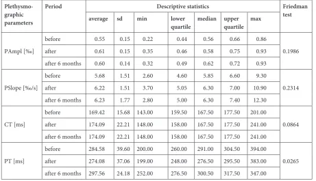
Table 5. Values of the studied parameters before, just after and 6 months after rehabilitation
Plethysmo-graphic parameters
Period Descriptive statistics Friedman
test
average sd min lower
quartile median upper quartile max
PAmpl [‰]
before 0.55 0.15 0.22 0.44 0.56 0.66 0.86
0.1986 after 0.61 0.15 0.35 0.46 0.58 0.75 0.93
after 6 months 0.60 0.14 0.32 0.49 0.62 0.72 0.93
PSlope [‰/s]
before 5.68 1.51 2.60 4.60 5.85 6.60 9.30
0.2314 after 6.22 1.51 3.70 5.05 6.30 7.00 10.90
after 6 months 6.23 1.77 2.80 5.00 6.30 7.40 12.30
CT [ms]
before 169.42 15.68 143.00 159.50 167.50 177.50 201.00
0.0864 after 174.09 22.21 148.00 158.00 167.50 177.50 241.00
after 6 months 174.09 22.21 148.00 158.00 167.50 177.50 241.00
PT [ms]
before 284.58 39.60 200.00 260.00 291.00 304.50 394.00
0.0265 after 274.08 37.06 199.00 248.00 276.50 295.50 383.00
aged 30–75 years. These studies demonstrated that despite worse baseline risk factors among women, less severe atherosclerosis in the coronary arteries was observed in women than in men of the same age [17].
All the patients in the present study had un-dergone myocardial revascularization or coronary artery bypass graft (CABG) surgery. CABG had been performed on 23 patients in Group 1, 19 pa-tients in Group 2 and 14 pa19 pa-tients in Group 3. Per-cutaneous coronary intervention (PCI) had been performed on 7 patients in Group 1, 11 in Group 2 and 16 in Group 3. Among the many treatments for heart disease in recent years, invasive methods are being used more and more frequently. This is indicated by the increasing number of centers per-forming open-heart procedures, as well as by the increase in the number of such procedures per-formed at cardiac centers in Poland [18]. In this situation, comprehensive cardiac rehabilitation plays an important role: Rehabilitation can con-tribute to sustaining the positive effects of expen-sive surgery.
During cardiac rehabilitation monitoring the functioning of the patients’ peripheral circulatory system is often forgotten. This problem is signifi-cant in that heart disorders may exacerbate pre-ex-isting dysfunctions of the blood flow in the lower limbs, eg in CABG patients.
Physical training, especially endurance train-ing, is associated with increased capillary densi-ty, improved muscle fiber composition and im-provement of the metabolism. The amount of type IIA muscle fiber increases, while type IIB fiber de-creases. This process leads to a substantial increase in the cross-sectional area of all types of muscle fi-bers and an increase in the ratio of fifi-bers to cap-illary vessels [19]. This is of vital importance, due to the fact that the muscle pump, integrally con-nected with the function of foot, lower leg and thigh muscles, is the main driving force caus-ing blood movement in the blood vessels of low-er limbs. The function of the joint pump, which is dependent on flexion and extension movements (mainly in the ankle joint) causing compression on the venous plexus of the foot, is also crucial [20]. Increased local blood flow intensifies the shear forces and therefore improves the reactivity of the vascular endothelium.
In comparison with baseline values, PAmpl in-creased in all three groups: in Group 1 by 4.2%, in Group 2 by 17% and in Group 3 by 2.5%. Like-wise, there were increases in PSlope in all three groups: in Group 1 by 2%, in Group 2 by 19% and in Group 3 by 3%. The CT on the other hand was reduced by 1.5% in Group 1, by 8% in Group 2 and by 5% in Group 3. The PT also decreased, by
1.2% in Group 1, by 6.5% in Group 2 and by 1% in Group 3. These changes indicate improvement in the vascular perfusion of the lower extremities. Only the results for Group 2 and the CT in the control group were significant and highly statisti-cally significant.
In the present study, the authors used imped-ance plethysmography to evaluate local blood flow in the calf. Barcroft and Dornhorst started studying this issue in the 1940s by introducing wrap pleth-ysmography as a research technique, analyzing blood flow in the soleus and gastrocnemius mus-cles of the calf during physical exercise involving contraction and relaxation of posterior calf muscle groups. During alternating dorsal and plantar flex-ion against resistance, Barcroft and Dornhorst ob-served a 40% increase in blood flow in the calf ar-ea [21]. Reading et al. confirmed a 20% increase in vascular perfusion in the calf area in patients par-ticipating in training sessions with a bicycle er-gometer with a 25-Watt load [22]. Goodman et al. analyzed the work of the central and peripheral cir-culatory system in 31 patients who had undergone coronary artery bypass grafting surgery. The pa-tients took part in a 12-week workout session that included walking and jogging, and the researchers observed an 18% increase in vascular perfusion in the calf area. Strain gauge plethysmography was used as a research method [23].
Possible reasons for the increase in local blood flow may be reflected in the changes taking place in the structure and function of the blood vessels. Post-workout changes in peripheral circulation may be due to the development of new collateral blood vessels, or increases in the diameter of exist-ing ones. The increase in local blood flow causes higher shear force influence on the vessels, which is a physiological agent of improving the reactivity of the vascular endothelium. Improvement in en-dothelial vasodilator function is caused by increas-es in the blood flow directly related to the reduc-tion in vascular resistance. It is also suggested that chronic increased shear forces on the vessel wall may stimulate the release of growth factors, result-ing in the stimulation of vascular adaptation pro-cesses and, consequently, an increase in vessel di-ameter [20].
ves-sel per unit of time, is proportional to the differ-ential pressure times the fourth power of the radi-us, and inversely proportional to the length of the artery and blood viscosity. It follows that the most important factor leading to improved blood circu-lation is an increase in vessel diameter. Thus, the more blood flow, the higher the differential pres-sure and the lower the resistance. This means that an increase in the blood supply can be achieved either by a pressure increase or by decreasing pe-ripheral resistance. It can therefore be conclud-ed that even a slight expansion of the vessel wall will cause a significant decrease in resistance and a marked increase in the flow [24]. Thus, improve-ment in the flexibility of the vascular wall could explain the increase in local blood flow in the study area. The observed increase in the elastici-ty of blood vessels in the present study indicated by an increase in the amplitude of the wave pulse and systolic slope of the pulse wave may be due to the previously mentioned increase in the reactivi-ty of the endothelium in the form of an increased propensity for mediated vasodilatation dependent on the flow. The explanation for this phenomenon lies mainly in increases in endothelial nitric oxide production [25].
Changes in the vascular perfusion in lower limbs as a result of physical training may be asso-ciated with blood flow redistribution during exer-cise from less working to more involved muscles. This view was presented by Tan et al., highlight-ing the reduced oxygen saturation measured in the femoral veins after training [26]. Better use of oxygen by the muscles of the lower limbs is not necessarily associated with better distribution of the circulating blood. This phenomenon can be the result of metabolic changes occurring due to an increase in mitochondrial enzymes and in-creased oxidative muscle fibers after a training program [27].
Summing up, the present study confirmed that physical activity leads to positive changes in blood flow in the peripheral vessels of the lower limbs in patients with ACS, which is shown by the signif-icant improvement in the plethysmographic pa-rameters in Group 2 after four weeks of controlled physical exercise. Impedance plethysmography al-lows non-invasive monitoring of local blood flow and is a precise and repeatable method for the as-sessment of peripheral circulation in patients dur-ing cardiac rehabilitation.
References
[1] Lloyd-Jones D, Adams R, Carnethon M: Heart disease and stroke statistics – 2009 update: A report from the American Heart Association Statistics Committee and Stroke Statistics Subcommittee. Circulation 2009, 119, 480–486.
[2] Piepoli MF, Davos C, Francis DP, Coats AJ: ExTra-MATCH Collaborative. Exercise training meta-analysis of trials in patients with chronic heart failure (ExTra-MATCH). B M J 2004, 328, 189.
[3] Rehabilitation after cardiovacular disease with special emphasis on developing countries. Geneva: WHO Technical Report Series 1993, No 831.
[4] Koch DW, Newcomer SC, Proctor DN: Blood flow to exercising limbs varies with age, gender and training status. Can J Appl Physiol 2005, 30, 554–575.
[5] Peake J, Nosaka K, Suzuki K: Characterization of inflammatory responses to eccentric exercise in humans. Exerc Immun Rev 2005, 11, 64–85.
[6] Green DJ, Spence A, Halliwill JR: Exercise and vascular adaptation in asymptomatic humans. Exp Physiol 2011, 96, 57–70.
[7] Ji JW, Mac Gabhann F, Popel AS: Skeletal Muscle VEGF gradients in peripheral arterial disease: Simulations of rest and exercise. Am J Physiol Heart Circ Physiol 2007, 293, 3740-3749.
[8] Higashi Y, Chayama K, Yoshizumi M: Angiotensin II type I receptor blocker and endothelial function in humans: Role of nitric oxide and oxidative stress. Curr Med Chem Cardiovasc Hematol Agents 2005, 3, 133–148
[9] Newcomer SC, Thijssen DH, Green DJ: Effects of exercise on endothelium and endothelium/smooth muscle cross talk: Role of exercise-induced hemodynamics. J Appl Physiol 2011, 111, 311–320.
[10] Much TI, Eklund KE, Hageman KS, Poole DC: Altered regional blood flow responses to submaximal exercise in older rats. J Appl Physiol 2004, 96, 81–88.
[11] Siebert J: Kardiografia impedancyjna. Via Medica, Gdańsk 2007, 1–3.
[12] Irzmańska E, Charłusz M, Kujawa J, Kowalski J, Pawlicki L, Irzmański R: Using impedance plethysmography to evaluate antidecubital underlay systems for chronically immobilized patients. Adv Clin Exp Med 2010, 19, 1–15.
[13] Jurczak J, Łukasik A, Charłusz M, Irzmański R: Pletyzmografia impedancyjna w ocenie krążenia obwodowego pod wpływem treningu fizycznego u chorych po CABG. Geriatria 2012, 6, 83–90.
[14] Kompleksowa rehabilitacja kardiologiczna. Stanowisko Komisji ds. Opracowania Standardów Rehabilitacji Kardiologicznej Polskiego Towarzystwa Kardiologicznego. http://www.rehabilitacjakardiologicznaptk.pl/wp-con-tent/uploads/2015/02/standardy.pdf (access date: April 2015).
[16] Matyjaszczak P, Hoffmann K, Bryl W: Epidemiologia wybranych czynników ryzyka chorób układu krążenia. Przegl Kardiodiab 2011, 6, 255–262.
[17] Nicholls SJ, Wolski K, Sipahi I, Schoenhagen P, Crowe T, Kapadia SR, Hazen SL, Tuzcu EM, Nissen SE: Rate of progression of coronary atherosclerotic plaque in women. J Am Cardiol 2007, 49, 1546–1551.
[18] KROK Raport Krajowego Rejestru Operacji Kardiochirurgicznych. Klub Kardiochirurgów Polskich 2009.
[19] Scarpelli M: Quantitative analysis of changes occuring in muscle vastus lateralis in patients with heart failure after low intensity training. Anal Quant Cytol Histol 1999, 21, 374–380.
[20] Clifford PS: Local control of blood flow. Adv Physiol Educ 2011, 35, 5–15.
[21] Barcroft H, Dornhorst AC: The blood flow through the human calf during rhythmic exercise. J Physiol 1949, 109, 402–411.
[22] Reading JR, Goodman J, McLaughlin PR: Design of a calf muscle ergometer for the study of local muscle blood flow. Can J Sport Sci 1992, 17, 91–93.
[23] Goodman JM, Pallandi DV, Reading JR, Piyiel MJ, Liu PP, Kavangh T: Central and peripheral adaptations after 12 weeks of exercise training in post-coronary artery bypass surgery patients. J Cardiopulm Rehabil 1999, 19, 144–150.
[24] Endemann DH, Schiffrin EL: Endothelial dysfunction. J Am Soc Nephrol 2004, 15, 1983–1992.
[25] Sarelius I, Pohl U: Control of muscle blood flow during exercise: Local factors and integrative mechanisms. Acta Physiol (Oxf) 2010, 199, 349–365.
[26] Tan KH, de Cossart L, Edwards PR: Exercise training and peripheral vascular disease. Br J Surg 2004, 87, 553–562.
[27] Clement M, Laurent M: Muscle (phosphocreatine) dynamics during exercise: Implication for understanding the regulation of muscle oxidative metabolism. J Physiol 2008, 13, 3027–3029.
Address for correspondence:
Ireneusz Jurczak
University of Social Sciences in Łódź ul. Gorkiego 10/12/11
92-525 Łódź Poland
Tel.: +48 600 047 067
E-mail: [email protected]
Conflict of interest: None declared