Cite as
Łaczmańska I, Stembalska A, Złocińska M, et al. Multiplex ligation-dependent probe amplification as a screening test in children with autism spectrum disorders. Adv Clin Exp Med. 2020;29(1):101–106. doi:10.17219/acem/112609
DOI
10.17219/acem/112609
Copyright
© 2020 by Wroclaw Medical University This is an article distributed under the terms of the Creative Commons Attribution 3.0 Unported (CC BY 3.0) (https://creativecommons.org/licenses/by/3.0/)
Address for correspondence
Agnieszka Stembalska
E-mail: [email protected]
Funding sources
This study was funded by Statutory Activities financed by the Ministry of Science and Higher Education as No. ST A.290.17.032.
Conflict of interest
None declared
Received on September 12, 2018 Reviewed on March 8, 2019 Accepted on September 25, 2019
Published online on January 28, 2020
Abstract
Background. Autism spectrum disorders (ASDs) are a heterogeneous group of neurodevelopmental dis-orders, characterized by the presence of various symptoms related to deficits in communication and social interactions as well as stereotyped and repetitive behavior. Increasing evidence indicates the contribution of genetic factors in the etiology of ASDs. Genetic diagnosis in ASDs is based on identifying chromosome aberrations, microaberrations and point mutations in specific genes. One of the diagnostic tools is multiplex ligase-dependent probe amplification (MLPA) with a set of probes dedicated to ASDs (SALSA MLPA P343 Autism-1; MRC-Holland BV, Amsterdam, the Netherlands) targeting the genes located in the regions 15q11-q13, 16p11 and the SHANK3 gene in the 22q13 region.
Objectives. Our study included 240 patients referred to the clinical genetics unit because of ASDs and/or de-velopmental delay and/or an intellectual disability. Before genetic testing, the patients underwent a com-prehensive medical work-up.
Material and methods. Multiplex ligase-dependent probe amplification was performed in 256 DNA samples from 240 probands and 16 family members using the SALSA MLPA P343 Autism-1 probe mix (MRC-Holland BV) according to the manufacturer’s protocol.
Results. We obtained 234 normal results and 22 abnormal results (15 probands and 7 abnormal results for probands’ parents or siblings). We diagnosed 1 16p11 microdeletion syndrome and 1 16p11 microduplication syndrome. We also found 3 deletions and 1 duplication in 15q13 region including 2 or 3 genes and 9 single probe alterations in the regions examined (1 duplication and 7 deletions).
Conclusions. Due to the low costs, MLPA test may be a good tool for the genetic screening of ASD patients. Key words: diagnostics, autism, MLPA
Multiplex ligation-dependent probe amplification
as a screening test in children with autism spectrum disorders
Izabela Łaczmańska
1,A–F, Agnieszka Stembalska
1,B,D–F, Magdalena Złocińska
1,B,C, Joanna Kozłowska
1,B,C, Paweł Skiba
1,B,C, Karolina Pesz
1,B–D,F,
Ryszard Ślęzak
1,B,C,E, Robert Śmigiel
2,B,C,E, Aleksandra Jakubiak
1,B,C, Błażej Misiak
1,B,C,E, Maria M. Sąsiadek
1,B,C,E,F1 Department of Genetics, Wroclaw Medical University, Poland
2 Department of Pediatrics and Rare Disorders, Wroclaw Medical University, Poland
A – research concept and design; B – collection and/or assembly of data; C – data analysis and interpretation; D – writing the article; E – critical revision of the article; F – final approval of the article
The prevalence of autism spectrum disorders (ASDs) has been estimated at 20−60/10,000 children, corresponding to 1–2% of the general population.1–3 Importantly, ASDs are highly heterogeneous neurodevelopmental disorders, characterized by the presence of various symptoms, in-cluding deficits in communication and social interactions as well as stereotyped and repetitive behaviors. Moreover, patients with ASDs present high rates of comorbid intel-lectual disability, sensory abnormalities or sleep distur-bances.2 The role of genetic factors and metabolic condi-tions in the development of ASDs is strongly suggested on the basis of family studies, especially twin studies.4 Although several genes have been identified and their association with ASD susceptibility is well-documented, a large group of genes is still suspected to be involved in this disease.2 Additionally, diagnoses of several ge-netic syndromes need to be excluded before a final diag-nosis of ASD is established.5 Diagnostic considerations are even more complex due to multifactorial inheritance and a number of environmental risk factors potentially involved in the etiology of ASDs.6
Over the last few years, the laboratory methods used for ASD diagnosis have moved from classical cytogenet-ics (banding analysis) or fluorescent in situ hybridization (FISH) to array comparative genomic hybridization (aCGH) and next-generation sequencing (NGS). Several single nucleotide variants (SNV) have been identified in many genes and confirmed by family-sequencing using whole-exome sequencing (WES) and whole-genome sequencing (WGS). These studies have revealed that ASDs may be caused by numerous alterations in genes acting in differ-ent molecular pathways with incomplete penetrance and variable expressivity.2,7
The WES and WGS methods make it possible to screen large parts or the entire genome and to identify all poten-tial genomic alterations, but their costs and laboratory equipment requirements are high. Taking into account these considerations, multiplex ligation-dependent probe amplification (MLPA) is an appealing alternative for initial screening in patients with ASD.5,8 The dedicated MLPA probe mix for ASD contains probes for 3 chromosomal regions: 15q11-q13, 16p11 and 22q13. Therefore, the aim of our study was to evaluate the rate of positive results in a large sample of ASD patients, using the MLPA mix dedicated to this disease.
Material and methods
Materials
The analyses were performed in 240 individuals with ASDs and 16 healthy family members. The patients’ group included 54 women and 202 men, with a mean age of 8.7 ±8.06 years. The patients had been referred to the clin-ical genetics unit with ASDs and/or developmental delay
and/or intellectual disability, with or without dysmorphic features or additional congenital abnormalities. The diag-nosis of ASD was established according to ICD-10 criteria.9 All the probands and/or their legal guardians signed a con-sent form. The MLPA test was performed after excluding genetic disorders that had been diagnosed based on clinical examinations or specific genetic tests (cytogenetics and fragile-X testing). The study was accepted by the Ethics Committee of Wroclaw Medical University (approval No. 320/2018).
DNA isolation
Genomic DNA was isolated from 200 μL of peripheral blood lymphocytes, using the Prepito DNA Blood kit and the chemagic Prepito-D isolator (both from PerkinElmer Inc., Waltham, USA) according to the manufacturer’s pro-tocol. DNA was diluted to 20 ng/μL and 5 μL (100 ng) was used in the MLPA reaction.
The MLPA analysis
The SALSA MLPA P343 Autism-1 probe mix (MRC-Holland BV, Amsterdam, the Netherlands) containing MLPA probes for 3 chromosomal regions 15q11-q13 (in-cluding, among others, UBE3A, GABRB3 and in 15q13
CHRNA7), 16p11 (including, among others, LAT, MAZ, MAPK3,and HIRIP3) and 22q13 (including SHANK3) was used to assess the deletions and duplications in these regions. The MLPA reaction was prepared using a TC512 thermocycler (Techne Inc., Burlington, USA). The pro-ducer’s protocol for MLPA was followed precisely.
The MLPA products were separated using the ABI 310 Genetic Analyzer with GeneScan Analysis v. 3.1.2 soft-ware, POP-4 Polymer and GeneScan™ 500 LIZ™ dye Size Standard (all from Thermo Fisher Scientific, Waltham, USA). The analysis of the results was prepared using GeneMarker v. 1.85 software (SoftGenetics LLC, State College, USA).
In the analysis of copy number variants (CNVs), a change in the peak values of over +0.3 was considered a duplication and –0.3 a deletion.
Microarray comparative
genomic hybridization analysis
(Agilent Technologies). The microarray design used for the analysis was SurePrint G3 CGH ISCA v. 2, 8 × 60K (Agilent Technologies). The design focuses on 498 regions in the human genome of high clinical significance, defined on the basis of the International Standards Cytogenomic Arrays (ISCA) Consortium.10 The approximate cover-age of the genome is about 60 kb, with median practical resolution for accurate detection of imbalances estimated at 300 kb (lower in ISCA regions). The data was analyzed using CytoGenomics software (Agilent Technologies) with the default CGH analysis method, and was interpreted in reference to available databases (DGV [last update: 2016-05-15], ClinVar [2018-06-01 03:26], DECIPHER v. 9.23 [2018-05-23] May, 2018]).
Results
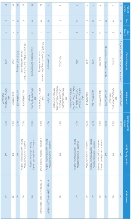
The MLPA analysis was performed on 256 DNA samples from patients and their families. We obtained 234 normal results and 22 abnormal results (in 15 patients and 7 par-ents or siblings of pati7 par-ents) (Table 1).
In the patients’ group, we diagnosed 1 16p11 micro-deletion syndrome and 1 16p11 duplication syndrome. We also found 1 duplication in the 15q13 region, 3 dele-tions in the 15q13 region, including KLF1 and CHRNA7
or ABPA2, NDNL1 and TJP1 genes, as well as 9 single-probe alterations in all the regions examined (2 duplication and 7 deletions) (Table 1).
15q11-13
In our study, alterations of region 15q11-13 were found in 11 out of 15 patients with a positive result: 4 out of 15 pa-tients had abnormal results for more than 1 probe in this region (3 deletions and 1 duplication) while 7/15 had an al-teration of 1 exon in this region (5 deletions and 2 dupli-cations). In 1 case, the deletion of 2 probes in the 15q13 region (KLF13ex2 and CHRNA7ex4) was confirmed using aCGH; it occurred as part of a 1.1 Mb deletion (arr 15q33.3[31516639_32635959]x1). In the group of indi-viduals with alterations in the 15q11-13 region, we observed ASDs with other clinical symptoms (6 cases) or without (4 cases); only 1 patient had developmental delay with epi-lepsy (Table 1).
In 5 patients we observed a deletion of 1 exon in the
GABRB3 gene (15q12), 4 in exon 9 (exon 6 according to new numbering) and 1 in exon 4. Three of these deletions were also present in 1 healthy parent (2 families were not ex-amined). In this group of patients, we observed the fol-lowing clinical picture: 1) ASDs without other clinical problems in 3 cases (1 with ASD family history); 2) ASD with developmental delay and epilepsy in 1 case; 3) other problems in 1 case; and 4) developmental delay with epi-lepsy in 1 case (Table 1).
We found 2 duplications and 2 deletions in the CHRNA7
gene (the probe for exon 4), 1 duplication and 3 altera-tions being parts of a more complex rearrangement. In this group of patients, we observed ASDs in all cases. The pa-tients with deletions presented with other comorbidities, such as intellectual disability in 1 case as well as develop-mental delay, speech delay and short stature in another 1 (Table 1). A single duplication of CHRNA7 exon 4 was also present in a healthy sister of the proband.
16p11.2
Out of 15 patients, 3 were carriers of 16p11.2 altera-tions. We found 2 deletions: a small deletion (represented by 1 probe for LAT4), confirmed with aCGH as a 187 kb deletion (arr 16p11.2[28843773_29031059]x1]; 1 deletion exposed by 9 out of 11 probes for this region in the MLPA analysis; and 1 duplication, also for 9 out of 11 probes. In this group of patients, we observed that patients with deletions presented with more clinical symptoms than patients with duplications (Table 1).
22q13
In 1 patient, 1 probe found a deletion of exon 15 of the
SHANK3 gene.
Discussion
Genetic testing is an important part of the diagnos-tic process in children with ASDs and/or developmen-tal delay and/or intellectual disability. Identification of a genetic cause of a disorder may elucidate the issue of etiology, enable assessment of the prognosis, facilitate care and management planning, and permit an estima-tion of the risk of recurrence. Using currently available standard laboratory methods (other than WES or WGS), it is possible to find a genetic cause in about 10% of ASD cases.11 According to the literature, the aCGH analysis is recommended as a first-tier diagnostic method for ASD patients.11 Additionally, single-gene tests, such as fragile-X syndrome testing, should be applied.11 Given that aCGH and WES are expensive procedures, it is reasonable to use more cost-effective methods for screening and select-ing patients for further analysis. One of these methods is MLPA – a method mainly used for the detection of small deletions and duplications. The SALSA MLPA P343 Au-tism-1 probe mix (MRC-Holland BV) is dedicated for ASD patients and allows deletions and duplications to be found in the 15q11-13, 16p11.2 and 22q13 chromosomal regions. Alterations identified in our study include deletions and duplications in regions 15q11-13, 16p11.2, and 22q13, as well as deletions and duplications in the GABRB3 and
Ta ble 1 . R es ult s o f t he M LP A a na lys is Pa tien t numb er Se x Ag e [ye ars ] Cli nic al d ia gn os is M LP A res ult Ch romo som al re gi on M LP A fa mi ly r es ult s aC G H res ult 1 M 10 Fra gil e X s yn dro m e s us pe cte d ( ex clu de d) de l SHAN K3e x15 22 q13 nd nd 2 M 5 ID , A SD de l KL F13 ex 2 , C H RN A7e x4 , TR PM1 ex 27 15 q13 nd nd 3 M 9 D D , ep ile ps y, ep ile ps y i n f am ily de l G AB R3 ex 9 ( 6) 15 q12 nd nd 4 F 15 D D , A SD du p CH RN A7e x4 15 q13 m oth er – n orm al r es ult , h ea lth y on e s ist er – d up lic ati on , h ea lth y nd 5 M 7 A SD de l G AB R3 ex 9(6) 15 q12 m oth er − d eleti on , h ea lth y fat he r – n orm al r es ult s, h ea lth y nd 6 F 5 A SD de l G AB R3 ex4 15 q12 m oth er – d ele tio n, h ea lth y nd 7 M 8 A SD du p H IR IP3 ex 3 , S EZ 6L 2e x1 , D O C2 Ae x4 , M AZe x5 , M VPe x5 , SP N ex 3 , M AZe x6 , H IR IP3 ex 4 , M AP K3e x5 16 p11 m oth er – d ele tio n, h ea lth y fat he r – n orm al r es ult s, h ea lth y nd 8 F 7 A SD , D D , ID del H IR IP3 ex 3 , S EZ 6L 2e x1 , D O C2 Ae x4 , M AZe x5 , M VPe x5 , SP N ex 3 , M AZe x6 , H IR IP3 ex 4 , M AP K3e x5 16 p11 nd nd 9 M 2 D D , d ys m or ph y de l LAT ex 4 16 p11 m oth er – d ele tio n, h ea lth y fat he r – n orm al r es ult s, h ea lth y arr 1 6p 11 .2(2 88 43 77 3_ 29 03 10 59 )x 1 10 M 11 D D , A D S, I D , s pe ec h d ela y, s ho rt s ta tu re , sh or t s ta tu re in f am ily de l KL F13 ex 2 , C H RN A7e x4 15 q13 3 s ib lin gs – h ea lth y, n orm al r es ult arr 1 5q3 3.3 (31 51 66 39_ 32 63 595 9)x 1 11 M 13 A SD , e pil eps y, g yn ec om as tia de l AB PA 2e x14 , N DNL 2e x1 , TJP 1in1 15 q13 m oth
er – n
Deletions and duplications
in region 15q11-13
Copy number variants in the 15q11-13 region are among the most common autosomal alterations in ASD patients.5 A substantially higher rate of ASDs has been found in in-dividuals with Prader–Willi syndrome, especially in those with the uniparental disomy (UPD) subtype.12
The proximal part of chromosome 15 is one of the most unstable regions in the human genome, because it con-tains 6 low copy repeat (LCR) elements that are grouped in 6 breakpoints involved in non-homologous recombina-tion. Microdeletions within this region are observed in pa-tients with Prader–Willi and Angelman syndromes, while microduplications are associated with ASDs, learning disabilities and seizures.13 Similarly, CNVs in the 15q13.3 region, located downstream to the 15q11-13 locus, are reported in patients with neuropsychiatric phenotypes, attention deficit hyperactivity disorder and ASDs.13
In 1 of our patients (No. 10), we found a 15q13 deletion not observed in 3 healthy siblings; using the aCGH meth-od, it was characterized as a 1.1 Mb deletion. Moreover, a smaller deletion in this region, covering genes ABPA2
(exon 14), NDNL2 (exon 1)and TJP1 (intron 1) was diag-nosed in patient No. 11. Although this alteration was also observed in the presumably healthy father, it may still be the cause of ASD in the proband if the penetrance of the al-teration is incomplete. To verify this hypothesis, further genetic testing in numerous family members is necessary.
Deletions and duplications
of
GABRB3
exons
Alterations in the GABRB3 gene reported in ASD pa-tients mainly involve variations of single or several nu-cleotides, and most of them are familial.14 Due to high incidence of this deletion (5 patients and 3 familial cases, with no data for 2 families) it is likely that this alteration is not causative of clinical features in these patients.
Deletions and duplications
of
CHRNA7
exons
The α7 subunit of the nicotinic acetylcholine receptor is encoded by CHRNA7 (15q13). This ion channel is report-ed to be expressis report-ed in the brain. A homopentameric form of the α7 subunit is involved in mediating signal transduc-tion at synapses, the regulatransduc-tion of neurotransmitter release, synaptic plasticity, learning, and memory. The CHRNA7
gene is located in the 15q13.3 region and has been associated with neurodevelopmental and neuropsychiatric disorders.13 Deletion of the whole CHRNA7 gene is observed in 1% of pa-tients with idiopathic generalized epilepsies, while duplica-tion of the gene occurs in patients with ASDs and cognitive impairment.13,15–17 For patients with CNVs in the 15q13.3 regions, incomplete penetrance (about 80%) and variable
expressivity have been reported.13 It is difficult to conduct a sequence analysis of the CHRNA7 gene because of the pres-ence of a large identical sequof the pres-ence in the CHRFAM7A gene, which is therefore sequenced together with CHRNA7, but no rare damaging variants were reported in the CHRNA7 gene in a group of 135 patients with ASD.17 It should be noted that because of the limitations of MLPA analysis, this alteration may also be caused by a single nucleotide polymorphism or mutation.The same change was also observed in a healthy sister, which may suggest that this alteration was not re-sponsible for ASD in the proband, or (as in previous reports) the penetrance of this mutation is incomplete.13
Deletion and duplication
in the 16p11.2 region
Deletion and duplication in the 16p11.2 region are among the most frequent CNVs in patients with ASDs and neu-rodevelopmental disorders.7 Several clinical characteris-tics, including speech articulation abnormalities, limb and trunk hypotonia with hyporeflexia, abnormalities of agil-ity, sacral dimples, macrocephaly, and epilepsy, have been observed in patients with 16p11.2 deletions. Additionally, the 16p11.2 duplication syndrome has been associated with speech articulation abnormalities, hypotonia, abnormali-ties of agility, sacral dimples, and epilepsy along with action tremor and microcephaly.7 In both deletion cases in our study, examination of the family members revealed a carrier status in mothers without any clinical features. As in cases of 15q11-13 microaberrations, incomplete penetrance and variable expressivity have been reported in 16p11.2 micro-deletions and especially in microduplications.7
Deletion and duplication
in the 22q13 region
Copy number variants in the 22q13 region are the cause of various neuropsychiatric disorders, including Phelan– McDermid syndrome (PMS), which is the result of a dele-tion of a critical region with the SHANK3 gene. A high rate of ASDs (as high as 20–50%) has been reported in children with 22q13 aberrations. Other highly characteristic fea-tures include speech and developmental delays.5
Microduplications in the 22q13 region, including the SHANK3 gene, are reported in patients with attention-deficit hyperactivity disorder (ADHD) and schizophrenia, while microdeletions and point mutations of the SHANK3
gene are present in patients with mild autism without intel-lectual disability. Incomplete penetrance is also reported in cases of SHANK3 gene alterations.5,18 In 1 patient in our study, we found a deletion of 1 probe for exon 15 of the
Conclusions
The MPLA analysis is an effective and low-cost tech-nique for screening genetic causes in ASD patients. It allows patients to be selected for further diagnostic consideration with more expensive methods. Because deletions and amplifications in the 15q11-13, 16p11.2 and 22q13 regions have also been described in healthy subjects, confirmation of the results of the MLPA analy-sis is recommended using another MLPA probe mix or another diagnostic method, e.g., FISH or aCGH. The importance of any detected changes should always be interpreted in relation to clinical data. Abnormal results of an MLPA analysis can also arise from the pres-ence of a point mutation or a single nucleotide poly-morphism within the sequence analyzed. Therefore, if sequencing is not applied, the parents of the patient should be examined in order to determine the origin of the change (inherited/de novo).18 Our analysis re-vealed that although MLPA can be an initial test in ASD patients, the majority of them will need further investi-gations, such as aCGH or WES.
Unraveling the genetic underpinnings of ASDs is an im-perative not only for the patients but also for the whole family. It provides a basis for genetic counseling and an ex-planation of observed neurodevelopmental and behavioral characteristics. Finally, it facilitates comprehensive medi-cal care for various comorbidities that might appear due to specific genetic alterations.
ORCID iDs
Izabela Łaczmańska https://orcid.org/0000-0003-2458-5755
Agnieszka Stembalska https://orcid.org/0000-0003-3528-6582
Magdalena Złocińska https://orcid.org/0000-0003-3626-0587
Joanna Kozłowska https://orcid.org/0000-0002-5473-9711
Paweł Skiba https://orcid.org/0000-0002-8811-4437
Karolina Pesz https://orcid.org/0000-0003-1482-1021
Ryszard Ślęzak https://orcid.org/0000-0001-6738-9565
Robert Śmigiel https://orcid.org/0000-0003-2930-9549
Aleksandra Jakubiak https://orcid.org/0000-0000-0000-0001
Błażej Misiak https://orcid.org/0000-0002-5392-6398
Maria M. Sąsiadek https://orcid.org/0000-0002-7599-7074
References
1. Bremer A, Giacobini M, Nordenskjöld M, et al. Screening for copy number alterations in loci associated with autism spectrum dis-orders by two-color multiplex ligation-dependent probe
amplifi-cation. Am J Med Genet Part B Neuropsychiatr Genet. 2009;153B(1):
280–285. doi:10.1002/ajmg.b.30954
2. An JY, Claudianos C. Genetic heterogeneity in autism: From
sin-gle gene to a pathway perspective. Neurosci Biobehav Rev. 2016;68:
442−453. doi:10.1016/j.neubiorev.2016.06.013
3. Baio J, Wiggins L, Christensen DL, et al. Prevalence of autism spec-trum disorder among children aged 8 years: Autism and Develop-mental Disabilities Monitoring Network, 11 Sites, United States, 2014. MMWR Surveill Summ. 2018;67(6):1−23. doi:10.15585/mmwr.ss6706a1 4. Ronald A, Hoekstra RA. Autism spectrum disorders and autistic traits:
A decade of new twin studies. Am J Med Genet Part B Neuropsychiatr
Genet. 2011;156(3):255−274. doi:10.1002/ajmg.b.31159
5. Cai G, Edelmann L, Goldsmith JE, et al. Multiplex ligation-dependent probe amplification for genetic screening in autism spectrum disor-ders: Efficient identification of known microduplications and
iden-tification of a novel microduplication in ASMT. BMC Med Genomics.
2008;1(1):50. doi:10.1186/1755-8794-1-50
6. Schaefer GB, Mendelsohn NJ; Professional Practice and Guidelines Committee. Clinical genetics evaluation in identifying the etiology
of autism spectrum disorders: 2013 guideline revisions. Genet Med.
2013;15(5):399−407. doi:10.1038/gim.2013.32
7. Steinman KJ, Spence SJ, Ramocki MB, et al; Simons VIP Consortium. 16p11.2 deletion and duplication: Characterizing neurologic
pheno-types in a large clinically ascertained cohort. Am J Med Genet Part A.
2016;170(11):2943−2955. doi:10.1002/ajmg.a.37820
8. Boggula VR, Shukla A, Danda S, et al. Clinical utility of multiplex liga-tion-dependent probe amplification technique in identification of aetiology of unexplained mental retardation: A study in 203 Indian
patients. Indian J Med Res. 2014;139(1):66−75.
9. World Health Organization. ICD-10: International Statistical
Classi-fication of Diseases and Related Health Problems. 10th revision, 2nd
ed. Geneva, Switzerland: World Health Organization; 2004. https:// www.who.int/classifications/icd/icdonlineversions/en/
10. International Standards for Cytogenomic Arrays (ISCA) Consortium – Submitter: ClinVar. https://www.ncbi.nlm.nih.gov/clinvar/submit-ters/500029/. Accessed May 23, 2019.
11. Shin S, Yu N, Choi JR, Jeong S, Lee K-A. Routine chromosomal micro-array analysis is necessary in Korean patients with unexplained devel-opmental delay/mental retardation/autism spectrum disorder. Ann Lab Med. 2015;35(5):510−518. doi:10.3343/alm.2015.35.5.510 12. Bennett JA, Germani T, Haqq AM, Zwaigenbaum L. Autism spectrum
disorder in Prader–Willi syndrome: A systematic review. Am J Med
Genet Part A. 2015;167(12):2936−2944. doi:10.1002/ajmg.a.37286 13. Gillentine MA, Schaaf CP. The human clinical phenotypes of altered
CHRNA7 copy number. Biochem Pharmacol. 2015;97(4):352–362.
doi:10.1016/j.bcp.2015.06.012
14. Papandreou A, McTague A, Trump N, et al. GABRB3 mutations: A new
and emerging cause of early infantile epileptic encephalopathy. Dev
Med Child Neurol. 2016;58(4):416−420. doi:10.1111/dmcn.12976 15. Moreira DP, Griesi-Oliveira K, Bossolani-Martins AL, et al.
Investiga-tion of 15q11-q13, 16p11.2 and 22q13 CNVs in autism spectrum
disor-der Brazilian individuals with and without epilepsy. PLoS One. 2014;
9(9):e107705. doi:10.1371/journal.pone.0107705
16. Stewart LR, Hall AL, Kang S-HL, Shaw CA, Beaudet AL. High frequen-cy of known copy number abnormalities and maternal duplication 15q11-q13 in patients with combined schizophrenia and epilepsy. BMC Med Genet. 2011;12(1):154. doi:10.1186/1471-2350-12-154 17. Bacchelli E, Battaglia A, Cameli C, et al. Analysis of CHRNA7 rare
variants in autism spectrum disorder susceptibility. Am J Med Genet
Part A. 2015;167(4):715−723. doi:10.1002/ajmg.a.36847
18. Peixoto S, Melo JB, Ferrão J, et al. MLPA analysis in a cohort of patients