different risk factors in patients with acute lymphoblastic leukemia
Saeed Yousefian MD PhD1
, Alireza Moafi MD2
, Maryam Khalilian MD3,*
1., Child Growth and Development Research Center, Research Institute for Primordial Prevention of Non- Communicable Disease, Isfahan University of Medical Sciences, Isfahan, Iran.
2. Hematology and oncology Research Center, Isfahan University of Medical Sciences, Isfahan, Iran. 3. Isfahan University of Medical Sciences, Isfahan, Iran.
*Corresponding author: Dr Maryam khalilian, Isfahan University of Medical Sciences, Isfahan, Iran. Email: medicine1381@gmail.com. Orchid ID: 0000-0003-0026-5220
Received: 09 May 2018 Accepted: 10 July 2019
Abstract
Background: Malignant disorder with B or T stem cell basis leads to development and continuation of acute lymphoblastic leukemia (ALL) due to aggregation of blast cells in bone marrow. The environmental, genetic, and demographic factors may influence the disease relapse. The objective of this study was to assess the relation between end of induction minimal residual disease and different risk factors in patients with ALL.
Materials and Methods: This analytic-descriptive study consisted of 91 patients with ALL who referred to Seyed Alshohada Hospital, Isfahan, Iran. The mean age of the patients was 4.91 3.07 years old. The patients were assessed in terms of demographic characteristics, socioeconomic status, and treatment protocol. Their treatment began with Prednisolon, Dexamethason, Vincristine, L-Asparginase (L.APS) or (PEG-ASP), and Anthracycline for 28 days. Then, the end of induction minimal residual disease was assessed in each patient. For data analysis, Spierman, Mann Whitney, and Kruskal wallis tests were applied.
Results: The monthly income level of the patients' families were poor, and we found a significant correlation between monthly income level of the patients' families and the incidence of minimal residual disease (P=0.03). None of the studied factors, including age, the mean of white blood cell count in the first complete blood count, hemoglobin level, platelet level, gender, central nervous system, mediastinal mass, splenomegaly, hepatomegaly, translocation, parents' education, and parents' occupation and response to corticosteroid treatment that might have had not any impacts on the studied disease(p>0.05).
Conclusion: In this study, it was found that assessing the effect of risk factors on the minimal residual disease in patients with leukemia could be a good solution for detecting and eliminating risk factors and increasing the relapse time.
Keywords: Acute Lymphoblastic Leukemia, Minimal Residual Disease, Risk Factors
Introduction
Widespread and epidemic distribution of
cancerous diseases among society,
especially among children is a formable phenomenon. Cancer is the second reason of death among children under 14 years old, and leukemia is the most prevalent one among children and teenagers (1, 2). Based on official reports in the U.S,
annually 2400 cases of acute
lymphoblastic leukemia (ALL) are
detected in individuals aged 0-20 years old (2).
Malignant disorder with T or B stem cells basis leads to ALL. This disorder is developed because of anemia induced following reduced hematopoiesis in bone marrow low hematopoiesis in anemia, thrombocytopenia and neutropenia, and an increase in aggregation of blast cells in bone marrow (3). Different low and high risk factors have been introduced by the researchers in this regard. The white globules count, patient's age, cytogenetic findings, immunephenotype, and early response to corticosteroids are the main known risk factors (4).
Appropriate treatment principles based on risk factors can reduce the toxicity rate of drugs in low risk patients and improve the high risk patients' status (5). The main problem is related to the disease relapse and applying clinical and biological information (6), primary response rate to treatment (7), and minimal residual disease or the exact estimation of leukemic cell count in the end of induction (8), which can predict relapse estimation or complete recovery in these cases.
To detect residual disease, the peripheral blood and bone marrow cell morphology method is adopted where the number of residual must be at least less than 5% of bone marrow cells' population. At this stage, the patient is considered to be in full recovery stage (9).
Previous studies have indicated that the end of induction phase, when a patient is without the minimal residual disease, can be satisfactory (10-12), where the advanced treatment for increasing survival of children with cancers, especially those with ALL is very important (13-15).
Flow cytometry of abnormal
immunephenotype, antigen receptor gens PCR, adjoint print gens PCR are some of the most significant diagnostic and treatment methods in minimal residual disease, where in 90% of flow cytometry or PCR analysis, there is a high possibility to detect minimal residual disease. One of the main features of these methods is their sensitivity to morphological bone marrow samples in a sense that a leukemic cell can be detected among 100000 or more BM normal cells (16). These methods provide appropriate leukocyte markers in detecting malignant cells, which cannot be done either by peripheral blood or bone marrow (17-20). Since 50-90% of patients with B-ALL precursore and almost all of the
patients with T-ALL had unusual
phenotype during diagnosis, they were good candidates for detection of residual leukemic cell (21-23).
Aghaei poor et al., (2004) studied patients with ALL who referred to blood
transfusion centers in Iran. They found that the minimal residual disease 28 days after ending induction, after initiating intensive treatment, and at the end of treatment was 2.7±0.4, 1.7±0.4 and 0.5±0.2, respectively, revealing a significant difference at these three levels (24).
. Consequently, attempt is made here to assess the relationship between the end of induction minimal residual disease and different risk factors in patients with ALL.
Materials and Methods
Adopting analytic-descriptive design, this study was performed in seyed alshohada Hospital, Isfahan, Iran, from 2014 to 2017. Before initiating the study, it was approved by the ethical committee of Isfahan University of Medical Sciences
(IR.mui.Rec.1395.3.078). The
statistical population of this investigation consisted of 91 patients
with ALLreferring to Seyed
Alshohada Hospital. Participants were selected with respect to our defined inclusion and exclusion criteria. Our inclusion criteria were as follows: 1. Being diagnosed with acute lymphoblastic leukemia
2. Not being under treatment for acute lymphoblastic leukemia
3. Being willing to take part in this research
4. Being younger than 16 years old.
Our only exclusion criterion was the impossibility of determining the minimal residual disease for different reasons such as not referring or death after ending induction.
Initially, the researcher introduced himself and recorded the primary information وsuch as age, sex, type of disease, the white blood cell (WBC) count in the first complete blood cell (CBC), central nervous system
(CNS)involvement, existence or nonexistence of mediastinal mass, existence or nonexistence of organomegaly, lymphadenopathy, or
translocation, socioeconomic
characteristics (job, address, parents
education, income), type of
treatment, hemoglobin (HBG) and platelets level, and response to corticosteroid in the first week. Treatment with prednisolon or dexamethason in the first week, and then withvincristine, L.APS or
PEG-ASP, and anthracycline
(Daunorubicin or Doxrubicin) is adopted as a four-drug treatment method.
The patients were assessed for minimal residual disease 28 days after induction treatment. The blasts' count in patients with ALL in bone marrow samples were assessed through three color flow cytometry method. The minimal residual disease was assessed based on risk factors. Finally, the data were analyzed using SPSS (version 23) and running correlation statistic tests, namely Pearson, Spearman, and Mann Whitney. P<0.05 was set as significant level.
Results
This study was run to evaluate the relation between end of induction minimal residual disease and different risk factors in patients with ALL. Their treatment began
with Prednisolon, Dexamethason,
Vincristine, L-Asparginase (L.APS) or (PEG-ASP), and Anthracycline. The bone marrow blast samples of patients were observed through flow cytometry 28 days after ending induction .The age range of patients was between 1 to 13 years old. With respect to sex, our findings showed that 50.5% were boys and 49.5% were girls.
Given that the mean ± SD of minimal residual disease was 0.26±0.24 % in our patients, it seems that patients' mean age (4.91±3.07 years old) had no significant relation with the minimal residual disease (p=0.47). The mean of WBC count in the
first CBC was 29769.34±60908.74,
revealing no significant relation with minimal residual disease (p=0.64). The relation between average of monthly income and minimal residual disease was weak, reverse, and significant (p=0.03) in a sense that a decrease in income led to an increase in the minimal residual disease. The mean of hemoglobin level was
8.06±2.87 (MG/DL), revealing no
significant relation with minimal residual disease in children (p=0.62). In addition, no significant relation was found between platelet level and the minimal residual disease (P=0.66). In Table I, these findings are depicted in details
The minimal residual disease mean was 0.31± 0.31 in boys and 0.21±0.13 in girls, demonstrating no significant different in terms of sex (p=0.22). The type of disease
(p=0.57), CNS-involvement (P=0.69),
mediastinal mass (p=0.18), splenomegaly (p=0.71), hepatomegaly (p=0.27), and translocation (p=0.48) were not detected as effective factors influencing minimal residual disease (Table II).
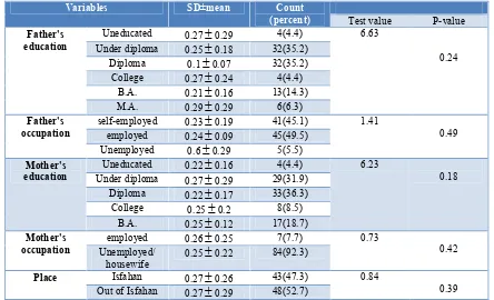
In this study, none of the socioeconomic characteristics such as father' education (p=0.24), father' occupation (p=0.49),
mother' education (p=0.18), mother'
occupation (p=0.42), and residential
address (p=0.39) had significant effect on the mean of minimal residual disease of children (Table III).
The results indicated that
lymphadenopathy (p=0.43) and response to corticosteroid treatment (p=0.11) had no significant effect on mean of minimal residual disease (Table IV).
As it is clear from Table IV,
lymphanopathy (p = 0.43) and response to treatment (p = 0.11) had no significant effect on minimal residual disease.
Table I: The relation among the minimal residual disease and age parameters, mean the WBC count in first CBC, income,
hemoglobin, and platelet level
Variables The SD±mean Relation with minimal residual disease
Test value P-Value
Minimal residual
disease (percent) 0.26
0.24 * *
Age (year-old) 4.913.07 0.47 0.07
The WBC count in
first CBC 29769.34
60908.74 0.64 0.05
Income in RLs 16444444.4414561.81 0.03 -0.22
Hemoglobin level
(MG/DL) 8.06
2.87 0.62 0.05
Platelet level
(number) 1011437.36
130.85 0.66 -0.04
Table II: The relationship between minimal residual disease and gender, type of disease, CNS involvement, mediastinal mass
organomegaly, lymphadenopathy, and translocation
Variables Relationship
with minimal residual disease
Number (percent)
Standard deviation±mean
P-VALUE Test-value
Gender Boy 0.31
0.31 46(50.5) 1.21 0.22Girl 0.21
0.13 45(49.5)Type of disease Pre BALL 0.26
0.27 52(57.8) 1.99 0.57Pro BALL 0.28
0.16 5(5.6) Early pre BALL 0.021
0.13 19(21.1)T cell 0.32
0.24 14(15.6)CNS-involvement
Yes 0.26
0.16 86(94.5) 0.39 0.69No 0.26
0.24 5(5.5) Mediastinalmass
Yes 0.3
0.16 84(93.3) 1.31 0.18No 0.25
0.24 6(6.7)Splenomegaly Yes 0.27
0.23 50(54.9) 0.36 0.71No 0.25
0.24 41(45.1)Hepatomegaly Yes 0.3
0.21 81(89) 1.09 0.27No 0.25
0.24 10(11)Translocation No 0.24
0.19 78(78.7) 0.69 0.48T(12.21) 0.37
0.43 12(13.3)Table III: The relation between mean of minimal residual disease and socioeconomic characteristics
Variables SD±mean Count
(percent) Test value P-value Father's
education
Uneducated 0.270.29 4(4.4) 6.63
0.24 Under diploma 0.25
0.18 32(35.2)Diploma 0.1
0.07 32(35.2) College 0.27
0.24 4(4.4)B.A. 0.21
0.16 13(14.3) M.A. 0.29
0.29 6(6.3) Father'soccupation
self-employed 0.23
0.19 41(45.1) 1.410.49 employed 0.24
0.09 45(49.5)Unemployed 0.6
0.29 5(5.5) Mother'seducation
Uneducated 0.22
0.16 4(4.4) 6.230.18 Under diploma 0.27
0.29 29(31.9)Diploma 0.22
0.17 33(36.3) College 0.25
0.2 8(8.5)B.A. 0.25
0.12 17(18.7) Mother'soccupation
employed 0.26
0.25 7(7.7) 0.730.42 Unemployed/
housewife
0.25
0.22 84(92.3)Place Isfahan 0.27
0.26 43(47.3) 0.840.39 Out of Isfahan 0.27
0.29 48(52.7)Table IV: The relationship between the mean of minimal residual disease and lymphadenopathy and response to
corticosteroid treatment in the first weak
Variables SD±mean Count
(percent)
Relationship with minimal residual disease
Test-value P-value
Lymphadenopathy Yes 0.31
0.24 75(82.4) 0.78 0.43No 0.25
0.24 16(17.6) Response to corticosteroidtreatment in first weak
0.25±0.23 87(95.6) 0.1 0.11
Discussion
Malignant disorder with B or T stem cell basis may lead to promotion of ALL due to the aggregation of blast cells in bone marrow. Moreover, the presence of genetic, demographic, and environmental factors is influential on the disease relapse. In this study, we aimed to assess the relation between end of induction minimal residual disease and different risk factors in patients with acute lymphoblastic leukemia. To best of our knowledge, no study has yet investigated this issue. The
findings of this study revealed that except the average of monthly income which had weak, reverse, and significant relation with minimal residual disease, none of the
studied factors, including
lymphadenopathy and response to
corticosteroid treatment had significant relationship with minimal residual disease. Aghaii pour et al. assessed 65 patients with ALLin blood transfusion center of Tehran, Iran. They revealed that the minimal residual disease in 28 days after ending induction, at the beginning of compact
treatment, and at the end of treatment was
2.7±0.4, 1.7±0.4, and 0.5±0.2,
respectively. In terms of minimal residual disease, there was a significant difference between these stages and the least rate belongs to the end of treatment (24).
Michael et al. run indicated that
Methotrexate, corticosteroid therapy, and patient's age affected the end of reduction minimal residual disease. There exist reports on significant difference between the mean of survival time and disease relapse based on the type of treatment and
treatment with Methotrexate (25).
Campana claimeds that more than 0.01% residual disease was effective on disease relapse. He also found a significant relation between residual disease rate and genetic and biological characteristics of tumor (26). Zareifar et al., sought to determine factors affecting 5-year-survival of patients with ALL. They found that the 5-year-survival of patients was 28.2 ±16.1 months and about 24.7% of patients passed away during this period. Based on their findings, platelet and the relapse cases' count were influential on survival rate (27).
In the present study, we found that assessment of effective risk factors on the minimal residual disease in patients with leukemia can be an appropriate choice for risk factors detection and elimination, thus the relapse time can be increased.
Conclusion
The relation between end of induction minimal residual disease and different risk factors in patients with ALL was assessed in the present study. The findings revealed that assessment of minimal residual disease effective risk factors in patients with leukemia would lead to their detection and elimination; thereby it can be an appropriate choice to increase the relapse time.
Acknowledgements
The present study was part of the doctoral dissertation on pediatrics. We are thankful
of the Deputy Director of the Research Department of Isfahan Medical School for financially supporting this study.
Conflict of interest
Author declared no conflict of interst.
References
1. Alijani H, Norouzi M, Aminasnafi
A, Latifi M. The effect of aromatherapy with orange essence on sleep quality of
school age children with acute
lymphoblastic leukemia 2015;1(1):112-115.
2. Walker AJ, Pongsing Y, Nail L,
Pedhiwala N, Leo M, Price J, et al. Sleep– Wake Patterns of School-Age Children and Adolescents Before Diagnosis and During Induction Chemotherapy for Acute
Lymphocytic Leukemia. J Pediatr Nurs
2011; 26(6):37-44.
3. Lichtman MA, Beutler E, Kipps
TJ, Seligsohn U, Kaushansky K. Acute lymphoblastic leukemia. 7th edition. New York:Saunders: Elsevier;2006.
4. Haghighi S, Rezvani H, Atarian H,
Isfahani F, Haghighi L, Ghadyani M,et al. The study of diettherapy on patient with
Hyper CVAD acute lymphoblastic
leukemia. JSBMU 2009; 4(1): 225-231.
5. Kliegman RM, Behrman RE,
Jenson H, Stanton BF. Nelson textbook of
pediatric. 18th ed. Philadelphia: Saunders
Elsevier;2007.
6. Allen HD, Driscoll DJ, Shaddy RE,
Feltes TF. Moss and Adam's heart disease
in infants, 7th ED. Philadelphia: Lippincott
Williams & wilkins; 2008.
7. Coustan-Smith E, Sancho J, Behm
FG, Hancock ML, Razzouk BI, Ribeiro RC, et al. Prognostic importance of measuring early clearance of leukemic cells by flow cytometry in childhood acute lymphoblastic leukemia. Blood 2002; 100(1):52-58.
8. Conter V, Bartram CR, Valsecchi
MG, Schrauder A, Panzer-Grümayer R, Möricke A, et al. Molecular response to treatment redefines all prognostic factors in children and adolescents with B-cell
results in 3184 patients of the AIEOP-BFM ALL 2000 study. Blood 2010; 115(16):3206-3214.
9. Chinkers M, Garbers DL. The
protein kinase domain of the ANP receptor is required for signaling. Science 1989; 245(4924):1392-1394.
10. Conter V, Aricò M, Basso G,
Biondi A, Barisone E, Messina C, et al.
Long-term results of the Italian
Association of Pediatric Hematology and Oncology (AIEOP) Studies 82, 87, 88, 91 and 95 for childhood acute lymphoblastic leukemia. Leukemia 2010; 24 (2):255-259.
11. Coustan-Smith E, Sancho J,
Hancock ML, Boyett JM, Behm FG, Raimondi SC, et al. Clinical importance of minimal residual disease in childhood acute lymphoblastic leukemia. Blood 2000; 96(8):2691-2696.
12. Dworzak MN, Fröschl G, Printz D,
Mann G, Pötschger U, Mühlegger N, et al. Prognostic significance and modalities of flow cytometric minimal residual disease detection in childhood acute lymphoblastic leukemia. Blood 2002 ;99(6):1952-1958.
13. Hochenberry , M. Wongs,D.
Nursing Care of Infants and children. 9th
ed. Ebook: Wilson; 2011.
14. Van Litsenburg RR, Huisman J,
Hoogerbrugge PM, Egeler RM, Kaspers GJ, Gemke RJ. Impaired sleep affects quality of life in children during
maintenance treatment for acute
lymphoblastic leukemia: an exploratory
study. Health Qual Life Outcomes
2011;9(1):25-35.
15. Kaleyias J, Manley P, Kothare SV.
Sleep disorders in children with cancer. Semin Pediatr Neurol 2012; 19(1): 25-34.
16. Pui CH, Campana D, Pei D,
Bowman WP, Sandlund JT, Kaste SC, et al. Treating childhood acute lymphoblastic
leukemia without cranial irradiation. N
Engl J Med 2009; 360(26):2730-2741.
17. Vora A, Goulden N, Mitchell C,
Hancock J, Hough R, Rowntree C, et al. Augmented post-remission therapy for a
subgroup of children and young people
with clinical standard-risk and
intermediate-risk acute lymphoblastic
leukaemia (UKALL 2003): a randomised controlled trial. Lancet Oncol 2014; 15(8):809-818.
18. Yeoh AE, Ariffin H, Chai EL,
Kwok CS, Chan YH, Ponnudurai K, et al. Minimal residual disease-guided treatment deintensilication for children with Acute Lyrnphoblastic Leukemia: results from the
Malaysia-Singapore Acuten
Lymphobiastic Leukemia 2003 Study. J clin Oncol 2012;30(19): 21-31.
19. Yamaji K, Okamoto T, Yokota S,
Watanabe A, Horikoshi Y, Asami K, et al. Minimal residual diseasebased augmented therapy in childhood acute lymphoblastic leukemia: A report from the Japanese Childhood Cancer and Leukemia Study
Group. Pediatric Blood Cancer 2010;
55(7):1287-1295.
20. Escherich G, Horstmann MA,
Zimmermann M, Janka-Schaub GE.
Cooperative study group for childhood acute lymphoblastic leukaemia (COALL. Leukemia 2010; 24(2):298-310.
21. Schmiegelow K, Forestier E,
Hellebostad M, Heyman M, Kristinsson J, Söderhäll S, et al. Long-term results of NOPHO ALL-92 and ALL-2000 studies of childhood acute lymphoblastic leukemia. Leukemia 2010; 24(2):345-349.
22. Marshall GM, Dalla Pozza L,
Sutton R, Ng A, Groot-Kruseman HA, Van der Velden VH, et al. High-risk childhood acute lymphoblastic leukemia in first remission treated with novel intensive
chemotherapy and allogeneic
transplantation. Leukemia
2013;27(7):1497-2503.
23. Weir EG, Cowan K, LeBeau P,
Borowitz MJ. A limited antibody panel
can distinguish B-precursor acute
lymphoblastic leukemia from normal B precursors with four color flow cytometry: implications for residual disease detection. Leukemia 1999;13(4):558-462.
24. Aghaei poor M, Vosough P, Nikougoftar M. minimal residual diseases with fleucitometery method among ALL disease: Thesis for obtaining of Msc degree, Blood transfusion association: Tehran University Med Sci 2004; 1-9.
25. Borowitz MJ, Wood BL, Devidas
M, Loh ML, Raetz EA, Salzer WL, et al.
Prognostic significance of minimal
residual disease in high risk B-ALL: a report from Children’s Oncology Group
study AALL0232. Blood 2015;
126(8):964-971.
26. Campana D. Minimal residual
disease in acute lymphoblastic leukemia.
ASH Education Program Book.
2010;4(1):7-12.
27. Zareifar S, Almasi-Hashiani A,
Karimi M, Tabatabaee SH, Ghiasvand R. Five-year survival rate of pediatric leukemia and its determinants. Koomesh 2012; 14(1):9-13.