S
Spprriinngg22001166,,VVooll55,,NNoo22
Mutation Screening of BRCA Genes in 10 Iranian Males
with Breast Cancer
Atieh Zorrieh Zahra1#, Sepideh Kadkhoda1#, Farkhondeh Behjati1, Fatemeh Aghakhani Moghaddam1, Azadeh Badiei1, Fereidoon Sirati2, Hossein Afshin Alavi3, Morteza Atri2, Ramesh Omranipour2, Elahe Keyhani1∗
1. Genetics Research Center-University of Social Welfare and Rehabilitation Sciences, Tehran, Iran.
2. Cancer Institute- Department of surgery- Tehran University of Medical Sciences, Tehran, Iran.
3. Pathology Ward-Day hospital,Tehran, Iran.
Male breast cancer is a rare disease with an increasing trend. Due to limited information especially about the genetic basis of the disease in Iran and the lower age of its onset, the disease requires more attention. The aim of this study was to screen the male patients with breast cancer for BRCA mutations as well as tissue markers of estrogen receptor (ER), progesterone receptor (PR), human epidermal growth factor receptor (HER-2) and cytokeratin 5/6 (CK5/6). Ten Iranian males with breast cancer were selected regardless of their histologic subtypes, age and family history from patients referred to Mehrad, Day and Parsian hospitals in Tehran, Iran, during a two-year period. Paraffin blocks of the tumoral regions were tested for ER, PR, HER-2 and CK5/6 immunostaining. DNA extraction was carried out on the EDTA blood samples followed by Sanger sequencing. Immunohistochemistry results for ER, and PR were negative in 2 out of 10 patients, while the results of HER-2 and CK5/6 were negative in all the cases. A missense mutation in exon 18 of BRCA1 and a nonsense mutation in exon 25 of in BRCA2 were detected in one patient each. Both patients belonged to luminal A subtype. Despite the low number of patients in this study, it could be concluded that mutations in BRCA1 and BRCA2 occur in male breast cancer patients of luminal A subtype. The negative status of the tissue markers could not be used for the prediction of BRCA mutations.
Key words: Male breast cancer, BRCA genes, human epidermal growth factor receptor, cytokeratin 5/6, estrogen
receptor, progesterone receptor
#
These authors had equal contribution.
∗
Corresponding author: Genetics Research Center-University of Social Welfare and Rehabilitation Sciences, Tehran, Iran. Email: ekeyhani1058@gmail.com
reast cancer is the most prevalent malignancy affecting women (1, 2). The disease is rare in males, accounting for 1% of all breast cancer cases (3-5) but recently a significant increasing rate from 0.86 to 1.08 cases per 100,000 populations has been
reported (5-7). There are many studies reported from Iran on female breast cancer (FBC) but male breast cancer (MBC) still needs to be studied (8, 9). Unfortunately, in Iran few researches and reports about the prevalence of the disease are available
B
Submmited 29 October 2015; Accepted 31 January 2016; Published 9 May 2016
(10). In Iran, MBC accounts for 0.65% of all cases of malignancy in men. There are 6674 incident cases of breast cancer diagnosed in Iran in 2007 of whom 3.26% were men (11). Due to its low incidence, there are few studies and limited information on this subject especially about the genetic etiology of the disease.
Furthermore, the average age of male breast cancer and female breast cancer in Iran is lower than developed countries (11), and men are usually diagnosed with breast cancer at more advanced stages often with lymph node metastasis (3, 12).
Several risk factors including advancing age, positive family history and mutations in susceptibility genes such as BRCA1 and especially BRCA2 are considered for the study of male breast
cancer (10).
BRCA1 and BRCA2 are tumor suppressor
genes, located on 17q21and 13q12 loci with 24 and 27 exons, respectively. These genes normally participate in the repair of damaged DNA by homologous recombination. Although BRCA2 is the most clearly associated gene, BRCA1 mutation is also considered in familial and even sporadic cases of male breast cancer (4, 5, 13). According to some studies, BRCA2 mutations occur in 4-16% of male breast cancer patients. However, BRCA1 mutations are much less common and 0–4% of men with breast cancer carry these mutations (14). BRCA1 mutations as a prognostic factor also have
great importance, as carriers of these mutations have a poorer prognosis compared to other patients. Mutation in BRCA2 also has a special significance as the mutation carriers can be diagnosed with breast cancer at lower age and has a shorter survival (13). In comparison with BRCA1 mutations, BRCA2 mutations have major significance in MBC patients (4, 5, 13). Patients with mutation in BRCA2 are predisposed to breast cancer and other malignancies such as pancreatic, prostate cancer and melanoma.
Molecular subtypes of MBC are similar to FBC (4, 11). According to several studies, MBC seems to be more hormone receptor positive (luminal A subtype) (3, 13, 14), than FBC. The main aim of this study was to screen the male patients with breast cancer for BRCA1 and BRCA2 mutations as well as the status of tissue tumor markers including ER, PR, HER-2 and basal marker (CK5/6). We also investigated the relationship between the BRCA1 and BRCA2 mutations and the tissue markers’ status.
Materials and methods
Patient selection and sampling
In this cross- sectional study, 10 Iranian males with breast cancer were selected from Mehrad, Day and Parsian hospitals in Tehran, Iran, regardless of age, family history, histopathological subtypes and occupation. Consent forms were obtained according to the guidelines of the research ethics committee of the University of Social Welfare and Rehabilitation Sciences. 5-10 ml blood sample was collected into EDTA tube from each patient, and paraffin-embedded tissue block samples were prepared from the tumoral regions. Information about patients’ age, involved breast side, tumor size, histologic subtypes, status of lymph nodes’ involvement, surgical technique, tumor stage, tumor grade and family history of cancer was obtained from the patents’ medical files in the related hospitals. The median age for the breast cancer patients was 52.7 years (range from 38 to 72). The average tumor size was 2.03 cm (ranges 0.9 to 3.5). The histological types of all the tumors were invasive ductal carcinoma. Only one patient had documented family history of breast cancer with an affected mother. Six of the ten patients, showed the involvement of the left side, and in the other four cases the right side was involved.
Histopathology
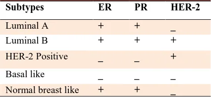
Tissue sections were prepared, stained by hematoxylin and eosin (H&E) and then were studied by a pathologist to confirm the diagnosis and characterization of the tumor. Immunostainig was performed on 4 µ m thickness sections. After incubation at 60°C and before proceeding with the staining protocol the slides were deparaffinized and rehydrated by xylene and ethanol (100%, 96% and 70%), respectively. Endogenous peroxidase activity was blocked with 5% hydrogen peroxide in methanol for 15 min. Heat- induced antigen retrieval with Tris-EDTA (pH=9) was done to break the methylene bridges and expose the antigenic sites in order to allow the antibodies to bind. Then slides were incubated with primary antibodies: 100 µl anti-human ER, PR, HER2, CK5/6 (Dako-Denmark). The secondary antibody or Envision was applied. The color was visualized by incubation with chromogen 3, 3’-Diaminobenzidine (DAB) for 5 min.. Then slides were placed in hematoxilin for 30 s and dehydrated in 70%, 96% and 100% (twice) ethanol and xylen (three times), one min. for each time, and finally were mounted. Positive and negative external controls were used to justify the test results. Interpretation of the results was performed using the classification method based on tissue tumor markers (Table 1).
Table 1. Classification of breast cancer based on tissue tumor markers.
Subtypes ER PR HER-2
Luminal A + + _
Luminal B + + +
HER-2 Positive _ _ +
Basal like _ _ _
Normal breast like + + _
For confirming HER-2 samples assessed by immunohistochemistry (IHC) as+2 score, FISH technique was performed (15). Routinely processed paraffin- embedded tissue sections (3 µm) were
deparaffinized in fresh xylene and were rehydrated by ethanol (100%, 85% and 70%) and washed with distilled water. Slides were then pretreated with 0.2 M HCl for 20 min, and washed with distilled water. Slides were placed in a preheated 80°C pretreatment reagent (8% sodium isothiocyanate) for 30 min, rinsed in 2×SSC for 3 min. Protease digestion was accomplished by placing the slides in a pre-warmed (37 °C) protease solution (0.025% pepsin) for 30 min. Samples were then rinsed in distilled water for one minute, washed in 2×SSC for 5 min. and dehydrated in ethanol (70%, 85% and 100%). All slides were hybridized under identical conditions and with appropriate control tissue. 10 µl of probe was applied to the region of interest on the slide. A cover slip was placed and sealed with rubber cement. The slides were denatured on a hot plate at 80 °C for 5 min. and incubated overnight at 37 ºC in a humidified chamber. After hybridization, the rubber cement was removed. When cover slips did not come off easily, slides were washed in 2×SSC / 0.1% igepal for 2 min, then washed in 0.4×SSC/ 0.3% igepal at 72 °C for 2 min and 2×SSC/ 0.1% igepal for 1 min. Slides were dehydrated in ethanol (70%, 85% and 100%) and counterstained by applying 10 µl of 4,6-diamidino-2- phenylindole dihydrochloride (DAPI) and a glass cover slip was placed on the slide. At least 20 cells were scored in each preparation, and the total number of HER2 and CEN17 signals in each cell was scored using a fluorescence microscope.
The basis of IHC scoring according to American society of clinical oncology/college of American pathologists (ASCO/CAP) guideline is as follows: As HER-2 is localized in membrane of tumor cells, IHC scoring of 3+, 2+, 1+ and 0 are defined as a strong complete, weak or moderate, faint or incomplete membrane, and no stain or membrane stain in less than %10 of tumor cells. The localization of ER or PR antigens is nuclear, in immunostaining. So staining in more than 1% of the tumor cell nuclei means positive expression and
Fig 1. Immunohistochemical staining for HER2. Different staining patterns are seen in two patients. a: positive control with strong
continuo-us membrane staining which is a good example of a 3+ positive tumor (3+);b: negative HER2 with no staining in membrane (0). Original
magnification × 400; scale bar: 100µm.
less than 1% staining reveals negative result. Concerning HER-2 staining, HER-2/CEN17 ratio< 1.8= negative, ratio= 1.8-2.2 means equivocal and ratio> 2.2 means positive.
Gene sequencing
Genomic DNA was extracted from the blood samples using salting out method. The quality and quantity of DNA was determined by reading the optical density of the sample at 260 nm and 280 nm. Primers for BRCA1 and BRCA2 genes were designed by primer 3 and UCSC genome browser. Self-dimers, heterodimers and the melting tempera-ture of the primers were also checked by oligo analyzer site (https:// www. idtdna.com/ calc/ analyzer).
In BRCA1, exon 10 and 24 were separated into 6 and 3 fragments, respectively. In BRCA2, exons11, 10, and 27 were separated into 9, 3, and 2 fragments respectively. DNA was amplified by polymerase chain reaction (PCR) technique.
Mutation analysis was performed by direct DNA sequencing of all the exons of BRCA1 and BRCA2. The partial flanking intronic sequences
were obtained using codon code aligner and Gene runner softwares
Results
Immunohistochemical analyzes
In this study, we screened 10 Iranian men with breast cancer in order to find any mutation in BRCA genes. On the other hand, tissue tumor markers (ER, PR, HER-2 and CK5/6) status, in related tissues of the patients was assessed. Analysis of the stained sections confirmed the presence of the tumoral tissue.
Results of IHC technique for CK5/6 were negative for all patients. For ER and PR markers 8 out of 10 cases were negative and 2 out of 10 had positive markers. The score of IHC for HER-2 marker was +2 for 3 patients, and FISH technique was carried out to confirm the results. Average ratio of HER-2/CEN-17 for these patients was 1.1. Therefore, the
amplification of HER-2 in these samples was negative. The 7 other patients had a score lower than +2 in IHC evaluation and were considered also as negative for Her 2. The results for IHC as well as FISH stainings for HER2, followed by IHC for CK5/6, are shown in Figures 1- 3.
Mutation analysis
Following PCR amplification and sequencing of BRCA1 and BRCA2, in 10 males with breast cancer, their variants and polymorphisms were detected. Further information, including the exon number, base changes and frequency of these variants in 10 patients, are shown in Tables 2 and 3 for the BRCA1 and BRCA2, respectively.
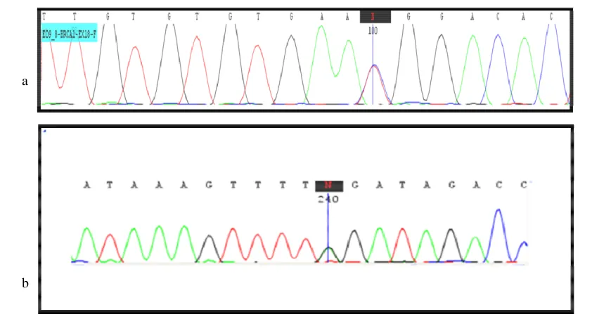
a b
Fig 4. BRCA sequencing results. a: sequence graph of the region surrounding the exon 18 of BRCA1 c.5158C>T mutation in the patient
(5158C/T heterozygous); b: DNA sequencing of BRCA2 exon 25 identified a heterozygous BRCA2 mutation (c.9317G>A) in the patient.
Fig 2. Immunohistochemical staining and FISH for HER2. a: HER2 with score 2 by IHC (borderline).There is a moderate membrane staining pattern. In this case, tumoral tissue was tested by FISH to confirm the diagnosis. Original magnification×400; scale bar: 100µm. b: FISH staining for evaluating IHC for HER2 score 2 patient revealed an absence of HER2 amplification. Red signal (Text-Red): HER-2, Green signal (FITC): chromosome17 centromere. For evaluating FISH results, 20 cells from 2 tumor regions were counted; scale bar: 10 µm.
Fig 3.
I
mmunohistochemical staining for CK5/6. a: positive control with cytokeratin 5/6 expression; b: negative CK5/6 with no expression of CK5/6. Original magnification × 400, scale bar: 100 µm.a b
a b
a
b
Table 2. Polymorphisms and variants in BRCA1 gene.
Variants Exon Base change Frequency n= 10 (100%)
rs1799949 10 C>T 0.7
rs16940 10 T>C 0.6
rs1799950 10 A>G 0.1
rs4986850 10 G>A 0.2
rs799917 10 C>T 0.5
rs16941 10 A>G 0.4
rs16942 10 A>G 0.5
rs80357396 10 A>T 0.1
rs8176318 24 G>T 0.5
rs12516 24 C>T 0.6
rs799923 7 G>A 0.2
rs1060915 12 T>C 0.7
rs1799966 16 A>G 0.6
Table 3. Polymorphisms and variants in BRCA2 gene.
Variants Exon Base change Frequency n=10 (100%)
rs17999432 10 G>A 0.2
rs766173 10 A>C 0.4
rs1801439 10 A>G 0.4
rs144848 10 A>C 0.3
rs1801499 11 T>C 0.4
rs1801406 11 A>G 0.2
rs1799944 11 A>G 0.4
rs543304 11 T>C 0.5
rs206075 11 A>G 1
rs80358755 11 A>G 1
rs206076 11 A>G 1
rs169547 14 T>C 1
rs1799955 14 A>G 0.2
r rs80359157 23 C>T 0.1
A missense mutation was detected in exon 18 of BRCA1 at position c.5158C>T (Figure 4a, Table 4). This mutation was evaluated by Mutation Taster, Mutation Assessor, SIFT, PROVEAN, POLY PHEN and ENSMBLE softwares and was confirmed as a cause of disease or pathogenic variant by BIC and HGMD databases. In another
patient a nonsense mutation was found in exon 25 of BRCA2 at position c.9317G>A (Figure 4b, Table 4).
This mutation was evaluated by BIC database, mutation taster site and HGMD database as a pathologic mutation Interestingly, in both patients with BRCA mutation, IHC results and tumor
markers status (ER, HER-2 and CK5/6) were identical to other patients, e.g. positive for ER and negative for the two other markers. Furthermore, none of the patients with BRCA mutation had a family history of the disease. The
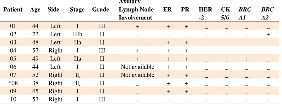
informations about patients including age, stage, lymph node status, degree and tumor markers’ status assessment along with the type of related mutation in BRCA genes are presented in Table 5.
Discussion
In this study, 10 males with breast cancer were screened for mutations in BRCA1 and BRCA2. Besides, the patients were characterized for immunohistochemical features such as ER, PR, Her-2, and CK5/6 tissue tumor markers. In one patient, mutation in exon 18 of BRCA1 was detected. Furthermore, another patient has a nonsense mutation in exon 25 of BRCA2. Moreover, several variants and polymorphisms associated with BRCA1 and BRCA2 were found, also some features correlated to those, such as the exon number, base changes and frequency of these variants, were summarized in Tables 3 and 4. As
the purpose of the current study was mutation screening in BRCA1 and BRCA2, associated variants were identified and assessed in order to note whether they were disease causing. Then mutation screening was followed until a mutation was found and confirmed by some known softwares and databases. Results of IHC technique for HER-2 and CK5/6 markers of all patients and ER and PR markers for two patients were negative.
Previous studies have indicated that mutation in BRCA2 in MBC is more common than BRCA1, but our findings could not conclude such results due to the small sample size. According to a similar study by Deb and his colleagues in Australia in Table 4. Results of the evaluation of BRCA sequencing
Gene Exon Mutation AA change Mutation type
BRCA1 18 c.5158C>T Arg1720Trp missense
BRCA2 25 c.9317G>A Trp3106Ter stop codon
Table 5. Pathologic features, tumor markers assessment and mutation screening of male breast cancer patients
Patient Age Side Stage Grade
Axillary Lymph Node Involvement
ER PR HER
-2
CK 5/6
BRC A1
BRC A2
01 44 Left І Ш + + + _ _ _ _
02 72 Left Шb Ц _ _ _ _ _ _ +
03 48 Left Цa Ц _ + + _ _ _ _
04 57 Right І Ш + + + _ _ _ _
05 49 Left Цa Ц + + + _ _ + _
06 44 Left І Ц Not available + + _ _ _ _
07 52 Right Ц Ц Not available + + _ _ _ _
*08 38 Right Ц Ц _ + + _ _ _ _
09 65 Right І Ц _ + + _ _ _ _
10 57 Right І Ш _ _ _ _ _ _ _
*The patient has a family history of breast cancer.
2012, most of MBC patients (89.7%) were classified in luminal subtypes and a minority were in HER-2 and basal categories. The rate of mutation in BRCA2 was higher and in BRCA1 was lower compared to FBC (6).
Based on other studies, positive status of ER and negative status of CK5/6 tissue markers followed by negative status of HER-2, concluded luminal A molecular subtype of MBC. The status of the studied tissue tumor markers could not predict the existence of mutation in BRCA genes.
Some studies have only focused on BRCA1 or BRCA2 or IHC features (16, 17), and therefore
concurrent review of BRCA1/2 mutational screening together with tumor markers have rarely been reported (17). Several studies based on concurrent investigations of BRCA genes and tumor markers performed only in FBC patients and outside Iran, showed results similar to the present study (18). While this project is a pioneer study based on genetic and histopathologic aspects of MBC in Iran, there is still a great need to study the genetic basis of MBC.
It should be mentioned that the main problems of this project were: “case finding” and “tiny tissue samples”. Considering the low prevalence of the disease around the world, especially in Iran, despite searching four hospitals to find males with breast cancer, and due to the problems such as lack of cooperation of the patients and their families, finally a small population of only 10 qualified cases were allocated for the study. According to the rarity of MBC and its increasing rate in the world, it seems to be necessary to perform studies with larger sample size. We believe that general education for the early diagnosis of breast cancer in males in order to increase their awareness of this disease, and also promotion of the quality of diagnostic methods in Iran are necessary.
Acknowledgements
The authors extend their thanks to the staff of Genetics Research Center for their assistance in this project:S. Abedini, S.Banihashemi, Kh. Jalalvand, I. Bahman, Dr. A. Mohammadzadeh, S. Biglari, M. Aghajanpour, Z. Mehrjou, M.Hosseini, M. Babanezhad, and M. Mohseni.
Conflict of interest:
The authors declared no conflict of interests.
References
1. Keyhani E, Muhammadnejad A, Behjati F, et al. Angiogenesis markers in breast cancer--potentially useful tools for priority setting of anti-angiogenic agents. Asian Pacific journal of cancer
prevention : APJCP 2013;14:7651-6.
2. Keyhani E, Muhammadnejad A, Karimlou M. Prevalence of HER-2-positive invasive breast cancer: a systematic review from Iran. Asian Pacific journal of cancer prevention : APJCP
2012;13:5477-82.
3. Vermeulen JF, Kornegoor R, van der Wall E, et al. Differential expression of growth factor receptors and membrane-bound tumor markers for imaging in male and female
breast cancer. PloS one 2013;8:e53353.
4. Korde LA, Zujewski JA, Kamin L, et al. Multidisciplinary meeting on male breast cancer: summary and research recommendations. Journal of clinical oncology : official journal
of the American Society of Clinical Oncology 2010;28:2114-22. 5. Weiss JR, Moysich KB, Swede H. Epidemiology of male breast cancer. Cancer epidemiology, biomarkers & prevention : a publication of the American Association for Cancer Research, cosponsored by the American Society of Preventive Oncology
2005;14:20-6.
6. Deb S, Jene N, Kconfab I, et al. Genotypic and phenotypic analysis of familial male breast cancer shows under representation of the HER2 and basal subtypes in BRCA-associated carcinomas. BMC cancer 2012;12:510.
7. Foerster R, Foerster FG, Wulff V, et al. Matched-pair analysis of patients with female and male breast cancer: a comparative
analysis. BMC cancer 2011;11:335.
8. Mousavi F, Noruzinia M, Keyhani E, et al. Methylation Analysis of 5’UTR Promoter Region of DBC2 as a Biomarker in
the Peripheral Bloods of Some Iranian Women with Sporadic
Breast Cancer, 2014. Iran J Pathol 2014;9:117-23.
9. Behjati F, Atri M, Najmabadi H, et al. Prognostic value of chromosome 1 and 8 copy number in invasive ductal breast carcinoma among Iranian women: an interphase FISH analysis.
Pathology oncology research : POR 2005;11:157-63.
10. Ruddy KJ, Winer EP. Male breast cancer: risk factors, biology, diagnosis, treatment, and survivorship. Annals of oncology : official journal of the European Society for Medical
Oncology / ESMO 2013;24:1434-43.
11. Salehi A, Zeraati H, Mohammad K, et al. Survival of male breast cancer in fars, South of iran. Iranian Red Crescent medical
journal 2011;13:99-105.
12. Tirgari F, Abdi Rad A, Mahjoub F, et al. Male Breast Carcinoma: An Immunohistochemical Study of 50 Cases from
Iran, 2008. Iran J Pathol 2008;3:94-9.
13. Mirmalek SA, Elhamkani F. Male Breast Cancer. Iranian
Journal of Surgery 2007;15:20-37.
14. Sharon H, Giordano A. Review of the Diagnosis and Management of Male Breast Cancer, 2005. Oncologist
2005;10:471-9.
15. Muhammadnejad A, Keyhani E, Mortazavi P, et al. Overexpression of HER-2/neu in Malignant Mammary Tumors Translation of Clinicopathological Features from Dog to Human,2012. Asian Pacific journal of cancer prevention :
APJCP 2012;13:6415-21.
16. Kuusisto KM, Bebel A, Vihinen M, et al. Screening for BRCA1, BRCA2, CHEK2, PALB2, BRIP1, RAD50, and CDH1 mutations in high-risk Finnish BRCA1/2-founder mutation-negative breast and/or ovarian cancer individuals. Breast cancer
research : BCR 2011;13:R20.
17. Kwong A, Ng EK, Wong CL, et al. Identification of BRCA1/2 founder mutations in Southern Chinese breast cancer patients using gene sequencing and high resolution DNA melting
analysis. PloS one 2012;7:e43994.
18. Lakhani SR, Reis-Filho JS, Fulford L, et al. Prediction of BRCA1 status in patients with breast cancer using estrogen receptor and basal phenotype. Clinical cancer research : an official journal of the American Association for Cancer Research
2005;11:5175-80.