email: [email protected] 4502
INCORPORATION OF AU AND AG NANOSTRUCTURES INSIDE SIO
2C. DIAZ
*,a, A. M. L. VALENZUELA
b, K. SOTO
aAND M. A. LAGUNA-BERCERO
caDe p a rt a me n to d e Qu í mi c a , Fac u l t ad de Qu í mi c a , Uni v e rsi dad d e Ch i l e. La Pa l me ra s 3 42 5 , N u ño a , ca si l la 6 53 , Sa nt i ag o de C h i le , C h i l e .
bUn i v e rsi d ad Au tó no ma d e C h il e , In st i t ut o d e C i en c ia s Qu í mic a s Ap l ic a da s, In o rga n ic C h e mi st ry a nd M o le c u la r M at e ri a l C e n t e r. A v . El Ll a n o S ub e rc a sea u x 2 80 1 . Sa n M ig u el . Sa n t ia go d e C h il e .
cIn st i t u to d e C i en c i a d e Ma t e ria l e s d e Ara g ón (IC M A), C S IC - Un i v e rsi d ad d e Za ra go za C / Pe d ro C e rb u na 12 , E -5 00 0 9, Zara g o za ,
S p a in .
ABSTRACT
Incorporation of Au° inside SiO
2 was achieved by a solid-state method from the pyrolysis of the composites (Chitosan)•(AuCl3)n•(SiO2)m and
(PS-co-4-PVP)•(AuCl3)n•(SiO2)m Similarly, the incorporation of Ag° inside SiO2 it was made from thermal treatment of the composites (Chitosan)•(AgNO3)n•(SiO2)m
and (PS-co-4-PVP)•(AgNO3)n•(SiO2)m. The nature of the polymer controls the particle size for the Au/SiO2 composite, while that for the Ag/SiO2 both, polymer
Chitosan and PS-co-4-PVP, produces similar particle size. In the case of the composite Ag/SiO2 the particle size as small as 5 nm were obtained.
The 1:1 or 1:5 metal/polymer ratios, as well as the nature of the polymer in the macromolecular precursors (Chitosan)•(AuCl3)n•(SiO2)m and
(PS-co-4-PVP)•(AuCl3)n•(SiO2)m influences the dispersion of the Au° nanostructures inside SiO2 matrix. The results are compared with those previously obtained
for bimetallic composites Au/Ag//SiO2. A formation mechanism of the Au°/SiO2 and Ag°/SiO2 composites involving the combustion of the organic matter and the
growth of the Au° and Ag° nanoparticles inside the holes, generated from the combustion process, is proposed.
Keywords: Gold nanoparticles, Silver nanoparticles, silica, composites.
1.- INTRODUCTION
Among the several practical applications of the metallic and metal oxides nanoparticles and nanostructured materials, are those in catalysis1,2.In this field,
the heterogeneous catalyst is one of the most important and most widely used in industrial processes. Is well known that the incorporation of these metal oxides on the inert supports materials, with high surface areas to form composites materials, could help to prevent their aggregations and improvement their reactivity and stability3,4. Specifically the nanocomposites Au°/SiO
2 are an
important catalyst for several oxidation processes5,6 . Since the finding by Haruta
et al7,8 of the high performance low temperature oxidation of CO by Au °/SiO 2,
several other oxidation processes, using this nanocomposites have been reported. Because of the usual solution method to prepare this catalyst composite, presents some problems due to the difficulty of depositing the Au° nanoparticles inside SiO2 the adequate preparation of Au°/SiO2, is a constant challenge9,10
Then, the new easily, fast and direct methods for to prepare nanoparticles and nanostructures anchored to a solid supporting is a constant challenge. The solution ways to synthesize this type of system, they have the difficulty that, on carry the nanoparticles, from solution to solid-state anchored over a solid support, they undergoes usually agglomeration with loss of the nanoscale properties11-14.
On the other hand, the interest of the Ag°/SiO2 composites mainly arises from
the antimicrobial properties15 of Ag° as well as from their catalysis16 and their
electronic devices practical applications17. These interesting properties of the
Ag° nanoparticles are they are altered by the uncontrollable release of Ag+ by
oxidation on O2 which cause toxicity in the antimicrobial properties, oxidation
to Ag+ in the high temperature catalyst applications decreasing the catalytic
efficiency and loss of the plasmon resonance, important in the electronic applications , also by the oxidation of Ag° to Ag+. All these disadvantage of the
use of Ag° in practical applications can be solved by their encapsulation in SiO2 18-21. Several solution methods to prepare Ag°/SiO
2 including the most usual, the
impregnation of previously formed SiO2 with some Ag+ salt afford in some cases
partial formation of Ag2O/SiO222. Here we present a route where phase pure
Ag°/SiO2 is obtained and further is general for the synthesis of both Au°/SiO2
and Ag°/SiO2. In contrast with the here reported method where, the pure phase
Ag° nanoparticles inside SiO2 was observed, in ours previous reports, the thermal
treatment the containing Ag+ [(O3SCF3)Ag(PPh3)(HOC5H4N)][SiO2]n
precursors affords Ag2O inside SiO2 nanoparticles22.
Previously we have reported a versatile and reliable solid-state method to prepare metallic and metal oxides nanostructures materials22-26 and their
incorporation in SiO2 matrices27,28. The method consists in the pyrolysis of the
macromolecular precursors (Chitosan)•(MLn)n•(SiO2)m and
(PS-co-4-PVP)•(MLn)n•(SiO2)m where the precursors (Chitosan)•(MLn)m and
(PS-co-4-PVP)•(MLn)m are generated according previous reports 22-26 .Now, we use this
approach to prepare Au° and Ag° inside SiO
2 to giveAu/SiO2 and Ag/SiO2
composites founding that the dispersion of the metal noble nanoparticles into the silica matrix can be controlled by the 1:1 or 1:5 metal/polymer ratio, as well as, by the nature of the polymer. The results are compared with those previously obtained27 for bimetallic composites Au/Ag//SiO
2. A preliminary work showing
some of these result have been reported27.
2.- EXPERIMENTAL
AuCl3 and Ag(NO3), PSP-co-4-PVP, Chitosan, TEOS (tetraethoxysilane) and
acetic acid were purchased from Aldrich and used as received. All the reactions were performed using CH2Cl2 as solvent and nanopure water.
2.1.- Precursors Synthesis: Chitosan•(MLn)n MLn=AuCl3 and Ag(NO3)
In a typical synthesis 22-26, the respective metallic salts AuCl
3 and Ag(NO3)
were added in a Schlenk tube using CH2Cl2 as solvent under magnetic stirring
and then the respective Chitosan amount was added according to the 1:1 (1),(5)
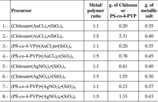
or 1:5 (2), (6) respectively for Au and Ag molar ratio (compound numeration according Table 1).
Precursor
Metal/ polymer
ratio
g. of Chitosan or PS-co-4-PVP
g. of metallic
salt
1.- (Chitosan•(AuCl3)n•(SiO2)n 1:1 0.20 0.35
2.- (Chitosan•(AuCl3)n•(SiO2)n 1:5 2.31 0.40
3.- (PS-co-4-VP)•(AuCl3)n•(SiO2)n 1:1 0.20 0.35
4.- (PS-co-4-PVP)•(AuCl3)n•(SiO2)n 1:5 0.78 0.45
5.- (Chitosan•(AgNO3)n•(SiO2)n 1:1 0.41 0.40
6.- (Chitosan•(AgNO3)n•(SiO2)n 1:5 1.55 0.30
7.- (PS-co-4-PVP)•(AgNO3)n•(SiO2)n 1:1 0.23 0.37
8.- (PS-co-4-PVP)•(AgNO3)n•(SiO2)n 1:5 1.33 0.43
Table 1. Experimental Details of the synthesis of the precursors 1-6
4503
2.3.- SiO2
SiO2 was prepared according a literature method 27, 28. Briefly, TEOS, ethanol,
acetic acid in molar ratios 1:4:4 and water nanopure were stirring for 3 days. The wet gels were further dried at 100 °C under reduced pressure in a vacuum furnace. Finally, the solids were calcined at 800 °C for 2 h.
2.4.- (Chitosan)•(MLn)n•(SiO2)m and (PS-co-4-PVP)•(MLn)n•(SiO2)m precursors
The (Chitosan)•(AgNO3)n•(SiO2)m and (PS-co-4-PVP)•(AgNO3)n•(SiO2)m
were prepared by simple solid-state mixture of the as prepared (Chitosan)•(MLn)n and (PS-co-4-PVP)•(MLn)m and SiO2 and subsequent
grinding of the solid mixture in a 1:10 molar ratio.
2.5.- Pyrolysis
The pyrolysis experiments were conducted by pouring a weighed portion (0.05–0.15 g) of the respective(Chitosan)•(AgNO3)n•(SiO2)m and
(PS-co-4-PVP)•(AgNO3)n•(SiO2)m precursors into aluminum oxide boats that were placed
in a furnace (Labtech.LEF-10 oven ) under a flow of air, heating from 25°C to upper temperature limits of 300°C, and then to 800 °C, followed by annealing for 2-4 h and at rates of 10 °C min-1 in each case.
2.6.- Characterization of the pyrolytic products
Solid pyrolytic samples were characterized by powder x-Ray diffraction (XRD) and scanning electron microscopy (SEM). SEM images were acquired with a JSM-6380LV, JEOL Ltda. microscope, with an acceleration potential of 20 keV. Energy dispersive X-ray analysis (EDS) was performed on a NORAN Instrument micro-probe attached to a JEOL 5410 scanning electron microscope. High-Resolution Transmission Electron Microscopy (HR-TEM) was performed using a JEOL 2000FX TEM microscope at 200 kV to characterize the average particle size, distribution and elemental and crystal composition. X-ray diffraction (XRD) was conducted at room temperature on a Siemens D-5000 diffractometer with θ-2θ geometry. The XRD data was collected using Cu-Kα radiation (40 kV, 30 mA). FTIR measurements were performed on a Perkin Elmer FTIR spectrophotometer model Spectrum BXII. UV-vis spectra were performed on a Shimadzu UV 2450 spectrophotometer.
3.- RESULT AND DISCUSSION
A pictorial drawing showing the methodology used for the preparation of the composites Au/SO2 and Ag//SiO2 is displayed in Figure 1.
Figure 1. Schematic representation of preparation of Au/SO2 and Ag//SiO2
composites.
The macromolecular complexes (PS-co-4-PVP)•(AuCl3)n and
(Chitosan•(AuCl3)n prepared as previously described (see experimental part and
ref. 22-26).and the SiO2 (prepared by sol-gel method see experiemental part and
ref. 27, 28) were mixture in solid state in a crucible and pyrolyzed under air at 800 °C. The composites were first characterized be powder XRD- ray diffraction. Figure 2 shows as a representative, the diffraction pattern of Au/SO2, from the
precursor (PS-co-4-PVP)•(AuCl3)n•(SiO2)n, as well as from
(Chitosan•(AuCl3)n•(SiO2)n in a pure phase. Clearly the (111), (200), (220) and
(311) main diffraction planes of Cubic Au are observed. A broad direction peak at 2θ= 23° is assigned to amorphous silica. Thus, these diffraction patterns are similar to that of reported for Au/SO2 composites using a several solution and
another miscellaneous methods29.
Figure 2. XRD diffraction pattern of the composite Au/SiO2 from the
(PS-co-4-PVP)•(AuCl3)n•(SiO2)m, 1:1 precursor (A) and from
(Chitosan•(AuCl3)n•(SiO2)m 1:1 (B).
Similar XRD pattern was found for the Au/SO2 prepared from the precursors
(Chitosan•(AuCl3)n•(SiO2)m see figure 2 (B).
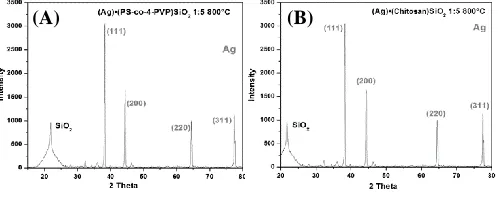
On the other hand the XRD patterns diffraction for the composites Ag//SiO2
from both polymer precursors , see figure 3 (A) and (B), exhibits a similar peaks patterns to that of Au/SiO2 due to both Au and Ag they have a similar unitary
cell27. Both XRD diffraction are in agreement with whose reported for another
Ag//SiO2 composite prepared from solution20,21.
Figure 3. XRD diffraction pattern of the composite Ag/SO2 from the
(PS-co-4-PVP)•(AgNO3)n•(SiO2)m, 1:1 precursor (A) and from
(Chitosan•(AgNO3)n•(SiO2)m 1:5 (B).
Notwithstanding the above the presence of Au and Ag inside the matrix SiO2
was corroborate by their SEM-EDS analysis as is shown in figure 4. The presence of Si, O and Au for the composite Au/SiO2 ,figure 4 (A) and (B) and
the presence of Si, O and Ag for the composite Ag/SiO2 , figure 4 (C) and (D)
was clearly confirmed from these images. Thus the morphology observed shows somewhat irregular shapes with exception in figure 4 (D) where some spheres of SiO2 surrounded of some Au nanoparticles.
Figure 4. SEM image together with their EDS analysis (inset) for of the composite Au/SO2 from the (PS-co-4-PVP)•(AuCl3)n•(SiO2)m, 1:1 precursor (A) and
from (Chitosan•(AuCl3)n•(SiO2)m 1:1 (B) as well as for composite Ag/SO2 from the (PS-co-4-PVP)•(AgNO3)n•(SiO2)m, 1:5 precursor (C) and from
(Chitosan•(AgNO3)n•(SiO2)m 1:5 (D).
(A)
(B)
4504 The TEM images, see figure 5, shows the TEM image for the Au/SiO2 and
Ag/SiO2 obtained from the precursors with both polymer Chitosan and
PS-co-4-PVP. For Au/SiO2 the TEM images shows the Au nanoparticles well dispersed
inside SiO2 for the nanocomposite prepared from (Chitosan•(AuCl3)n•(SiO2)m
precursor with average size of 5.7 nm (see histogram inside ) but with some agglomerates nanoparticles into SiO2 composite from the
(PS-co-4-PVP)•(AuCl3)n•(SiO2)m, with average size of 108 nm (see histogram inside
figure).
Similar TEM images were obtained for Au/SiO2 composites obtained by
other methods 9.10.
On the other hand well dispersed Ag nanoparticles inside SiO2 where observed
for the Ag/SiO2 composites obtained from (Chitosan•(AgNO3)n•(SiO2)m figure
5d and (PS-co-4-PVP)•(AgNO3)n•(SiO2)m,, figure 5c, with particle size 5 nm
and 6 nm respectively obtained from the respective inset histograms. Similar TEM images were obtained for Ag/SiO2 composites prepared by other methods. 21,30.
Figure 5. TEM image with their histogram (inset) for of the composite Au/SO2 from the (PS-co-4-PVP)•(AuCl3)n•(SiO2)m, 1:1 precursor (A) and from
(Chitosan•(AuCl3)n•(SiO2)m 1:1 (B) as well as for composite Ag/SO2 from the (PS-co-4-PVP)•(AgNO3)n•(SiO2)m, 1:5 precursor (C) and from
(Chitosan•(AgNO3)n•(SiO2)m 1:5 (D).
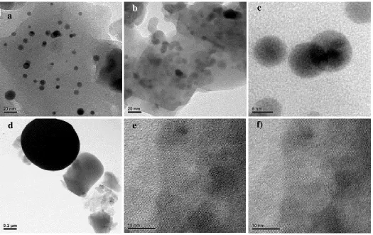
Some most detailed features are observed by HRTEM. For instance from figure 6 two Au overlapping nanoparticles and other two isolates both with size near to 5 nm as observed in the histogram from the TEM , se figure 6 a,b,c.These were obtained from the precursor (PS-co-4-PVP)•(AuCl3)n•(SiO2)m. From Au
nanoparticles from the Chitosan•(AuCl3)n•(SiO2)m precursor most rather large
nanoparticles were observed as is observed from figure 5c,f. Interplanar distances corresponding to the plane typical of Au were observed as is showed in figure 5 e, f.
Figure 6. HRTEM images of Au nanoparticles from (PS-co-4-PVP)•(AuCl3)n•(SiO2)m (a), (b) ,(c) and from Chitosan•(AuCl3)n•(SiO2)m (d), (e) and (f).
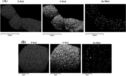
Most detailed information of the dispersion of the Au nanoparticles inside SiO2
was obtained from SEM-EDS mapping by element. Figure 7 shows Au
nanoparticles well dispersed inside SiO2 for the nanocomposite prepared from
1:1 and 1:5, as well as from (Chitosan•(AuCl3)n•(SiO2)m 1:1and 1:5 precursor.
d
)
f)
e
)
c
)
b
)
4505
Figure 7. EDS mapping by elements of the Au/SiO2 nanocomposite from the precursor (Chitosan•(AuCl3)n•(SiO2)m in 1:1 (A) and 1:5 (B) molar metal polymer
ratio. Similar well dispersed Au into silica was observed for the composite Au/SiO2 prepared from precursors 1:5 (Chitosan•(AuCl3)n•(SiO2)m see figure 6 (B) but
with minor content of Au nanoparticles dispersed into silica.
Figure 8. EDS mapping by elements of the Au/SiO2 nanocomposite from the precursor 1:1 (A) and 1:5 (B) (PS-co-4-PVP)•(AuCl3)n•(SiO2)m.
Similar results were obtained from both, 1:1 and 1:5, (PS-co-4-PVP)•(AuCl3)n•(SiO2)m precursors, although in this later case with
less amount of Au nanoparticles incorporated into Silica.
For these systems the macromolecular complexes 1:5 metal/polymer ratio induce, a most well dispersed distribution of the Au° nanoparticles inside silica
than the 1:1 metal/polymer ratio precursor. Using a similar solid-state method, the composites Au/Ag//SiO2 from the pyrolysis of the macromolecular
precursors PSP-4-PVP•(AuCl3/AgSO3CF3)n•SiO2 and Chitosan
•(AuCl3/AgSO3CF3)n•SiO2 both in 1:1, and 1:5 molar ratios have been
obtained27. For this system, a similar tendency was observed.
In basis of previous studies about the formation mechanism of M° and metal oxides nanoparticles using the solid state method, and macromolecular metal-polymer complexes as precursors31 a tentative formation of the formation of the
Au// SiO2 and Ag//SiO2 nanocomposites can be proposed.
(A)
(B)
(A)
4506 The first step on heating the macromolecular metal-polymer complexes
involves the formation of a 3D network32 to produce a thermal stable matrix,
shown in Fig. 9. In our system, the first heating step likely involves a cross-linking of the Chitosan and PSP-4-PVP polymer precursors to give a 3D matrix containing ●-O-M-O-●, ●-H2N-M-NH2-● as well as ●-O-M- NH2-● links M=
Au3+ and Ag+ in the case of Chitosan and ●-C
5H4N-M-N C5H4-● M= Au3+ and
Ag+ in the case of PSP-4-PVP with --● the respective polymer backbone.This
step precludes a physical evaporation of the respective polymer on heating.The following steps involves the initiation of the organic carbonization, where in presence of air and of incomplete combustion can produce some CO which reduce the Au3+ and Ag+ to Au° and Ag° 33 respectively. Following this, holes
on voids of graphitic materials are produced where the nanoparticles of Au° and Ag° are able to nucleate. Finally, at 800 °C the carbonaceous material disappears, almost for complete and remaining the Au° and Ag° nanoparticles, which as a part of the second step are included in the simultaneous formed on heating, amorphous SiO2. A schematic representation of the Au° nanoparticles inside
the amorphous SiO2 is shown in Figure 10.
Figure 9. Proposed formation mechanism of the composites Au/SO2 and
Ag//SiO2.
Figure 10. A schematic representation of the Au° nanoparticles inside the amorphous SiO2.
4.- CONCLUSIONS
The composites Au/SO2 and Ag/SiO2 have been conveniently prepared form
pyrolysis of the precursors (Chitosan•(AuCl3)n•(SiO2)m and
(PS-co-4-PVP)•(AuCl3)n•(SiO2)m and (Chitosan•(AgNO3)n•(SiO2)m and
(PS-co-4-PVP)•(AgNO3)n•(SiO2)m in 1:1 and 1:5 molar ratios The nanoparticles of Au
and Ag are uniformly distributed inside the Silica matrix and the content of the correspondent Au or Ag can be controlled by the molar metal polymer ratio. A high ratio 1:1 produces a major metal content than the 1:5 ratio metals: polymer. These results are similar to that of bimetallic system Au/Ag//SiO228. The solid
state template Chitosan induce Au ° particle size most smaller (57 nm) than the PS-co-4-PVP template (108 nm) while that for the Ag° nanoparticles no polymer template effect was observed affording nanoparticles as small as 5 nm. A formation mechanism involving the combustion of the organic matter where and reduction of the Au3+ and Ag+ with the CO (in small amounts produced by
incomplete combustion) is proposed.
ACKNOWLEDGEMENTS
The authors acknowledge Fondecyt Projects 1160241 for financial support. This research has also received funding from Consejo Superior de Investigaciones Científicas, Spain under grant I-COOP LIGHT 2015CD0013. The use of Servicio General de Apoyo a las Investigación (SAI, University of Zaragoza) is also acknowledged.
REFERENCES
1. Xiao-Feng Yang, A. Wang, B. Qiao, J. Li, J.Liu, T. Zhang, Acc. Chem Res. 46, 1740, (2013).
2. J.M. Campelo, D. Luna, R. Luque, J.M. Marinas, A.A. Romero, ChemSusChem, 2, 18, (2009).
3. Sh.Liu, M-Y Han, Chem. Asian J., 5, 36-45, (2010).
4. J.R.A. Sietsma, J.D. Meeldijk, J.P. den Breejen, M.Versluijs-Helder, A.Jos van Dillen, P.E. de Jongh, K.P. De Jong, Ang.Chem Int. Ed., 46, 4547, (2007).
5. G.Schmid, B. Corain Eur. Inorg. Chem. 3081, (2003).
6. Robin J. White, Rafael Luque,* Vitaliy L. Budarin, James H. Clark, Chem. Soc. Rev., 38, 481, (2009).
7. M. Haruta, T. Kobayashi, H. Sano, N. Yamada, Chem. Lett., 2, 405, (1987). 8. Masatake Haruta, Catalysis Today, 36, 153, (1997).
9. G. Martra, L. Prati, C. Manfredotti, S. Biella, M. Rossi, S. Coluccia, J. Phys. Chem. B, 107, 5453, (2003).
10. Rodolfo Zanella, Alberto Sandoval, Patricia Santiago, Vladimir A. Basiuk, Jose M. Saniger, J. Phys. Chem. B, 110, 8559, (2006).
11. Boks of American Scientific Publishers , Encyclopedia of Nanoscience and Nanotechnology , H.S Nalwa Editor, American Scientific Publishers, Volume 16, Chapter 232, C.Díaz and M.L. Valenzuela Chapter 2010, 16, 239-256.
12. M.P. Pileni, Accounts Chem. Res., 40, 685, (2007). 13. M. P. Pileni, J. Mat. Chem. 21, 16748, (2001).
14. Y. F.,Wan, N. Goubet, P. A, Albouy, M.P. Pileni, Langmuir, 29, 7456, (2013).
15. Catalina Marambio-Jones • Eric M. V. Hoek, J Nanopart Res, 12, 1531, (2010).
16. Kelly de O. Santos, Welman C. Elias, Aline M. Signori, Fernando C. Giacomelli, Hong Yang, Josiel B. Domingos, J. Phys. Chem. C, 116, 4594−4604, (2012).
17. Yuning Li, Yiliang Wu, and Beng S. Ong, J. Am. Chem. Soc. , 127, 3266, (2005).
18. Michele Pittol , Daiane Tomacheski , Douglas Naue Simões, Vanda Ferreira Ribeiro, Ruth Marlene Campomanes Santana, Braz. Arch. Biol. Technol. 61, e18160667, (2018).
19. Zhong-Jie Jiang, Chun-Yan Liu, Lu-Wei Sun, J. Phys. Chem. B, 109, 1730, (2005).
20. H. Granbohm, J. Larismaa, S., L.-S. Johansson and S-P. Hannula, Materials, 11, 80, (2018).
21. S. Thomas, S. K. Nair, E. Muhammad Abdul Jamal, S. H. Al-Harthi, M. Raama Varma M. R. Anantharaman, Nanotechnology 19, 075710 (7pp), (2008).
22. C. Díaz, M. L. Valenzuela,D. Carrillo, J. Riquelme , R. Díaz, J. Inorg Organomet Polym 22,1101–1112,(2012).
23. C Díaz, M.L.Valenzuela, M.A Laguna-Bercero, A. Orera, D. Bobadilla, S. Abarca, O. Peña, RSC Advances, 7, 27729 ,(2017).
24. C. Diaz, L. Barrientos, D. Carrillo, J. Valdebenito, M.L. Valenzuela, P. Allende, H. Geaneye C.O'Dwyer, New Journal of Chemistry 40 ,6769-6776. (2016).
25. C. Diaz, M.L. Valenzuela,R. Baez, M. Segovia, J. Chil. Chem.Soc. 60, 2986, (2015).
26. C. Diaz, M.L. Valenzuela,C. Rios, M. Segovia, J. Chil. Chem.Soc. 61, 3014, (2016).
27. C. Diaz, M.L. Valenzuela, Daniela Bobadilla M.A. Laguna-Bercer, J.Cluster Science , 28, 2809 ,(2017).
28. C. Diaz, M. L.Valenzuela, M. Segovia, K. Correa, R. de la Campa, A. Presa Soto, J.Cluster Science, 29, 251, (2018).
29. C. Wang, L. Chen, Zhiwen Qi, Catal. Sci. Technol., 3, 1123, (2013) 30. S. Pal, P.De, Material Research Bulletin 44,355 (2009).
31. C. Díaz, M.L. Valenzuela,V. Lavayen, C. O’Dwyer Inorganic Chem., 51, 6228, (2012).
32. C. Díaz, M.L. Valenzuela L. Zuñiga, C. O’Dwyer J Inorg and Organometallic Polym 19, 507, (2009).