MAGNETIC RESONANCE IMAGING OF ACUTE ISCHEMIC STROKE
Dupinder Kaur*1, Dr. L. K. Gotecha and Dr. Ashok Uppal
1M.R.I.T., PhD. (
Scholar
), Senior Radiographer (MRI/CT Scan), Department of Radiology, Uppal Neuro HospitalAmritsar (143001), India.
2Professor, Dept. of Radiology NIMS University Jaipur, Rajasthan – 303121, India.
Article Received on 22/07/2019 Article Revised on 12/08/2019 Article Accepted on 02/09/2019
INTRODUCTION
Stroke is a serious and common disorder. Stroke is a leading cause of death and disability worldwide, especially young adults and lifelong disability is common in those who survive.
Acute ischemic stroke (AIS) has varied morbidity in surviving patients. The major causes of AIS is overweight, family history of stroke, less active in daily routine, chronic alcoholic smoker. Acute ischemic stroke in 18 years of age and above, high blood pressure and diabetes is very common young and up to 80yrs of age, the cause behind is frequent and fast mobility for education and purpose of job and business. It is estimated that in the USA, approximately 40 percent of people die from stroke are male and 60 percent of death occurring in females. AIS are the leading killer and disabler of young adults and old age. As a first step the Government of India has started the National Programme for Prevention and Control of Cancer, Diabetes, Cardiovascular Diseases & Stroke (NPCDCS). The government is focusing on early diagnosis, management,
infrastructure, public awareness and capacity building at different levels of health care for all the non-communicable diseases including stroke. An organized effort from both the government and the private sector is needed to tackle the stroke epidemic in India.
Acute ischemic stroke requires immediate, quick diagnosis for the early management to show the incidence of mortality and morbidity can be minimized. MRI Scan is the primary most important diagnostic modality for stroke, it is superior to CT for the diagnosis of acute ischemic stroke.[4] It has got limitation:
Beam hardening effect there by not suitable for the posterior fossa of brain.
The age of the hemorrhage cannot be evaluated by the CT Scan.
The follow-up and complications are not visualized.
Due to radiation effect cannot be utilized in pregnant women. Superior to MRI for the diagnosis of acute ischemic stroke.
WJPMR
AND MEDICAL RESEARCH
www.wjpmr.com*Corresponding Author: Dupinder Kaur
M.R.I.T., PhD. (Scholar), Senior Radiographer (MRI/CT Scan), Department of Radiology, Uppal Neuro Hospital Amritsar (143001), India.
ABSTRACT
Objective: To make effective comparison between the images of magnetic resonance imaging (MRI) is stroke is superior to computed tomography for the diagnosis of acute ischemia. To make effective analysis and comparison between the patients to treated the stroke with rt-PA and not treated with rt-PA. To explore the role advanced neuro-Imaging in acute stroke treatment. Methods: The Present study was conducted with 70 patients, age between 18years and above presenting to emergency department of Uppal neuro hospital Amritsar Punjab with a history of acute ischemic from October 2018 to March 2019. All patients were examined using 32 slices CT and 1.5T MRI Scanner also. Results: acute ischemic stroke patients caused by various reasons like acute ischemic stroke caused by high blood pressure (HBP) 65.0%, and diabetes (DIA) 20.0%,over weight 8%,heart disease 5% in above 70 case. Conclusion: acute ischemic stroke leading cause of death and disability worldwide, especially young adults and lifelong disability is common in those who survive. MRI and CT is well characterized of the extent and various types of stroke and hemorrhages in AIS patients. The present study data is indicated 65.0 % majority of AIS patients is suffered by high blood pressure and high sugar and alcoholism. To make effective analysis and comparison between the patients to treat the stroke with t-PA and not treat with t-PA. There is a role between advanced imaging in acute stroke treatment
MRI has got some advantage
No radiation hazard.
Age of the acute ischemic stroke can be better evaluated.
It is better modality of choice compare to CT scan to see the AIS complications and follow up.
Absence of beam hardening effect, multisectional imaging make it better modality for posterior fossa pathology.
MRI is better than CT Scan in the detection of Acute ischemic stroke A T2* FLAIR, Gradient Refocused Echo (GRE) sequence is used to detect acute stroke and chronic stroke or hemorrhagic stroke.
MATERIAL AND METHOD
In the present study was done by diagnosis of 70 patients of Acute stroke and positive findings on head MDCT and MRI scanning between October 2018 and March 2019.
Including Criteria (i) Age 18 years & above.
(ii) It includes the facts that include, No evidence of intracranial hemorrhage.
(iii)Time of onset (when patient was last seen as normal) less than 6 hours before treatment.
(iv)Taking complete history of all stroke patients. (v) General examination of the patients was done by the
Emergency department of Uppal Neuro Hospital Amritsar.
(vi)CT scan of head using MDCT scanner and MRI 1.5T Scanner with using intravenous contrast media.
Study Area
The study will be carried out in the Department of Radiology, Uppal Neuro Hospital Amritsar.
Multidetector Computed Tomography Technique The diagnosis of AIS was performed using a Multi detector computed tomography scanner, Siemens somatom sensation. Axial section images (1.25 to 5 mm slice thickness and image interval of 5 mm), with a high standard frequency reconstruction algorithm. On 32 slice siemens somatom sensation scanner, CT head data sets
were performed in the supine position. For adequate Multi planar reconstruction, scanning was performed to cover the area from orbito-meatal line to the vertex of head. Then makes the thin slice of whole data we acquired and load to MMWP work station, where MPR images were obtained in axial, coronal and sagittal
planes whenever need, volume rendering technique (VRT) are used to obtained three dimensional image according to the findings from the original image.
Magnetic Resonance Imaging Techniques
The experimental data has been generated by using SEIMENS 1.5 T MAGNETOM ESSENZA. Fast spin echo (FSE) T1 and T2 weighted FLAIR (Fluid Attenuated Inversion Recovery) and sequence are used in the evaluation of Acute ischemic stroke FSE T2 weighted sequence are sensitive in the detection of non-hemorrhagic lesions because of the sensitivity of these sequences to the presence of extracellular free water content. A T2 gradient-refocused echo (GRE) sequence with sensitivity to magnetic susceptibility effects will allow the detection of acute and chronic hemorrhagic lesions that may not be well visualized on FSE T2 weighted sequence. Diffusion –weighted image (DWI) sequences are also helpful in the evaluation of acute stroke. MR Perfusion is a tool for brain stroke .the use of MR perfusion can allow integration the neuro imaging protocol.in MR perfusion we can use contrast enhanced technique for acute stroke management. MR perfusion, DWI /FLAIR and /T2 has proven capable of detecting hyperacute /acute stroke.
The multiplanar imaging capability and superior contrast resolution of MRI are advantages over CT Scan, allowing more accurate localization and characterization of intracranial strokes. Image parameters for T1- weighted images-Repetition time (TR) = 500msec, echo time (TE) = 20msec, number of excitations (NEX) = 2
For FLAIR images- TR=9000 msec,
TE=155msec,inversion time (TI)=2200, NEX=1 For FSE T2-weighted images- TR=3640msec, TE= 80msec, NEX=1 For T2 GRE- TR=500msec, flip angle -20degrees For DWI- TR=5700msec, TE=95msec and NEX=1.
RESULTS
In the present study, total 70 Acute ischemic stroke patients were diagnosed in which 55 males and 15 females patients, with male and female ratio of 8:2. Their age ranges from 18 year to 90 year, with a mean age of 36 years. The peak age was the third decades including 24 patients with average of 30.0% from the total no. of patients (Table 1, Fig-1). The majority of the 70 patients studied, who have acute ischemic stroke caused by high blood pressure (HBP) 65.0%, and diabetes (DIA) 20.0%, (Table 2).
Table 1: Age and sex distribution among the studied 70 patients with acute ischemic stroke. S. N. Age in years Male Female Total
1 20 to 40 5 01 6
2 40 to 60 18 03 21
3 60 to 80 25 8 33
4 80 to 90 07 03 10
Many examined that victims presented 45 patients of the seventy patients reported Acute stroke. 11 patients had hyper acute infarct, 8 patients had subacute infarcts, 6 patients had subacute infarct with the hemorrhagic transformation. (Table 4).
Table 2: Causes of acute ischemic stroke among the study population (70 patients).
S. NO. Causes of AIS Number of patients
1- High BP 33
2- Diabetes 24
3- Overweight 8
4- Heart disease 5
The total no. of patients presented with different clinical presentation. 12% of AIS patients were complained Loss of consciousness, 33% high blood pressure/ RBS 6% facial palsy and 19 irrelevant talk 13% speech difficulty
and 7% suffered vision loss (Table 3).
Table 3: Clinical representations among the studied (70 patients) with acute ischemic stroke.
S.NO. Clinical Presentation No. of Patients (%) 1 Loss of consciousness 12(%) 2 High BP/ diabetes 33 (%)
3 Facial palsy 6 (%)
4 Irrelevant talk 19 (%)
5 speech 13 (%)
5 vision 7 (%)
Most of the examined patients showed more than one lesion. 45 (%) of the seventy patients reported Acute stroke. 11 patients had hyper acute infarct (%), 8 patients had subacute infarcts (%), 6 patients had subacute infarct with hemorrhagic transformation (%). (Table 4).
Table 4: MRI findings among the 70 studied patients with ischemic stroke.
S. NO. MRI Findings No. of Patients
1- Hyperacute Infarct 11
2- Acute infarct 45
3- Acute to subacute infarct 8
4- Subacute with hem.transformation 6
DISCUSSION
Discussion images provides information which is useful for acute ischemic stroke treatment like t-pa patients. There is a role of MRI sequences like DWI FLAIR GRE and PWI. The MRI images are more helpful for t-pa and non t-pa patients. role of MRI imaging are able to diagnose the different brain stroke.
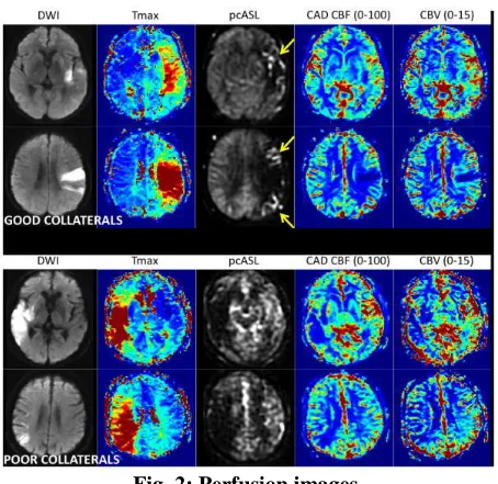
Fig. 2: Perfusion images.
In MR perfusion, MRI evaluate that the detection of acute infarct. MR perfusion is performed by Gadodiamide injection through the IV. It involves 1.36 seconds during dynamic of a small (10cc) high flow
contrast, in fig 2. one patient is with good collateral and either is poor collateral.
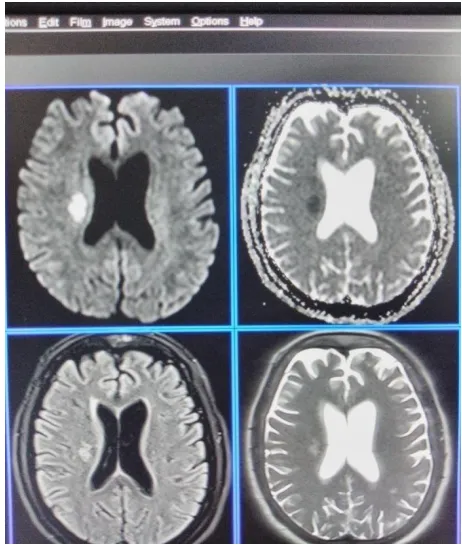
Fig. 3: Hyperacute Infarct.
I present AIS patient cases were observed under MRI imaging. 18%, in above case hyper acute infarct showed as large left MCA infarct of acute ischemic patient subtle hyperintense on FLAIR images, (fig 3.)
Cortical effacement loss of insular ribbon and attenuating large vessel. These signs are associated with a worse prognosis and poor functional outcome. But are not contraindication for treatment. The MCA is produced by an intra-arterial thrombus but it seen very rarely, it is difficult to choose t-pa injection
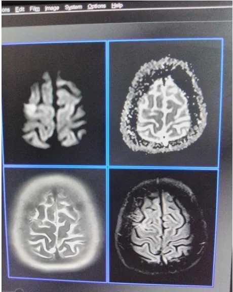
Fig. 4: Subacute infarct with hem transformation. Faint.
Fig. 5: Acute to subacute with hem Transformation.
In the present study, it is revealed that 11% patients of Acute ischemic stroke due to high BP and sugar. MRI is very useful in detecting of acute ischemic stroke and hemorrhages Figure (4,5). In fig 5 patients was loss of vision and unconscious.
Fig. 6: 3D Acute infarct.
Fig. 7: MRI shows acute to subacute infarct.
Fig. 6: 3D Acute infarct.
the detection of hyperacute infarct acute infarct hemorrhagic transformation in infarct. MRI is the modality of choice in easily assessment of hyperacute and acute infarct (time duration). FLAIR sequence are used in detection of hyperacute infarct. Figure (3). The Multiplanar Imaging capability and superior contrast resolution of MRI are the key advantage over CT Scan.
Fig. 7: MRI shows acute to subacute infarct.
CONCLUSION
Above all discussions we can say that, the fast modern life style increasing incidents of acute ischemic stroke has increase the risk infarct. The present study indicated that 65.0% patients suffered with AIS by. The main cause of AIS occurs due to Alcoholism, high BP and diabetes. The role of MRI imaging are able to diagnose the different brain stroke, their outcome and follow up. In the modern medical science these two modalities are indispensible for neurosurgery and other medical field.
ACKNOWLEDGEMENT
We gratefully acknowledge the managing director of Uppal Neuro Hospital, who supported well in this study and also emergency room doctors of Uppal neuro hospital Amritsar Punjab.
Financial Support and Sponsorship Nil.
Declaration of interest
The present study was performed with support from Uppal Neuro Hospital radiology department Amritsar Punjab.
REFERENCES
1. Thrombolytic therapy with t-PA for the treatment of acute stroke. The Standard Treatment with Alteplase to Reverse Stroke study].[Ital Heart J Suppl. 2000], 112: 123.
2. Prevention of venous thromboembolism after acute ischemic stroke.[J ThrombHaemost. 2005], 112: 121.
3. Prevention of venous thromboembolism after acute ischemic stroke.[J ThrombHaemost. 2005], 146-147. 4. Prevention of venous thrombosis in patients with acute intracerebral hemorrhagic.[Neurology. 2005] [Ann Pharacother. 2011], 56-67.
5. The efficacy and safety of enoxaparin versus unfractionated heparin for the prevention of venous thromboembolism after acute ischaemic stroke (PREVAIL Study): an open-label randomised comparison [Lancet. 2007], 123.
6. Role of tissue plasminogen activator in acute ischemic stroke. Hatcher MA, Starr JA.Ann Pharmacother, 2011 Mar; 321-322.
7. Statins for primary prevention of venous thromboembolism.[Cochrane Database Syst Rev. 2014], 22-34.
8. DW. Magnetic resonance imaging in acute ischemic stroke treatment. Journal of stroke. Sep; 16Kim BJ, Kang HG. 49-50A randomized trial of intra-arterial treatment for acute ischemic stroke. New England Journal of Medicine. 2015 Jan 1Berkhemer OA, Fransen PS., 2014; 121.