C A S E R E P O R T
Open Access
Rectal laterally spreading tumors successfully
treated in two steps by endoscopic submucosal
dissection and endoscopic mucosal resection
Italo Stroppa
1, Giovanni Milito
2*, Raffaella Lionetti
1, Giovanni Palmieri
3, Federica Cadeddu
2, Francesco Pallone
1Abstract
Background:Endoscopic submucosal dissection (ESD) is an advanced technique of therapeutic endoscopy
alternative to endoscopic mucosal resection (EMR) for superficial gastrointestinal neoplasms >2 cm. ESD allows for the direct dissection of the submucosa and large lesions can be resected en bloc. ESD is not limited by resection size, increases histologically complete resection rates and may reduce the local recurrence.
Nevertheless, the technique is time-consuming, technically demanding and associated with a high complication rate. To reduce the risk of complications, different devices and technical advances have been proposed with conflicting results and, still, ESD en bloc resections of huge lesions are associated with increased complications. Case Presentation:We successfully used a combined ESD/EMR technique for huge rectal laterally spreading tumors (LSTs). ESD was used for circumferential resection of 2/3 of the lesion followed by piecemeal resection (2-3 pieces) of the central part of the tumour.
In all three patients we obtained the complete dissection of the polyp and the complete histological evaluation in absence of complications and recurrence at 6 months’follow up.
Conclusions:In the treatment of rectal LSTs, the combined treatment - ESD/EMR resection may be considered a
suitable therapeutic option, indicated in selected cases as an alternative to surgery, in which the two techniques are neither reliable nor safe separately. However, to confirm our results, larger trials with longer follow up are required together with improvement of the technique and of the technical devices.
Background
Endoscopic mucosal resection (EMR) have become increasingly accepted and regularly used for the treat-ment of early gastric cancer and of superficial intestinal carcinomas with no risk of lymph node metastasis [1].
EMR can be performed using suction or lift technique such as EMRL/EMRC or EMR-strip biopsy [2-4]. Never-theless, this method depends on the diameter of the lesions; if the neoplasm is larger than 2 cm, the method cannot always be feasible and is associated high recur-rence rate, ranging between 2% and 35% [5].
Endoscopic submucosal dissection (ESD) is a new devel-opment in therapeutic endoscopy which allows for en bloc resection of sessile lesions regardless of the diameter, using special“devices”[6-8]. Briefly, the technique is performed
as follows: 1) following 0,25% indigo carmine chromoendo-scopy to evidence the margin of the tumour, the deeper submucosal tissue plane is opened using solutions such as saline, glycerol, sodium hyaluronate, or fructose; 2) the mucosa outside the markings is incised circumferentially using a flex-knife; 3) the connective tissue of the submuco-sal plane is dissected from the muscular layer [9].
However, this new technique, it is still associated with a higher incidence of complications than standard EMR procedure and requires a high level of endoscopic skill [10,11].
Case Presentation
We herein report 3 cases of patients with rectal LSTs endoscopically treated at the Gastroenterology Unit of Tor Vergata University Hospital, Rome, between October 2006 and March 2007.
* Correspondence: giovanni.milito@virgilio.it
2Department of Surgery, Tor Vergata University, Rome, Italy
Full list of author information is available at the end of the article
All patients presented with rectal LSTs larger than 3 cm (diameter was defined by positioning an open poly-pectomy snare close to the lesion); an en bloc resection was planned to ensure the accurate histopathologic assessment of the resected specimens and a sub-mucosal injection of hyaluronic acid was initially applied around the lesion. Nevertheless, in consideration of the impossi-bility to safety complete ESD, we decided to convert this technique into EMR- strip biopsy, performing a piece-meal resection of the centre of the polyps (two-three large pieces not less than 3 cm in diameter). Total pro-cedure times were 45 minutes, 60 minutes, and 30 min-utes respectively. All patients were hospitalized and discharged after 24-72 hours.
Only one intraoperative haemorrhage was observed. Given the ESD/EMR combined technique, patients were followed up at 3, 6 and 12 months for the first year then they were followed once a year.
Patient 1
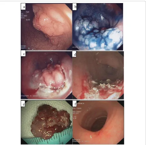
A 56-year-old male presented with a LST, 8-9 cm in diameter, located on the posterior wall of the rectum, 10 cm far from the anal verge and occupying 2/3 of the colonic lumen at colonoscopy (Figure 1a). The patient stated abdominal pain and lower gastrointestinal bleed-ing for 2 years.
Ecoendoscopy (Olympus GFUE160, Milan, Italy) showed a mucosal lesion with an area involving the superficial part of the submucosal layer (sm1), in absence of lymph node involvement.
The ESD/EMR combined procedure was carried out using a double-channel colonoscope (Olympus CF2T160I, Olympus, Milan, Italy) with the patient dee-ply sedated by propofol. Given the position and size of the lesion, a gastroscope (Olympus Gif Q 165, Olympus, Milan, Italy) retroflexed into the colon was used to resect the distal margin of the polyp.
The sub-mucosal injection of hyaluronic acid was per-formed around the lesion, previously enhanced with 1% indigo carmine (Figure 1b), followed by pre-cutting the surrounding mucosa of the tumor (Flex-knife Olympus, Tokio, Japan) (Figure 1c). The resection of the biggest part of the lesion was achieved using two knives (Hook-knife and insulation-tipped diathermic (Hook-knife IT-(Hook-knife; Olympus, Tokio, Japan). Nevertheless, even using a dou-ble-channel colonoscope (with the simultaneous use of forceps and knives), the excision of the central zone of the polyp was not completed. The remnant lesion was, therefore, removed by means of EMR-strip biopsy, with the “piecemeal” technique (Figure 1d). Post-operative course was uneventful. Histological examination revealed a tubulo-villous adenoma with high grade focal dysplasia. At 3 and 12 months’follow-up, no recurrence was detected in the excised area (Figure 1e, f).
Patient 2
A 74-year-old male presented with a LST, 10 cm in dia-meter, located on the posterior wall of the rectum and 8 cm far from the anal verge at colonoscopy (Figure 2a). The patient stated haematochezia and mucorrea for 12 months. The eco-endoscopy excluded involvement of the submucosa,
ESD was planned; the polyp spray with 1% indigo carmine was carried out, followed by the injection of hyaluronic acid at the base of the lesion and by the pre-cutting of surrounding mucosa of the polyp with the Flex-Knife (Figure 2b).
The circumferentially excision was then performed, from the margin to the central area. Despite the use of dual channel colonoscopy, hook knife and repeated injec-tions of hyaluronic acid, the complete en bloc resection was impossible due to the size of the lesion already excised (Figure 2c, d). The procedure was completed by EMR -strip biopsy- using the“piecemeal”technique (Figure 2e). Histological examination revealed tubulo-villous adenoma with low grade dysplasia. At 6 months’follow up endo-scopy showed lack of recurrence (Figure 2f).
Patient 3
A 59-year-old female presented with a LST approxi-mately 8 cm in diameter, located at the recto-sigmoid junction at colonoscopy. The patient stated postdefeca-tory abdominal pain and rectal bleeding for few months. The ecoendoscopy, performed upon admission, excluded submucosa involvement.
ESD was planned (Figure 3a, b, c). ESD en bloc resection was not possible in consideration of the size and the site of the lesion. Thus, the resection was completed with EMR-strip biopsy, using piecemeal technique (Figure 3d), which allowed for complete removal (Figure 3e). During the pro-cedure, a severe haemorrhage occurred at the base of the polyp and was successfully treated with the injection tech-nique. At histology, a tubulo-villous adenoma with low grade dysplasia was detected. Three months’endoscopy showed intact mucosa in the procedure site (Figure 3f).
Conclusions
EMR has been increasingly accepted for the treatment of gastrointestinal lesions at an early stage. Nonetheless, difficulty in correctly assessing the depth of tumour invasion and an increase in local recurrence have been reported for tumours larger than 2 cm, since such lesions are often resected piecemeal [12-14].
Furthermore, local recurrence after EMR is particu-larly difficult to treat, due to the submucosal fibrosis with an increased risk of complications [15-17].
ESD has been developed to remove the lesions in an en bloc fashion regardless of size, shape, coexisting ulcer, and location. ESD is, therefore, considered the Stroppaet al.BMC Gastroenterology2010,10:135
http://www.biomedcentral.com/1471-230X/10/135
treatment of choice to achieve en bloc resection of lesions > 2.0 cm and correct histological evaluation minimizing recurrence risk [18-20].
Although technically demanding, endoscopic submuco-sal dissection is quite effective and safe in experienced
perforation was high when an insulated-tip diathermic knife was used; practicing this technique was insufficient to reduce the rate of perforation. The average duration of follow-up was over 20 months, and no case of local persis-tence and recurrence or metastasis was observed [21].
The purpose of the present case series is to describe technical difficulties of ESD resection of rectal LST lar-ger than 2 cm.
In all patients of our series ESD was planned, given the lesion size and depth. After the excision of the external part, we were unable to reach the centre of the LST removing the lesions en bloc [22]. This is due to the limited vision of the cutting area for huge lesions with difficulty in assessing the correct deeper submuco-sal tissue plane during the resection from the margins to the centre of the lesion.
Figure 2Patient 2 ESD/EMR combined procedure.aSpraying with 1% indigo carmine and lesion marking with Flex-Knife.bInfiltration of submucosa and removal of the anterior part of the lesion with Hook-Knife.cAnterior and lateral excision.dRemoval of a large flap of lesion.e
Complete polyp removalfFollow up at 6 weeks.
Stroppaet al.BMC Gastroenterology2010,10:135 http://www.biomedcentral.com/1471-230X/10/135
Except for the excision of the distal part, achieved by a gastroscope retroflexed in the colon [23]. ESD treatment was performed with the dual channel colonoscope. The inconvenience of this device is the presence of both accessories emerging from the distal tract of the instru-ment, in a parallel vertical or transversal fashion, with-out the possibility of a co-ordinated action of the two equipments, effective both in lifting and cutting.
without the straps, to make a dissection deeper than with the hood. An attempt was also made to position a polypectomy flap (25 mm wide and 50 mm long) on the residual base of the lesions, but the size of the pre-viously excised flaps made correct placement of the snare for“en bloc”excision impossible.
In our case series, ESD was planned and we subse-quently decided to perform a combined ESD/EMR pro-cedure considering both the polyp bulk and the limited number and the size of the fragments obtained from the “piecemeal” excision, suitable for correct histopathologi-cal evaluation (Figure 4a, b, c).
The accurate histopathologic assessment of the speci-mens is essential because the depth of the invasion and lymphovascular infiltration of the tumor is associated with considerable risk for lymph node metastasis. In our experience, no recurrence was detected during the fol-low up period.
Most possible node-negative epithelial neoplasms can be resected en bloc by ESD, when they are treated by very experienced hands. This does not mean that all endoscopic resection should be performed as ESD. Poly-pectomy or endoscopic mucosal resection (EMR) are beneficial for patients with pedunculated neoplasms, small neoplasms or large flat-elevated colorectal
tumours because of the little invasiveness. If the lesions are apparently pre-malignant neoplasms, piecemeal resection by using EMR may be permissible with the best balance of risks and benefits [25].
Accordingly, Lishi and coworkers [26] removed 56 sessile colorectal polyps 2 cm or greater in diameter in 56 patients and reported a cure rate of 83% in patient submitted to piecemeal resection. They concluded that endoscopic piecemeal resection after submucosal saline injection with an intensive follow-up program is a safe and effective treatment for large, sessile colorectal polyps.
Therefore, although ESD en bloc resection, if possible, remains the treatment of choice [27,28] our experience shows the feasibility of this combined ESD/EMR treat-ment to overcome the difficulty in dissecting central area of massive lesions, also considering the absence of neoplastic recurrence at follow-up and the post-opera-tive complications detected.
Accordingly Smith et al [29] stated the efficacy and safety of the combined ESD-EMR using the Olympus KD-630 L insulation-tipped knife even for the resection of an adenoma-like mass in chronic ulcerative colitis in 67 patients. The authors reported at a median of 18 months follow-up, an overall cure rates for the
Figure 4Patient 4.aHistological examination of the case 1 showing a tubulo-villous adenoma with high grade focal dysplasia.bmicroscopic view of the case 2 revealing tubulo-villous adenoma with low grade dysplasia.chistological examination of the patientdtubulo-villous adenoma with low grade dysplasia.
Stroppaet al.BMC Gastroenterology2010,10:135 http://www.biomedcentral.com/1471-230X/10/135
ESD-assisted EMR cohort of 98% Bleeding complica-tions occurred in 7/67 (10%) of cases in absence of bowel perforations.
Besides this, a new double-channel endoscope ("R-scope”) seems promising to solve this technical pro-blem [30,31]; it allows for the vertical lifting of the mucosal layer with a forceps and the horizontal move-ment of a knife to cut the submucosal layer. Further studies are mandatory to confirm the validity of this new double-channel endoscope which would allow en bloc resection of complicated lesions on account not only of the large size, but also of the site, without ESD/ EMR combined procedure.
In addition Uraoka and co-workers recently success-fully treated 3 colorectal LSTs using the thin endo-scope-assisted (TEA) ESD, a new traction system for improving submucosal cutting line visualization [32].
Hopefully, these devices will be forthcoming allowing to reach the central base of the lesions [31,33]. Mean-while, this combined ESD/EMR treatment could be pro-posed for rectal giant polyps’resection, since it allows endoscopic removal of lesions reducing the risk of com-plications, particularly perforation and recurrence, con-sidering the size and number of polyp fragments that allow for correct histopathological evaluation and low recurrence risk.
In conclusion, depending on the clinicopathological characteristics of the colorectal tumor, the colorectal ESD is indicated for lesions difficult to remove en bloc with a snare EMR [25]. In our experience, in the treat-ment of huge rectal polyps, shows that the combined treatment - ESD/EMR may be considered a suitable therapeutic option, indicated in selected cases as an alternative to surgery, in which the two techniques are neither reliable nor safe separately.
Abbreviations
ESD: Endoscopic submucosal dissection; EMR: Endoscopic mucosal resection; LTS: laterally spreading tumors; TEA: thin endoscope-assisted.
Acknowledgements
Written consent was obtained from each patient for publication of these case reports.
Author details 1
Gastrointestinal Unit, Department of Internal Medicine, Tor Vergata University, Rome, Italy.2Department of Surgery, Tor Vergata University, Rome,
Italy.3Pathology Unit, Department of Biopathology, Tor Vergata University, Rome, Italy.
Authors’contributions
IS: manuscript preparation and critical review.GM: critical review.RL: data collection and manuscript preparation.GP: critical review.FC: literature review and manuscript preparation.FP: critical review. All authors read and approved the final manuscript.
Competing interests
The authors declare that they have no competing interests.
Received: 2 February 2010 Accepted: 17 November 2010 Published: 17 November 2010
References
1. Gotoda T, Yamamoto H, Soetikno RM:Endoscopic submucosal dissection of early gastric cancer.J Gastroenterol2006,41:929-42.
2. Karita M, Tada M, Okita K, Kodama T:Endoscopic therapy for early colon cancer: strip biopsy resection technique.Gastrointest Endosc1991,
37:128-132.
3. Ponchon T:Endoscopic mucosal resection.J Clin Gastroenterol2001,
32:6-10.
4. Higaki S, Hashimoto S, Harada K, Nohara H, Saito Y, Gondo T, Okita K: Long-term follow up of large flat colorectal tumors resected endoscopically.
Endoscopy2003,35:845-849.
5. Oka S, Tanaka S, Kaneko I, Mouri R, Hirata M, Kanao H, Kawamura T, Yoshida S, Yoshihara M, Chayama K:Endoscopic submucosal dissection for residual/local recurrence of early gastric cancer after endoscopic mucosal resection.Endoscopy2006,38:996-1000.
6. Watanabe K, Ogata S, Kawazoe S, Watanabe K, Koyama T, Kajiwara T, Shimoda Y, Takase Y, Irie K, Mizuguchi M, Tsunada S, Iwakiri R, Fujimoto K:
Clinical outcomes of EMR for gastric tumors: historical pilot evaluation between endoscopic submucosal dissection and conventional mucosal resection.Gastrointest Endosc2006,63:776-782.
7. Onozato Y, Ishihara H, Iizuka H, Sohara N, Kakizaki S, Okamura S, Mori M:
Endoscopic submucosal dissection for early gastric cancers and large flat adenomas.Endoscopy2006,38:980-986.
8. Imagawa A, Okada H, Kawahara Y, Takenaka R, Kato J, Kawamoto H, Fujiki S, Takata R, Yoshino T, Shiratori Y:Endoscopic submucosal dissection for early gastric cancer: results and degrees of technical difficulty as well as success.Endoscopy2006,38:987-990.
9. Lee I-L, Lin PY, Tung S-Y, Shen CH, Wei KL, Wu CS:Endoscopic submucosal dissection for the treatment of intraluminal gastric subepithelial tumors originating from the muscularis propria layer.Endoscopy2006,
38:1024-1028.
10. Kakushima N, Fujishiro M, Kodashima S, Muraki Y, Tateishi A, Omata M:A learning curve for endoscopic submucosal dissection of gastric epithelial neoplasms.Endoscopy2006,38:991-995.
11. Fujishiro M, Yahagi N, Kakushima N, Kodashima S, Muraki Y, Ono S, Kobayashi K, Hashimoto T, Yamamichi N, Tateishi A, Shimizu Y, Oka M, Ogura K, Kawabe T, Ichinose M, Omata M:Successful nonsurgical management of perforation complicating endoscopic submucosal dissection of gastrointestinal epithelial neoplasms.Endoscopy2006,38:1001-1006.
12. Hurlstone DP, Sanders DS, Cross SS, Adam I, Shorthouse AJ, Brown S, Drew K, Lobo AJ:Colonoscopic resection of lateral spreading tumors: a prospective analysis of endoscopic mucosal resection.Gut2004,
53:1334-1339.
13. Saito Y, Fujii T, Kondo H, Mukai H, Yokota T, Kozu T, Saito D:Endoscopic treatment for laterally spreading tumors in the colon.Endoscopy2001,
33:682-86.
14. Tanaka S, Haruma K, Oka S, Takahashi R, Kunihiro M, Kitadai Y, Yoshihara M, Shimamoto F, Chayama K:Clinicopathological features and endoscopic treatment of superficial spreading colorectal neoplasms larger than 20 mm.Gastrointest Endosc2001,54:62-66.
15. Hurlstone DP, Sanders DS, Cross SS, George R, Shorthouse AJ, Brown S:A prospective analysis of extended endoscopic mucosal resection for large rectal villous adenomas: an alternative technique to transnasal endoscopic microsurgery.Colorectal Dis2005,7:339-44.
16. Walsh RM, Ackroyd FW, Shellito PC:Endoscopic resection of large sessile colorectal polyps: technical implications and results over eight years.Dis Colon Rectum1986,29:831-35.
17. Seewald S, Ang TL, Omar S, Groth S, Dy F, Zhong Y, Seitz U, Thonke F, Yekebas E, Izbicki J, Soehendra N:Endoscopic mucosal resection of early esophageal squamous cell cancer using the Duette mucosectomy kit.
Endoscopy2006,38:1029-1031.
18. Fujishiro M, Yahagi N, Omata M:Successful outcomes of a novel endoscopic treatment for GI tumor: endoscopic submucosal dissection with a mixture of high-molecular-weight hyaluronic acid, glycerin, and sugar.Gastrointest Endosc2006,63:243-249.
20. Fujishiro M:Endoscopic submucosal dissection for stomach neoplasm.
World J Gastroenterol2006,12:5108-5112.
21. Tanaka S, Oka S, Kaneko I:Endoscopic submucosal dissection for colorectal neoplasia: possibility of standardization.Gastrointestinal Endoscopy2007,66:100-107.
22. Repici A, Conio M, De Angelis Sapino A, Malesci A, Arezzo A, Hervoso C, Pellicano R, Comunale S, Rizzetto M:Insulated-Tip Knife endoscopic mucosal resection of large colorectal polyps unsuitable for standard polypectomy.Am J Gastroenterol2007,102:1-7.
23. Hurlstone DP, Sanders DS, Thomson M, Cross SS:“Salvage”endoscopic mucosal resection in the colon using a retroflexion gastroscope dissection technique: a prospective analysis.Endoscopy2006,38:902-906. 24. Imaeda H, Iwao Y, Ogata H, Ichikawa H, Mori M, Hosoe N, Masaoka T,
Nakashita M, Suzuki H, Inoue N, Aiura K, Nagata H, Kumai K, Hibi T:A new technique for endoscopic submucosal dissection for early gastric cancer using an external grasping forceps.Endoscopy2006,38:1007-1010. 25. Fujishiro M:Perspective on the practical indications of endoscopic
submucosal dissection of gastrointestinal neoplasms.World J Gastroenterol2008,14:4289-4295.
26. Iishi H, Tatsuta M, Iseki K, Narahara H, Uedo N, Sakai N, Ishikawa H, Otani T, Ishiguro S:Endoscopic piecemeal resection with submucosal saline injection of large sessile colorectal polyps.Gastrointest Endosc2000,
51:697-700.
27. Reavis KM, Melvin WS:Advanced endoscopic technologies.Surgical Endoscopy2008,22:1533-46.
28. Kakushima N, Fujishiro M:Endoscopic submucosal dissection for gastrointestinal neoplasms.World J Gastroenterol2008,14:2962-7. 29. Smith LA, Baraza W, Tiffin N, Cross SS, Hurlstone DP:Endoscopic resection
of adenoma-like mass in chronic ulcerative colitis using a combined endoscopic mucosal resection and cap assisted submucosal dissection technique.Inflamm Bowel Dis2008,14:1380-6.
30. Yonezawa J, Kaise M, Sumiyama K, Goda K, Arakawa H, Tajiri H:A novel double-channel therapeutic endoscope ("R-scope”) facilitates endoscopic submucosal dissection of superficial gastric neoplasms.Endoscopy2006,
38:1011-1015.
31. Neuhaus H, Costamagna G, Devière J, Fockens P, Ponchon T, Rösch T, (ARCADE Group):Endoscopic submucosal dissection (ESD) of early neoplastic gastric lesions using a new double-channel endoscope (the
“R-scope”).Endoscopy2006,38:1016-1023.
32. Uraoka T, Kato J, Ishikawa S, Harada K, Kuriyama M, Takemoto K, Kawahara Y, Saito Y, Okada H:Thin endoscope-assisted endoscopic submucosal dissection for large colorectal tumors.Gastrointestinal Endosc
2007,66:836-9.
33. Akahoshi K, Honda K, Akahane H, Akiba H, Matsui N, Motomura Y, Kubokawa M, Endo S, Higuchi N, Oya M:Endoscopic submucosal dissection by using a grasping-type scissors forceps: a preliminary clinical study.Gastrontest Endosc2008,67:1128-1133.
Pre-publication history
The pre-publication history for this paper can be accessed here: http://www.biomedcentral.com/1471-230X/10/135/prepub
doi:10.1186/1471-230X-10-135
Cite this article as:Stroppaet al.:Rectal laterally spreading tumors successfully treated in two steps by endoscopic submucosal dissection and endoscopic mucosal resection.BMC Gastroenterology201010:135.
Submit your next manuscript to BioMed Central and take full advantage of:
• Convenient online submission
• Thorough peer review
• No space constraints or color figure charges
• Immediate publication on acceptance
• Inclusion in PubMed, CAS, Scopus and Google Scholar
• Research which is freely available for redistribution
Submit your manuscript at www.biomedcentral.com/submit Stroppaet al.BMC Gastroenterology2010,10:135
http://www.biomedcentral.com/1471-230X/10/135