Aptamer-Based Surface Plasmon Fibre Sensor for Thrombin Detection
T. Allsop, D. Nagel
1, R. Neal
3, E. M. Davies, C. Mou, P. Bond
4, S. Rehman
6, K. Kalli
2, D.J. Webb
P. Calverhouse
3,
M. Mascini
5, I. Bennion
Authors’ Affiliations
Photonics Research Group, Aston University, Aston Triangle, Birmingham, B47ET, UK.
1Dept. Life and Health Sciences, Aston University, Aston Triangle, Birmingham, B47ET, UK.
2
Cyprus University of Technology, 31 Archbishop Kyprianos, Lemessos 3036, Cyprus
3Dept of Maths and Computing, and the Faculty of Science and Technology, University of
Plymouth, Plymouth, PL4 8AA, U.K.
4
Plymouth Electron Microscopy Unit, University of Plymouth, Plymouth, PL4 8AA, U.K
5Dipartimento di Chimica, Università di Firenze, Via della Lastruccia 3, 50019 Sesto Fiorentino,
Italy
6
STR Fiber Technologies, Rickmansworth, Herts, UK
Abstract
A series of surface plasmonic fibre devices were fabricated using multiple coatings deposited on a lapped section of a single mode fibre and post-fabrication UV laser irradiation processing with a phase mask, producing a surface relief grating structure. These devices showed high spectral sensitivity in the aqueous index regime ranging up to 4000 nm/RIU for wavelength and 800 dB/RIU for intensity. The devices were then coated with human thrombin binding aptamer. Several concentrations of thrombin in buffer solution were made, ranging from 1nM to 1μM. All the concentrations were detectable by the devices demonstrating that sub-nM concentrations may be monitored.
Keywords: Bio-sensing, surface plasmons, fibre optic sensors, thrombin
Introduction
As biotechnology progresses, aptamers have come to the fore as attractive diagnostic reagents and potential antibody replacements for the development of biomolecular nanosensors due to their high affinity, specificity, and stability1-4 and the fact that they can be synthesized in vitro. One application for aptamers is the measuring the levels thrombin,
important in the blood coagulation process. Several types of tests have been developed to quantify thrombin, such as end-point assays measuring the formation of clots or chromogenic and fluorescent substrates5 that allow thrombin activity to be detected through the use of spectrophotometers. Fibre optic evanescent wave sensors based upon fluorescent coagulation6 and other evanescent wave sensors, such as surface plasmon resonance sensors7-9, have been developed recently. These have been used to detect other biochemical compounds by utilising immobilized bio-recognition molecular coatings on a metallic surface.
(LRSP)11, the short range surface plasmon (SRSP)12 and the localised surface plasmon13. It has been found that SPR generation is very sensitive to the polarisation of the illuminating light, its wavelength and its angle of incidence on the metal surface. This polarisation sensitivity can be used to detect index changes in biochemical/chemical reactions14. SPR biosensors offer the opportunity for real-time and label-free monitoring of biomolecular interactions15. The plasmons exist at a metal–dielectric interface and depending on the topology of supporting metal coating the dispersion relation can change13; the LRSP and SRSP obey the following dispersion relation for two homogeneous semi-infinite media:
⎟ ⎟ ⎠ ⎞ ⎜
⎜ ⎝ ⎛
+ ⋅ =
2 2
s m
s m
n n k
ε ε
β (1)
where k is the free space wave number, εm is the dielectric constant of the metal (εm = εmr+ i εmi) and ns is the refractive index of the dielectric sample to be tested.
Fabrication and Characterisation of SPR Devices
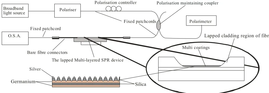
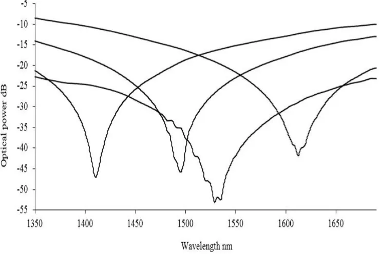
The fibre devices were constructed in three stages15. Firstly, a standard single mode fibre (Corning, SMF-28e) was mechanically lapped down to within 10μm of the core-cladding interface. Secondly, using RF sputtering, a series of coatings were deposited upon the flat of the lapped fibre. These coatings consisted of germanium (48nm), silicon dioxide (48nm) and gold (38nm),. The coated fibre was then exposed to a UV light interference pattern produced with a uniform phase mask of period 1.018μm, via laser beam scanning and multi-exposures. This produced a surface relief structure with dominant spatial periods close to 0.5μm, 1μm, 2μm and 4μm. The devices were characterised by measuring the changes in their response as a function of the polarisation properties of the illuminating light and the changes in the surrounding medium’s refractive index. Light from a broadband light source, was passed through a polariser and a polarisation controller before illuminating the sample, with the transmission spectrum being monitored using an optical spectrum analyser. The change in polarisation of the illuminating light is monitored with a polarimeter (Tektronix, PAT 9000B) through a polarisation maintaining coupler (fig. 1). Polarisation sensitivity results are shown in figure 2.
Polarisation controller Broadband
light source Polariser
O.S.A.
Fixed patchcord
Fixed patchcords
Bare fibre connectors
The lapped Multi-layered SPR device
Lapped cladding region of fibre
Multi coatings
Silver
Germanium Silica
Polarisation maintaining coupler
Polarimeter
Figure 1. Scheme used for the characterisation of the devices
I
-10
-15
-20
-30
-40
-45
-50
1350 1400 1450 1500 1550 1600 1650
Wavelength rim
Figure 2 The observed surface plasmon resonances obtained at different wavelength by varying the polarisation of the illuminating light for the device consisting of Au-SiO2-Ge with a surrounding refractive index of 1.36.
Aptamer thrombin assay procedure
Coated fibres bearing the thrombin aptamer 5’ – SH(CH2)6 - GGT TGG TGT GGT TGG - 3’ imobilised to the fibre by a thiol mediated linkage (Marco could give this a proper name I presume it is a malamide coupling but don’t want to guess) were a gift from / were obtained from ---- (there probably is a paper reference for this asptamer which could be included in the introduction). Fibres were stored in potassium phosphate buffer (100mM pH7.4). Alpha thrombin from human plasma was obtained from Sigma (Sigma T6884). Binding buffer (50mM Tris 140mM NaCl 1mM MgCl2) pH 7.4, was used for all binding experiments.
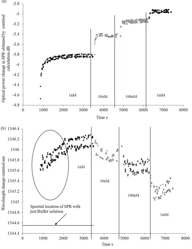
Detection of thrombin
(a) 2.8
4.8
0 1000 2000 3000 4000
Time
5000 6000 7000 8000
-o -3
b) 1346.4
1346.2
1346
1345.8
1345.6
-0 0 0
bfj 1345.4
1345.2
-
13451344.8
1344.6
-1344.4 0
Spectral location of SPR with just Buffer solution
in:' i
1000 2000 3000
.ss'$9%ss 4st £
4 £ I
1 OnM
4000
Time
A
iOOnM
5000 6000
huM
7000 8000
0.0001 0.001 0.01 0.1 10 100 1000 Thrombin concentration nlvI
(b) 1.4
1.2
1
0.8
0.6
0.4
0.2
0.001 0.01 0.1 1 10 100 1000
Thrombin concentration nM
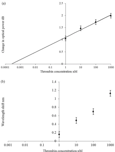
Figure 4 The spectral sensitivity of the aptamer coated Multilayered SPR fibre device as a function of thrombin concentration (a) the change in optical coupling strength (b) the change in centroid wavelength
be showing the ingression of just the buffer solution into the coating. Secondly the wavelength response is not as clear as the optical power change, though we believe this can be improved by obtaining a more well-defined SPR coupling spectral feature. Assuming that the red wavelength shift is due to buffer ingression into the aptamer coating we can estimate the spectral and intensity sensitivity of the SPR device, see figure 4. Working to the intensity detectability associated with this interrogation scheme (0.1dB) and from the experimental data using linear regression, the empirical relationship ΔΙ(dB)=0.1338In(Concen)+1.098 was used to estimate the minimum detectable concentration of thrombin which is 500fM. The same resolution allows us to detect a change in concentration of around 5%, i.e. 50pM at 1nM. We believe that these devices are creating localised surface plasmons (LSP), with decay length of the order of ~20nm (work to be published). For the following reason an adsorbed film of biomolecules (typically <10 nm) will occupy a larger fraction of the total sensing volume of a LSP sensor compared with a typical SPR sensor. The expected peak shift, induced by a molecular film with thickness t and effective refractive index nf can be obtained from
(
)
⎟⎟ ⎠ ⎞ ⎜⎜ ⎝ ⎛ ⎟ ⎠ ⎞ ⎜ ⎝ ⎛− − − ⋅ ⎟ ⎠ ⎞ ⎜ ⎝ ⎛ = L t n n dn d s film bulkp 1 exp
λ
λ where
bulk dn d ⎟ ⎠ ⎞ ⎜ ⎝
⎛ λ is the bulk sensitivity and ns is the refractive index of the surrounding
medium and L is the decay length19. Thus the LSP will be affected more by the aptamer coating. Comparing these results with those given in reference 20 (detection limits in the order of ~1nM) this SPR device shows good detection sensitivity.
Conclusion
Surface plasmonic fibre devices fabricated with multiple coatings deposited on a lapped section of a single mode fibre, have suggested detection limits of sub-pM concentrations of thrombin in a sodium phosphate buffer solution, which would outperform other types of fibre optic thrombin sensors.
References
[1] D. Ellington, J. Szostak, “In vitro Selection of RNA Molecules that Bind Specific Ligands” Nature, Vol. 346, pp.818–822, 1990
[2] C. Tuerk, L. Gold, “Systematic evolution of ligands by exponential enrichment: RNA ligands to bacteriophage T4 DNA polymerase”, Science, Vol. 249, pp.505–510. 1990
[3] N. O. Fischer, T. M. Tarasow, J. B. Tok, “Aptasensors for biosecurity applications” Curr. Opin. Chem. Biol., Vol. 11, pp.316–328.2007 [4] G. Szakacs, et al, “Targeting multidrug resistance in cancer”, Nat. ReV. Drug DiscoVery, Vol. 5, pp.219–234, 2006.
[5]Ramjee, M.K, “The use of fluorogenic substrates to monitor thrombin generation for the analysis of plasma and whole blood coagulation”, Anal. Biochem. Vol. 277, pp.11–18. 1999.
[6] S. R. Garden, et al, “A fluorescent coagulation assay for thrombin using a fibre optic evanescent wave sensor”, Biosensors and Bioelectronics, Vol. 19, pp.737–740, 2004
[7] A. Leung, P. M. Shankar, R. Mutharasan, “A review of fiber-optic biosensors”, Sensors and Actuators B, Vol. 125, pp. 688–703, 2007 [8]T.M. Battaglia et al, “Quantification of cytokines involved in wound healing using surface plasmon resonance”, Anal. Chem., Vol.77, No.21, pp.7016–7023. 2005
[9] J.F. Masson, et al, “Sensitive and real-time fiber-optic-based surface plasmon resonance sensors for myoglobin and cardiac troponin” I, Talanta Vol. 62, No. 5, pp.865–870. 2004
[10] S. Patskovsky et al, “Properties and sensing characteristics of surface plasmon resonance in infrared light”, J. Opt. Soc. Am. A, Vol.20, No. 8, pp.1644-1650, 2003.
[11] M. Piliarik et al, “Surface plasmon resonance sensor based on a single-mode polarisation-maintaining optical fiber”, Sensors and Actuators B, Vol. 90, pp.236-242, 2004
[12] R. Slavík, “Ultrahigh resolution long range surface plasmon-based sensor”, Sensors and Actuators B, Vol.123, No. 1, pp.10-12, 2007. [13] A. J. Haes et al, “A unified view of propagating and localized surface plasmon resonance biosensors”, Anal. Bioanal. Chem. Vol. 379, pp.920, 2004
[14] ‘‘Surface Plasmons on smooth and Rough Surfaces and on Gratings’’, H. Raether,, eds. (Academic, New York, 1997),
[15] J. M. Brockman et al, “Surface Plasmon Resonance Imaging Measurement of Ultra-thin Organic Films”, Annu. Rev. Phys. Chem., Vol. 51, pp.41-63, 2000.
[16] S. Tombelli, M. Minunni, M. Mascini, “A surface plasmon resonance biosensor for the determination of the affinity of drugs for nucleic acids”, Anal. Lett., Vol.35, pp.599 – 613, 2002
[17] E. J. Cho, et al, “Applications of Aptamers as Sensors, Annual Review of Analytical Chemistry, Vol. 2, pp. 241-264, 2009
[18] E. Torres-Chavolla, E. C. Alocilja, “Aptasensors for detection of microbial and viral pathogens”, Biosensors and Bioelectronics, Vol.24, pp.3175–3182, 2009
[19] M. P. Jonsson, et al, “Nanoplasmonic biosensing with focus on short-range ordered nanoholes in thin metal films: Review”, Biointerphases Vol. 3, No. 3, pp.30-40