transition
Steinestel
et al.
R E V I E W
Open Access
Clinical significance of epithelial-mesenchymal
transition
Konrad Steinestel
1,2*, Stefan Eder
1, Andres Jan Schrader
3and Julie Steinestel
3Abstract
The concept of epithelial-mesenchymal transition (EMT), a process where cells change their epithelial towards a mesenchymal phenotype, has gained overwhelming attention especially in the cancer research community. Thousands of scientific reports investigated changes in gene, mRNA and protein expression compatible with EMT and their possible correlation with tumor invasion, metastatic spread or patient prognosis; however, up to now, a proof of clinical significance of the concept is still missing. This review, with a main focus on the role of EMT in tumors, will summarize the basic molecular events underlying EMT including the signaling pathways capable of its induction as well as changes in EMT-associated protein expression and will very briefly touch the role of microRNAs in EMT. We then outline protein markers that are used most frequently for the assessment of EMT in research and diagnostic evaluation of tumor specimens and depict the link between EMT, a cancer stem cell (CSC) phenotype and resistance to conventional antineoplastic therapies. Furthermore, we evaluate a possible correlation between EMT marker expression and patient prognosis as well as current therapeutic concepts targeting the EMT process to slow down or prevent metastatic spread of malignant tumors.
Keywords:Epithelial-mesenchymal transition; Invasion; Metastasis; Prognosis; Therapy
Introduction
Epithelial-mesenchymal transition (EMT) is a central element of embryonic development, wound healing and tumor cell migration, and has thus obtained much atten-tion by the research community since Greenburg and Hay firstly described a mesenchymal-like transformation of epithelial cells when suspended in collagen gels [1]. Basically, the term describes a process in which cells lose epithelial and gain mesenchymal characteristics; this is accompanied by a loss of cell-cell cohesiveness, leading to enhanced migratory capacity [2]. Multiple genes as well as proteins that seem to play a central role in EMT have so far been identified and are either up- or down-regulated during the process, thus serving as possible markers in the assessment of EMT. Since it seems to be a key element in wound healing and tumor cell migration, there is also great interest in EMT as a pharmaceutical tar-get; recent publications even proposed vaccination against
drivers of EMT as an immunotherapeutic approach against tumor progression [3].
However, since many studies on EMT are based on in vitro results and not all findings could be confirmed in vivo, the clinical significance of the concept remains unclear [4]. This review lays the main focus on EMT in tumor cells and aims at recapitulating what is known about the molecular basis of EMT. Furthermore, we will summarize current markers of EMT that are in clinical and/or diagnostic use and, finally, evaluate EMT from a translational point of view and in the context of clinical feasibility.
Review
The molecular basis of EMT
Basically, EMT stands for a loss of epithelial and a gain of mesenchymal cellular characteristics that enhance mi-gration and invasion by the cell [5]. This process in-cludes loss of cell cohesiveness as well as fundamental reorganization of the cytoskeleton inducing a switch from apical-basal to front-rear polarity, and may further-more be associated with the acquisition of invasive prop-erties through the secretion of lytic proteases as well as
* Correspondence:konrad@steinestel.com
1
Bundeswehr Institute of Radiobiology, Neuherbergstrasse 11, Munich 80937, Germany
2
Institute of Pathology and Molecular Pathology, Bundeswehrkrankenhaus Ulm, Oberer Eselsberg 40, Ulm 89081, Germany
Full list of author information is available at the end of the article
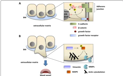
resistance to senescence and apoptosis [6]. EMT is under tight control of multiple regulatory pathways; first and foremost, transforming growth factorβ(TGF-β) sig-naling activity is enhanced in many physiological and pathological conditions in which EMT is observed, such as organogenesis, inflammation and tumor invasion [7,8]. In canonical TGF-βsignaling, binding of TGF-βto its cell surface receptors (type I-III) activates complex formation of Smad family transcription factors, which translocate to the nucleus and cooperate with transcription factors from the Snail and Twist family, so-called “EMT master genes” [9,10]. Non-Smad signaling molecules down-stream TFG-βand supportive of EMT include activated Rho-like GTPases, Phosphatidylinositol-3-kinase (PI3K) and mitogen-associated protein kinase (MAPK; the vari-ous signaling pathways mediating TGF-β signaling in EMT are excellently reviewed in [6]). Taken together, these effectors mediate transcriptional repression of genes that are involved in cell polarity and cell-cell adhe-sion, such as RhoA and E-cadherin (Figure 1A) [11,12]. The latter is mediated by the recruitment of histone
deacetylases (HDACs) and other repressors to E-box ele-ments in the E-cadherin promoter, leading to chromatin condensation and transcriptional repression [13]. At the same time, the expression of N-cadherin, another member of the cadherin family that allows for enhanced adhesion between mesenchymal cells, is upregulated; this balanced change in cadherin expression has thus been designated
“cadherin switch”and is regarded a hallmark of EMT [14]. Not only the expression, but also specific membraneous targeting of E-cadherin is repressed in EMT via loss of the epithelial-specific intermediate filament keratin; therefore, loss of keratin immunostaining is widely regarded as a marker for ongoing EMT [15,16]. Further mechanisms that lead to degradation of cell-cell junctions include a repression of claudin and occludin expression, while zonula occludens 1 (ZO-1) is subsequently lost in a post-transcriptional manner [17-19]. This repression is main-tained throughout further progression of EMT [20]. Since protein complexes (such as partitioning defective –PAR) that define the apical compartment of the cell are nor-mally associated with intercellular junctions, degradation
of the junctions also weakens the apical-basal polarity cellular phenotype [6]. Moreover, the TGF-β-facilitated signaling along the MAPK axis exerts pro-proliferative and anti-apoptotic effects on the cell, while Ras/MAPK activity alone–without TGF-βinduction - has also been linked to enhanced EMT [21-23]. After losing cohesive-ness due to degradation of cell-cell junction complexes, mesenchymal-like tumor cells are able to invade through the basement membrane into underlying tissue by the se-cretion of lytic enzymes such as matrix metalloproteinases (MMPs) and MAPK-mediated reorganization of the actin cytoskeleton which is enhanced by the expression of Vimentin (Figure 1B) [24]. In detail, migration and inva-sion of moving cells is facilitated by specialized cellular protrusions, such as filopodia, lamellipodia and invadopo-dia. While filopodia, consisting of actin filaments arranged in a parallel fashion, seem to sense changes to the cellular microenvironment and act as a “guide” through the sur-rounding matrix, lamellipodia are built upon a branched actin network and allow for actin-myosin interactions as a prerequisite for cellular movement [25,26]. Both filopodia and lamellipodia have been linked to an EMT-like pheno-type in migrating tumor cells [27,28]. Invadopodia are closely related to lamellipodia in a sense that they also consist of a branched network of actin filaments, but have the ability to degrade the extracellular matrix (ECM) through the secretion of lytic proteases, such as MMP-1, MMP-7 and MMP-9 (Figure 1B) [26]. Invadopodia forma-tion has been linked to activity of the EMT transcripforma-tion factor Twist1 cancer, and own results showed high expres-sion of invadopodia-associated proteins, such as Cortactin and Abelson interactor 1 (Abi1), in a colorectal carcinoma cell line with an EMT-like phenotype shown by loss of E-cadherin [29,30]. Accordingly, TGF-βsignaling activates small GTPases that enhance local reorganization of the actin cytoskeleton as a prerequisite for lamellipodia and filopodia formation, such as Rho, Rac and Cdc42 [31]. Vimentin, which is frequently upregulated in cells with an EMT-like phenotype, is then required for the further maturation of invadopodia [32]. Besides clearing the way for migrating tumor cells, MMPs that are released during tumor cell invasion are themselves further fueling the EMT process; the same effect is achieved via liberated TGF-βfrom the ECM [33-35]. In a mouse model of gas-tric cancer, it could be shown that EMT cooperates with MMP activity to gain access to lymph vessels and to spread distant metastases [36]. Accordingly, blood and lymph vessel infiltration by triple-negative breast cancer cells is associated with the expression of EMT transcrip-tion factor Zeb1 in surrounding stroma [37]. Alteratranscrip-tions in MMP expression are linked to changes in the integrin repertoire with downregulation of some (epithelial) and upregulation of other integrins that facilitate interaction with extracellular matrix components such as collagen [6].
Targeting transmembrane proteins - like E-cadherin - or increasing the levels of intracellular reactive oxygen spe-cies via enhanced activation of Rac1b are further mecha-nisms of MMP-induced EMT [38,39].
Upon arrival at the site of metastasis, it seems a pre-requisite for metastatic colonization that tumor cells undergo a partial reversal of the EMT, the so-called
“mesenchymal-epithelial transition” (MET) [40,41]. Dur-ing that process, tumor cells regain the expression of epi-thelial markers, such as E-cadherin, while the expression of EMT-associated transcription factors, such as Twist1, is repressed [41]. Thus, EMT can be seen as a reversible and transient process that enables epithelial tumor cells to gain access to the vasculature, allowing for the formation of distant metastasis.
Besides TGF-β, other signaling pathways have also been implied in the activation of EMT; for example, hypoxia-inducible factor (HIF) contributes to EMT in tissue fibrosis and cancer cell invasion by modulating the activity of pro-EMT transcription factors Notch and β-catenin [42-44]. HIF1α induces Twist and Snail ex-pression in endothelial as well as ovarian carcinoma cells [45-47]. Additionally, activation of several receptor tyro-sine kinases (RTKs) may result in induction of EMT; in these scenarios, growth factor binding to RTKs as well as activating mutations in oncogenes downstream of the receptors leads to enhanced signaling along the Ras/ MAPK or Akt/mTOR axis, resulting in upregulation of Snail expression [6]. Finally, it has been shown that en-hanced wnt signaling activity as well as an upregulation of chemokine receptors (such as CXCR-1) also support the process of EMT [48,49]. Here, wnt signaling leads to an inhibition of glycogen synthase kinase 3β (GSK3β )-mediated phosphorylation of β-catenin; the resulting decrease in proteosomal degradation and cytoplasmic accumulation ofβ-catenin supports its translocation to the nucleus, where it acts as a transcriptional co-activator of EMT-associated gene expression [50].
and their clinical significance would lie beyond the scope of this text, where we would like focus on the role of well-characterized proteins in EMT.
Tissue markers of EMT
Unlike the various mechanisms that are known to initiate or repress EMT, the observed hallmarks of established or ongoing EMT are quite consistent. As previously men-tioned, loss or degradation of proteins associated with epi-thelial homeostasis, cell polarity and cell adhesion, such as E-cadherin, RhoA or Plakophilin 2 is frequently observed in EMT (Figure 1A); some proteins that play key roles in cell-cell adhesion when attached to the membrane, such asβ-catenin, are redistributed to the cytoplasm [11,12,17]. Moreover, cells undergoing EMT show decreased expres-sion of epithelial cytokeratin filaments, such as keratins 8 and 18 [53]. On the other hand, the intermediate filament protein Vimentin is frequently overexpressed and contrib-utes to cell migration as well as invasion-associated gene expression by stabilizing the phosphorylated state of MAPK and is thus regarded as a stable marker of EMT; moreover, its presence is a prerequisite for the maturation of invadopodia which are indispensable for cell invasion [32,54,55]. Dysregulated expression of transcription fac-tors, such as Notch1, Slug, Snail, Twist or Zeb1 has been described in invasive tumors displaying EMT; these
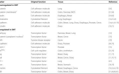
markers are therefore designated as“EMT master genes”. Table 1 provides an overview over selected dysregulated protein markers that have been and still are frequently used in the assessment of EMT.
EMT, tumor invasion and metastasis
The highest clinical significance of the EMT process is linked to its role in tumor cell invasion and metastasis. In a transgenic mouse model of pancreatic beta-cell car-cinogenesis, the switch from noninvasive adenoma to in-vasive carcinoma is associated with a loss of E-cadherin expression [82]; moreover, it has been shown that loss of membraneous β-catenin is associated with tumor cell budding, a morphologic hallmark of invasive tumor phenotype and tumor aggressivity in colorectal cancer tissue specimens [83-85]. In samples from 49 breast can-cer patients, the single-cell infiltration pattern that is ob-served in some lobular carcinomas has been linked to protein truncation mutations in the CDH1 gene encod-ing for E-cadherin [86], and hypoxia-induced upregula-tion of Slug and Snail is associated with increased breast cancer cell migration and invasion in vitro[77]. Accord-ingly, expression of Vimentin can be found in many ag-gressive breast cancer cell lines [87]. As mentioned above, to allow for tumor cell invasion into the vasculature as a prerequisite for metastatic seeding, EMT cooperates with
Table 1 Frequently used protein markers for epithelial-mesenchymal transition (EMT)
Marker Original function Tissue Reference
Downregulated in EMT
α-catenin Cell adhesion molecule Lung [56]
β-catenin (membrane)1 Cell adhesion molecule Colon, Pancreas (NET) [57,58]
Claudin Cell adhesion molecule Esophagus, Breast [59,60] Cytokeratins Cytoskeletal filament Lung, Esophagus [16,61,62] E-cadherin Cell adhesion molecule Colon, Breast, Lung, Ovary, Esophagus, Prostate, Cervix [16,61,63-70] Occludin Cell adhesion molecule Ovary [18,71]
Upregulated in EMT
Brachyury Transcription factor Pancreas, Breast, Lung [72]
β-catenin (cytoplasm/ nucleus)1 Transcription factor Breast, Cervix [73]
EGFR Tyrosine kinase receptor Cervix [70] N-cadherin Cell adhesion molecule Ovary, Prostate [68,74] Notch-1 Transcription factor Prostate [75]
p16INK4a Cell cycle regulator Colon, Urothelium [23,76]
Slug Transcription factor Breast, Ovary [11,77] Snail Transcription factor Breast, Cervix, Ovary [11,70,77] TTF-1 Transcription factor Lung [61] Twist Transcription factor Breast, Stomach [78,79] Vimentin Cytoskeletal filament Breast, Esophagus, Cervix [16,55,62,70] ZEB1 Transcription factor Colon, Breast, Ovary [51,68,80,81]
1
Membraneous depletion, but cytoplasmic accumulation/nuclear translocation.
invadopodia formation and MMP activity [36,37]; circulat-ing tumor cells (CTCs) obtained from peripheral blood of breast cancer patients frequently show an EMT-like phenotype [88,89]. In human and murine malignant mel-anoma cells, metastatic dissemination is enhanced and
ac-celerated via Snail-induced EMT [90], and bone
metastases of human prostate carcinomas show significant overexpression of Notch-1 compared to the primary tu-mors [75]. In lung carcinoma surgical specimens, tumor dedifferentiation as well as lymphogenous metastasis are also associated with reduced E-cadherin expression [91].
However, as mentioned above, some authors also reported reexpression of epithelial markers, such as E-cadherin, along with loss of EMT-associated tran-scription factors in established metastases [41]. This apparent reversal of EMT, often referred to as mesenchymal-epithelial transition (MET), has been described for metastases of colorectal carcinoma, non-small cell lung cancer and transitional cell carcinoma [92-94]. There is an ongoing debate regarding the extent to which these findings reflect a basic mechanism in the establishment of metastases or if they are restricted to certain tumor entities or reflect distinct circumjacent conditions [4,41]. There are also critical voices that doubt the role of EMT in invasion at all, since in most histopathologic speci-mens, many tumors invade and metastasize by cohesive and multicellular rather than single-cell migration, and histopathologists rarely see abundant mesenchymal-like tumor cells in routine surgical specimens [4,95,96]. This apparent contradiction might in part be explained by re-garding EMT as a transient state of a small proportion of migrating tumor cells, with only single tumor cells or small clusters of cells obtaining the ideal dynamic configuration for different stages of invasion and me-tastasis; this reasonable compromise has been referred to as “spatial and temporal heterogeneity of EMT” by Voulgari et al. (Figure 1) [97,98].
Notably, there is another controversy regarding the point whether the EMT program is associated with en-hanced or attenuated proliferative activity of the cell. While under normal circumstances TGF-βsignaling ex-erts an anti-proliferative and pro-apoptotic effect, there is experimental evidence that tumor cells having under-gone EMT do in fact show enhanced proliferation and resistance to apoptosis [99,100]. This apparent contradic-tion might also be explained by a possible heterogeneity in the course and the extent of EMT, with specialized cell populations exerting different roles during invasion and metastasis; this is in line with findings that highly meta-static breast cancer cells in fact show strong activity of the TGF-βsignaling pathway [101]. It has also been proposed that the two oppositional endpoints of TGF-β signaling might be distinguished by loss of Smad4 in tumor tissue, which promotes TGF-β-mediated tumorigenesis, while in
parallel abolishing its tumor-suppressive functions [102]. Additionally, as described above, signaling along various non-TGF-β-dependent pathways might be capable of overcoming the original anti-tumorigenic effect of TGF-β in the course of an“unfriendly takeover”of central TGF-β signaling nodes and target genes; concurrent PI3K/AKT signaling, for example, thwarts the pro-apoptotic effect of TGF-β, thus selectively allowing for the pro-metastatic ef-fects of the pathway to occur [13,49,50,77].
EMT, cancer stem cells and therapy resistance
urinary bladder, head and neck, pancreas, and colorectal carcinoma; here, increased resistance to anti-epithelial growth factor receptor (EGFR)-directed therapy is also associated with an EMT-like phenotype of the tumor cells [115].
EMT and patient prognosis
Since the metastatic spread of malignant tumors accounts for the majority of cancer-specific deaths [116-118], pos-sible correlations between EMT markers and patient prognosis have been intensely studied in multiple tumor entities. However, there is still controversy regarding the impact of the EMT concept on the actual situation in hu-man malignancies [119]. Therefore, much effort has been put into linking the expression of EMT markers to data on patient survival. In colon cancer, the upregulation of genes involved in EMT/matrix remodeling defines a mo-lecularly distinct subtype with very unfavorable prognosis; downregulation of E-cadherin in patient samples, on the other hand, seems to be associated with high TNM stages and distant metastasis [120,121]. Accordingly, basal-like, triple-negative breast cancers that show upregulation of Vimentin have a poor prognosis [54,87]. In a meta-analysis of 1107 breast cancer samples, Tobin et al. showed reduced recurrence free survival in tumors dis-playing increased gene expression of EMT markers SNAI2, TWIST1 and VIM, and decreased levels of CDH1(encoding for E-cadherin) [122]. In contrast, only recently Lee et al. were unable to confirm an impact of the tissue expression of EMT markers on disease-free survival or overall survival in breast cancer patients [123]. In prostate cancer, expression levels of EMT markers Twist and Vimentin - as assessed by immuno-histochemistry in radical prostatectomy specimens - are independent predictors for biochemical recurrence as defined by a resurgence in serum prostate-specific anti-gen (PSA) levels following surgery [124]. Additionally, loss of membraneous E-cadherin staining seems to be associated with increased Gleason score, advanced clin-ical stage, and poor prognosis in prostate cancer [125]. In tissue samples from 354 primary tumors and 30 metas-tases of endometrial carcinomas, Tanaka et al. reported that EMT status (E-cadherin-negative/ Snail-positive im-munostaining) correlated with histological type, FIGO stage, myometrial invasion and positive peritoneal cytology while it was inversely associated with both progression-free survival (HR = 0.443) and overall sur-vival (HR = 0.366) [126].
Taken together, numerous studies in a variety of tumor entities show statistical correlations between patient prognosis and alterations of various markers compatible with EMT. However, it may be difficult to yield reliable prognostic information for an individual patient from the expression pattern of EMT markers in surgical
specimens; this is in part due to high variability of marker expression patterns in different tumor areas in a heterogeneous sample [127,128]. Moreover, artificial in-duction of EMT in vitro (under certain cell culture conditions) as well asin vivo(in surgical specimens sub-jected to ischemia) has been shown [129,130]. Another key problem is the lack of a standardized diagnostic definition of which gene or which extent of expression changes is suf-ficient to determine EMT; in many reports, expression changes of one or two genes are already referred to as EMT or “partial EMT”, thus impairing the comparability of studies [4,131]. Furthermore, as has already been dis-cussed above, it is still unclear whether the gene expression changes observed in EMT reflect “passenger mutations” caused by genetic instability during tumor dedifferentiation rather than a real mesenchymal transdifferentiation state of the cell [4]. From this point of view, the expression of EMT markers simply represents a more primitive differen-tiation state of the cancer cell that is associated with onco-genic activation of a variety of signaling molecules [132].
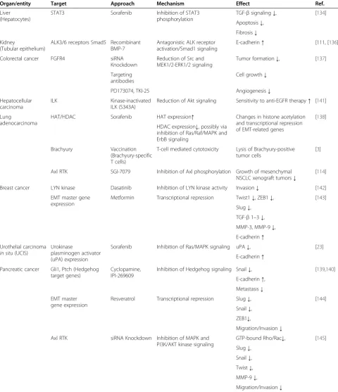
Table 2 Therapeutic approaches targeting EMT in benign and malignant processes
Organ/entity Target Approach Mechanism Effect Ref.
Liver (Hepatocytes)
STAT3 Sorafenib Inhibition of STAT3 phosphorylation
TGF-βsignaling↓, [134] Apoptosis↓,
Fibrosis↓ Kidney
(Tubular epithelium)
ALK3/6 receptors Smad5 Recombinant BMP-7
Antagonistic ALK receptor activation/Smad1 signaling
E-cadherin↑ [111, [136] Colorectal cancer FGFR4 siRNA
Knockdown
Reduction of Src and MEK1/2-ERK1/2 signaling
Tumor formation↓, [137] Targeting
antibodies
Cell growth↓ PD173074, TKI-25 Angiogenesis↓ Hepatocellular
carcinoma
ILK Kinase-inactivated ILK (S343A)
Reduction of Akt signaling Sensitivity to anti-EGFR therapy↑ [141] Lung
adenocarcinoma
HAT/HDAC Sorafenib HAT expression↑ Changes in histone acetylation and transcriptional repression of EMT-related genes
[138] HDAC expression↓, possibly via
inhibition of Ras/Raf/MAPK and ErbB signaling
Brachyury Vaccination (Brachyury-specific T cells)
T-cell mediated cytotoxicity Lysis of Brachyury-positive tumor cells
[3]
Axl RTK SGI-7079 Inhibition of Axl phosphorylation Growth of mesenchymal NSCLC xenograft tumors↓
[114] Breast cancer LYN kinase Dasatinib Inhibition of LYN kinase activity Invasion↓ [142]
EMT master gene expression
Metformin Transcriptional repression Twist1↓, ZEB1↓, [143] Slug↓,
TGF-β1–3↓, MMP-3, MMP-9↓, E-cadherin↑ Urothelial carcinoma
in situ(UCIS)
Urokinase
plasminogen activator (uPA) expression
Sorafenib Inhibition of Ras/MAPK signaling uPA↓, [23] E-cadherin↑
Pancreatic cancer Gli1, Ptch (Hedgehog target genes)
Cyclopamine, IPI-269609
Inhibition of Hedgehog signaling Snail↓, [139,140] E-cadherin↑,
Metastasis↓ EMT master
gene expression
Resveratrol Transcriptional repression Slug↓, [144] Snail↓,
ZEB1↓,
Migration/Invasion↓ Axl RTK siRNA Knockdown Inhibition of MAPK and
PI3K/AKT kinase signaling
GTP-bound Rho/Rac↓, [145] Slug↓,
Snail↓, Twist↓, MMP-9↓,
Migration/Invasion↓
From the knowledge of the diverse kinase-dependent signaling pathways that are activated during EMT, it is not surprising that the application of multi-kinase inhibitors such as Sorafenib is capable of reversing the process to a certain extent. Up to now, concepts that are directly tar-geting EMT master genes or their effectors are rare. Inter-esting new approaches include the previously mentioned vaccination against Brachyury-positive tumor cells and the transcriptional repression of EMT master gene expression by the anti-diabetic drug Metformin (Table 2) [3,72,143]. Resveratol, a dietary polyphenol, downregulated expres-sion of EMT master genes Zeb1, Snail and Slug and im-paired CSC self-renewal capacity, tumor growth and invasion in a mouse model of pancreatic ductal adenocar-cinoma [144]. However, despite the abundance of litera-ture on effectors of EMT, there is a lack of studies that show a solid effect of a specific compound in anin vivo system additionally to cell culture data, and to our best knowledge, a study that rescued the EMT phenotype after application of a certain compound - for example by over-expressing an EMT-inducing transcription factor - has so far not been conducted. Therefore, most of the data on drugs targeting EMT has to be regarded as prelimin-ary, and further research is needed to identify valuable pharmacologic targets during the induction or progres-sion of the EMT process.
Conclusions
Taken together, the concept of EMT is a valuable model for the morphologic and molecular changes observed in tumor cell invasion as well as tissue fibrosis. However, it is still unclear whether or to which extent cells in fact do undergo a complete conversion of cell type or show only transient changes in cellular morphology and pro-tein expression patterns that are supportive of a migra-tory phenotype. Despite the controversies dealing with the definition and extent of EMT, the association be-tween an EMT-like cellular phenotype - as shown by changes in marker protein expression - and tumor ag-gressivity has been well-proven in a variety of malignan-cies. In recent years, first promising results have been reported concerning a possible use of the EMT process as a pharmacological target, especially with multi-kinase inhibitors such as Sorafenib. However, since most of these results are actually derived from in vitrodata and definite proof of druggable EMTin vivo is still missing, the clinical utility of these approaches remains to be elu-cidated in future studies.
Abbreviations
AJ:Adherens junction; CXCR-1: CXC motif chemokine receptor 1/Interleukin-8-receptor alpha; ECM: Extracellular matrix; EMT: Epithelial-mesenchymal transition; FGFR: Fibroblast growth factor receptor; FIGO: Fédération internationale de Gynécologie et d’Obstétrique; GSK3β: Glycogen synthase kinase 3β; HDAC: Histone deacetylase; HIF: Hypoxia-inducible factor; MAPK: Mitogen-associated protein kinase; MEK: Mitogen-associated protein
kinase kinase; MET: Mesenchymal-epithelial transition; MMP: Matrix metalloproteinase; NSCLC: Non-small cell lung cancer; PAR: Partitioning defective; PDGFR: Platelet-derived growth factor receptor;
PI3K: Phosphatidylinositol-3-kinase; RTK: Receptor tyrosine kinase; TGF-β: Transforming growth factorβ; TNM: Tumor/Nodes/Metastasis (clinical classification system for tumor spread); uPA: Urokinase plasminogen activator; ZO-1: Zonula occludens 1.
Competing interests
Prof AJ Schrader receives compensation as a consultant for Bayer Healthcare AG, which manufactures Sorafenib (Nexavar®) for clinical application.
Authors’contributions
All authors participated in the design of this review. KS, SE and JS reviewed literature on the molecular basis of EMT, on EMT markers and on EMT in tumor invasion and metastasis; KS and AJS reviewed literature on the association between EMT and cancer stem cells, therapy resistance and patient prognosis as well as on EMT as a pharmaceutical target. All authors read and approved the final manuscript.
Author details
1
Bundeswehr Institute of Radiobiology, Neuherbergstrasse 11, Munich 80937, Germany.2Institute of Pathology and Molecular Pathology,
Bundeswehrkrankenhaus Ulm, Oberer Eselsberg 40, Ulm 89081, Germany.
3Department of Urology, Ulm University Medical Center, Prittwitzstrasse 43,
Ulm 89075, Germany.
Received: 7 April 2014 Accepted: 27 June 2014 Published: 2 July 2014
References
1. Greenburg G, Hay ED:Epithelia suspended in collagen gels can lose polarity and express characteristics of migrating mesenchymal cells. J Cell Biol1982,95:333–339.
2. Guarino M, Rubino B, Ballabio G:The role of epithelial-mesenchymal transition in cancer pathology.Pathology2007,39:305–318.
3. Palena C, Fernando RI, Hamilton DH:An immunotherapeutic intervention against tumor progression: targeting a driver of the epithelial-to-mesenchymal transition.Oncoimmunology2014,3:e27220. 4. Chui MH:Insights into cancer metastasis from a clinicopathologic
perspective: epithelial-mesenchymal transition is not a necessary step. Int J Cancer2013,132:1487–1495.
5. Kalluri R, Weinberg RA:The basics of epithelial-mesenchymal transition. J Clin Invest2009,119:1420.
6. Lamouille S, Xu J, Derynck R:Molecular mechanisms of epithelial– mesenchymal transition.Nat Rev Mol Cell Biol2014,15:178–196. 7. Zhang H, Liu L, Wang Y, Zhao G, Xie R, Liu C, Xiao X, Wu K, Nie Y, Zhang H,
Fan D:KLF8 involves in TGF-beta-induced EMT and promotes invasion and migration in gastric cancer cells.J Cancer Res Clin Oncol2013,
139:1033–1042.
8. Vittal R, Fan L, Greenspan DS, Mickler EA, Gopalakrishnan B, Gu H, Benson HL, Zhang C, Burlingham W, Cummings OW:IL-17 induces type V collagen overexpression and EMT via TGF-β-dependent pathways in obliterative bronchiolitis.Am J Physiol Lung Cell Mol Physiol2013,304:L401–L414. 9. Massagué J:TGFβin cancer.Cell2008,134:215–230.
10. Shi Y, Massagué J:Mechanisms of TGF-βsignaling from cell membrane to the nucleus.Cell2003,113:685–700.
11. Elloul S, Bukholt Elstrand M, Nesland JM, Tropé CG, Kvalheim G, Goldberg I, Reich R, Davidson B:Snail, slug, and smad-interacting protein 1 as novel parameters of disease aggressiveness in metastatic ovarian and breast carcinoma.Cancer2005,103:1631–1643.
12. Thiery JP, Huang R:Linking epithelial-mesenchymal transition to the well-known polarity protein Par6.Dev Cell2005,8:456–458. 13. Singh A, Settleman J:EMT, cancer stem cells and drug resistance: an
emerging axis of evil in the war on cancer.Oncogene2010,29:4741–4751. 14. Hazan RB, Qiao R, KEREN R, BADANO I, SUYAMA K:Cadherin switch in
tumor progression.Ann N Y Acad Sci2004,1014:155–163.
16. Lorenz KJ, Kraft K, Graf F, Pröpper C, Steinestel K:The role of reflux-induced epithelial-mesenchymal transition in periprosthetic leakage after prosthetic voice rehabilitation.Head Neck2014, Advance online publication 9 April 2014.
17. Huang RY-J, Guilford P, Thiery JP:Early events in cell adhesion and polarity during epithelial-mesenchymal transition.J Cell Sci2012,125:4417–4422. 18. Ikenouchi J, Matsuda M, Furuse M, Tsukita S:Regulation of tight junctions during the epithelium-mesenchyme transition: direct repression of the gene expression of claudins/occludin by Snail.J Cell Sci2003,
116:1959–1967.
19. Ohkubo T, Ozawa M:The transcription factor snail downregulates the tight junction components independently of E-cadherin downregulation. J Cell Sci2004,117:1675–1685.
20. De Craene B, Berx G:Regulatory networks defining EMT during cancer initiation and progression.Nat Rev Cancer2013,13:97–110.
21. Pickup M, Novitskiy S, Moses HL:The roles of TGF [beta] in the tumour microenvironment.Nat Rev Cancer2013,13:788–799.
22. Mulholland DJ, Kobayashi N, Ruscetti M, Zhi A, Tran LM, Huang J, Gleave M, Wu H:Pten loss and RAS/MAPK activation cooperate to promote EMT and metastasis initiated from prostate cancer stem/progenitor cells. Cancer Res2012,72:1878–1889.
23. Steinestel J, Cronauer MV, Müller J, Al Ghazal A, Skowronek P, Arndt A, Kraft K, Schrader M, Schrader AJ, Steinestel K:Overexpression of p16INK4a in urothelial carcinoma in situ is a marker for MAPK-mediated epithelial-mesenchymal transition but is not related to human papillomavirus infection.PLoS One2013,8:e65189.
24. Bourboulia D, Stetler-Stevenson WG:Matrix metalloproteinases (MMPs) and tissue inhibitors of metalloproteinases (TIMPs): positive and negative regulators in tumor cell adhesion.InSeminars in cancer biology.
Amsterdam: Elsevier; 2010:161–168.
25. Gerhardt H, Golding M, Fruttiger M, Ruhrberg C, Lundkvist A, Abramsson A, Jeltsch M, Mitchell C, Alitalo K, Shima D:VEGF guides angiogenic sprouting utilizing endothelial tip cell filopodia.J Cell Biol2003,161:1163–1177. 26. Ridley AJ:Life at the leading edge.Cell2011,145:1012–1022. 27. Chen Y-S, Huang W-L, Chang S-H, Chang K-W, Kao S-Y, Lo J-F, Su P-F:
Enhanced filopodium formation and stem-like phenotypes in a novel metastatic head and neck cancer cell model.Oncol Rep2013,30:2829–2837. 28. Gulhati P, Bowen KA, Liu J, Stevens PD, Rychahou PG, Chen M, Lee EY,
Weiss HL, O’Connor KL, Gao T:mTORC1 and mTORC2 regulate EMT, motility, and metastasis of colorectal cancer via RhoA and Rac1 signaling pathways.Cancer Res2011,71:3246–3256.
29. Eckert MA, Lwin TM, Chang AT, Kim J, Danis E, Ohno-Machado L, Yang J:
Twist1-induced invadopodia formation promotes tumor metastasis. Cancer Cell2011,19:372–386.
30. Steinestel K, Brüderlein S, Lennerz JK, Steinestel J, Kraft K, Pröpper C, Meineke V, Möller P:Expression and Y435-phosphorylation of Abelson interactor 1 (Abi1) promotes tumour cell adhesion, extracellular matrix degradation and invasion by colorectal carcinoma cells.Mol Cancer2014,
13:145.
31. Kardassis D, Murphy C, Fotsis T, Moustakas A, Stournaras C:Control of transforming growth factorβsignal transduction by small GTPases. FEBS J2009,276:2947–2965.
32. Schoumacher M, Goldman RD, Louvard D, Vignjevic DM:Actin, microtubules, and vimentin intermediate filaments cooperate for elongation of invadopodia.J Cell Biol2010,189:541–556.
33. Deryugina E:Experimental approaches for understanding the role of matrix metalloproteinases in cancer invasion.InMatrix Proteases in Health and Disease.Edited by Behrendt N. Weinheim: Wiley-VCH Verlag; 2012:181–211.
34. Lin CY, Tsai PH, Kandaswami CC, Lee PP, Huang CJ, Hwang JJ, Lee MT:
Matrix metalloproteinase-9 cooperates with transcription factor Snail to induce epithelial–mesenchymal transition.Cancer Sci2011,102:815–827. 35. Shah PP, Fong MY, Kakar SS:PTTG induces EMT through integrin
αVβ3-focal adhesion kinase signaling in lung cancer cells. Oncogene2011,31:3124–3135.
36. Yoo YA, Kang MH, Lee HJ, B-h K, Park JK, Kim HK, Kim JS, Oh SC:Sonic hedgehog pathway promotes metastasis and lymphangiogenesis via activation of Akt, EMT, and MMP-9 pathway in gastric cancer.Cancer Res
2011,71:7061–7070.
37. Karihtala P, Auvinen P, Kauppila S, Haapasaari K-M, Jukkola-Vuorinen A, Soini Y:Vimentin, zeb1 and Sip1 are up-regulated in triple-negative and
basal-like breast cancers: association with an aggressive tumour phenotype.Breast Cancer Res Treat2013,138:81–90.
38. Nisticò P, Bissell MJ, Radisky DC:Epithelial-mesenchymal transition: general principles and pathological relevance with special emphasis on the role of matrix metalloproteinases.Cold Spring Harb Perspect Biol2012,
4:a011908.
39. Radisky DC, Levy DD, Littlepage LE, Liu H, Nelson CM, Fata JE, Leake D, Godden EL, Albertson DG, Nieto MA:Rac1b and reactive oxygen species mediate MMP-3-induced EMT and genomic instability.Nature2005,
436:123–127.
40. Ocaña Oscar H, Córcoles R, Fabra Á, Moreno-Bueno G, Acloque H, Vega S, Barrallo-Gimeno A, Cano A, Nieto MA:Metastatic colonization requires the repression of the epithelial-mesenchymal transition inducer Prrx1. Cancer Cell2012,22:709–724.
41. Tsai Jeff H, Donaher Joana L, Murphy Danielle A, Chau S, Yang J:
Spatiotemporal regulation of epithelial-mesenchymal transition is essential for squamous cell carcinoma metastasis.Cancer Cell2012,22:725–736. 42. Higgins DF, Kimura K, Bernhardt WM, Shrimanker N, Akai Y, Hohenstein B,
Saito Y, Johnson RS, Kretzler M, Cohen CD, Eckardt KU, Iwano M, Haase VH:
Hypoxia promotes fibrogenesis in vivo via HIF-1 stimulation of epithelial-to-mesenchymal transition.J Clin Invest2007,117:3810–3820.
43. Sahlgren C, Gustafsson MV, Jin S, Poellinger L, Lendahl U:Notch signaling mediates hypoxia-induced tumor cell migration and invasion.Proc Natl Acad Sci U S A2008,105:6392–6397.
44. Kaidi A, Williams AC, Paraskeva C:Interaction between beta-catenin and HIF-1 promotes cellular adaptation to hypoxia.Nat Cell Biol2007,
9:210–217.
45. Yang F, Sun L, Li Q, Han X, Lei L, Zhang H, Shang Y:SET8 promotes epithelial–mesenchymal transition and confers TWIST dual transcriptional activities.EMBO J2012,31:110–123.
46. Luo D, Wang J, Li J, Post M:Mouse snail is a target gene for HIF. Mol Cancer Res2011,9:234–245.
47. Imai T, Horiuchi A, Wang C, Oka K, Ohira S, Nikaido T, Konishi I:Hypoxia attenuates the expression of E-cadherin via up-regulation of SNAIL in ovarian carcinoma cells.Am J Pathol2003,163:1437–1447.
48. Bates RC, DeLeo Iii MJ, Mercurio AM:The epithelial–mesenchymal transition of colon carcinoma involves expression of IL-8 and CXCR-1-mediated chemotaxis.Exp Cell Res2004,299:315–324.
49. Wu Y, Ginther C, Kim J, Mosher N, Chung S, Slamon D, Vadgama JV:
Expression of Wnt3 activates Wnt/β-catenin pathway and promotes EMT-like phenotype in trastuzumab-resistant HER2-overexpressing breast cancer cells.Mol Cancer Res2012,10:1597–1606.
50. Vincan E, Barker N:The upstream components of the Wnt signalling pathway in the dynamic EMT and MET associated with colorectal cancer progression.Clin Exp Metastasis2008,25:657–663.
51. Hur K, Toiyama Y, Takahashi M, Balaguer F, Nagasaka T, Koike J, Hemmi H, Koi M, Boland CR, Goel A:MicroRNA-200c modulates epithelial-to-mesenchymal transition (EMT) in human colorectal cancer metastasis. Gut2013,62:1315–1326.
52. Ward A, Balwierz A, Zhang JD, Kublbeck M, Pawitan Y, Hielscher T, Wiemann S, Sahin O:Re-expression of microRNA-375 reverses both tamoxifen resistance and accompanying EMT-like properties in breast cancer.Oncogene2013,32:1173–1182.
53. Fortier A-M, Asselin E, Cadrin M:Keratin 8 and 18 loss in epithelial cancer cells increases collective cell migration and cisplatin sensitivity through Claudin1 Up-regulation.J Biol Chem2013,288:11555–11571.
54. Vuoriluoto K, Haugen H, Kiviluoto S, Mpindi JP, Nevo J, Gjerdrum C, Tiron C, Lorens JB, Ivaska J:Vimentin regulates EMT induction by Slug and oncogenic H-Ras and migration by governing Axl expression in breast cancer.Oncogene2011,30:1436–1448.
55. Sohal SS, Soltani Abhari A, Weston S, Wood-Baker R, Walters E:Intermediate filament vimentin and potential role in epithelial mesenchymal transition (EMT).InVimentin Concepts and Molecular Mechanisms.
Edited by de Mello RA. New York: Nova Publishers; 2013:37–61. 56. Hirano S, Kimoto N, Shimoyama Y, Hirohashi S, Takeichi M:Identification
of a neural alpha-catenin as a key regulator of cadherin function and multicellular organization.Cell1992,70:293–301.
58. Galván JA, Astudillo A, Vallina A, Fonseca PJ, Gómez-Izquierdo L, García-Carbonero R, González MV:Epithelial-mesenchymal transition markers in the differential diagnosis of gastroenteropancreatic neuroendocrine tumors.Am J Clin Pathol2013,140:61–72.
59. Lioni M, Brafford P, Andl C, Rustgi A, El-Deiry W, Herlyn M, Smalley KS:
Dysregulation of claudin-7 leads to loss of E-cadherin expression and the increased invasion of esophageal squamous cell carcinoma cells. Am J Pathol2007,170:709–721.
60. Kominsky SL, Argani P, Korz D, Evron E, Raman V, Garrett E, Rein A, Sauter G, Kallioniemi OP, Sukumar S:Loss of the tight junction protein claudin-7 correlates with histological grade in both ductal carcinoma in situ and invasive ductal carcinoma of the breast.Oncogene2003,22:2021–2033. 61. Shi Y, Wu H, Zhang M, Ding L, Meng F, Fan X:Expression of the
epithelial-mesenchymal transition-related proteins and their clinical significance in lung adenocarcinoma.Diagn Pathol2013,8:89.
62. Kagalwalla AF, Akhtar N, Woodruff SA, Rea BA, Masterson JC, Mukkada V, Parashette KR, Du J, Fillon S, Protheroe CA:Eosinophilic esophagitis: epithelial mesenchymal transition contributes to esophageal remodeling and reverses with treatment.J Allergy Clin Immunol2012,
129:1387–1396. e1387.
63. van Roy F:Beyond E-cadherin: roles of other cadherin superfamily members in cancer.Nat Rev Cancer2014,14:121–134.
64. Pichler M, Ress AL, Winter E, Stiegelbauer V, Karbiener M, Schwarzenbacher D, Scheideler M, Ivan C, Jahn SW, Kiesslich T, Gerger A, Bauernhofer T, Calin GA, Hoefler G:MiR-200a regulates epithelial to mesenchymal transition-related gene expression and determines prognosis in colorectal cancer patients.Br J Cancer2014,110:1614–1621. 65. Zheng H, Li W, Wang Y, Xie T, Cai Y, Wang Z, Jiang B:miR-23a inhibits
E-cadherin expression and is regulated by AP-1 and NFAT4 complex during Fas-induced EMT in gastrointestinal cancer.Carcinogenesis2014,
35:173–183.
66. Shah P, Gau Y, Sabnis G:Histone deacetylase inhibitor entinostat reverses epithelial to mesenchymal transition of breast cancer cells by reversing the repression of E-cadherin.Breast Cancer Res Treat2014,143:99–111. 67. Jin L, Chen J, Li L, Li C, Chen C, Li S:CRH suppressed TGFβ1-induced
epithelial-mesenchymal transition via induction of E-cadherin in breast cancer cells.Cell Signal2014,26:757–765.
68. Huang RYJ, Wong MK, Tan TZ, Kuay KT, Ng AHC, Chung VY, Chu YS, Matsumura N, Lai HC, Lee YF, Sim WJ, Chai C, Pietschmann E, Mori S, Low JJH, Choolani M, Thiery P:An EMT spectrum defines an anoikis-resistant and spheroidogenic intermediate mesenchymal state that is sensitive to e-cadherin restoration by a src-kinase inhibitor, saracatinib (AZD0530). Cell Death Dis2013,4:e915.
69. Sun Y, Wang B-E, Leong KG, Yue P, Li L, Jhunjhunwala S, Chen D, Seo K, Modrusan Z, Gao W-Q, Settleman J, Johnson L:Androgen deprivation causes epithelial–mesenchymal transition in the prostate: implications for androgen-deprivation therapy.Cancer Res2012,72:527–536. 70. Lee M-Y, Chou C-Y, Tang M-J, Shen M-R:Epithelial-mesenchymal transition
in cervical cancer: correlation with tumor progression, epidermal growth factor receptor overexpression, and snail up-regulation.Clin Cancer Res
2008,14:4743–4750.
71. Zhu Y, Nilsson M, Sundfeldt K:Phenotypic plasticity of the ovarian surface epithelium: TGF-β1 induction of epithelial to mesenchymal transition (EMT) in vitro.Endocrinology2010,151:5497–5505.
72. Fernando RI, Litzinger M, Trono P, Hamilton DH, Schlom J, Palena C:The T-box transcription factor Brachyury promotes epithelial-mesenchymal transition in human tumor cells.J Clin Invest2010,120:533.
73. Li J, Zhou BP:Activation ofβ-catenin and Akt pathways by Twist are critical for the maintenance of EMT associated cancer stem cell-like characters.BMC Cancer2011,11:49.
74. Tanaka H, Kono E, Tran CP, Miyazaki H, Yamashiro J, Shimomura T, Fazli L, Wada R, Huang J, Vessella RL, An J, Horvath S, Gleave M, Rettig MB, Wainberg ZA, Reiter RE:Monoclonal antibody targeting of N-cadherin inhibits prostate cancer growth, metastasis and castration resistance. Nat Med2010,16:1414–1420.
75. Sethi S, Macoska J, Chen W, Sarkar FH:Molecular signature of epithelial-mesenchymal transition (EMT) in human prostate cancer bone metastasis.Am J Transl Res2011,3:90.
76. Dawson H, Koelzer VH, Karamitopoulou E, Economou M, Hammer C, Muller D-E, Lugli A, Zlobec I:The apoptotic and proliferation rate of tumour budding cells in colorectal cancer outlines a heterogeneous population
of cells with various impacts on clinical outcome.Histopathology2014,
64:577–584.
77. Chen J, Imanaka N, Griffin JD:Hypoxia potentiates Notch signaling in breast cancer leading to decreased E-cadherin expression and increased cell migration and invasion.Br J Cancer2009,102:351–360.
78. Lo H-W, Hsu S-C, Xia W, Cao X, Shih J-Y, Wei Y, Abbruzzese JL, Hortobagyi GN, Hung M-C:Epidermal growth factor receptor cooperates with signal transducer and activator of transcription 3 to induce epithelial-mesenchymal transition in cancer cells via up-regulation of TWIST gene expression.Cancer Res2007,67:9066–9076.
79. Yang Z, Zhang X, Gang H, Li X, Li Z, Wang T, Han J, Luo T, Wen F, Wu X:
Up-regulation of gastric cancer cell invasion by Twist is accompanied by N-cadherin and fibronectin expression.Biochem Biophys Res Commun
2007,358:925–930.
80. Sánchez-Tilló E, de Barrios O, Siles L, Amendola PG, Darling DS, Cuatrecasas M, Castells A, Postigo A:ZEB1 promotes invasiveness of colorectal carcinoma cells through the opposing regulation of uPA and PAI-1. Clin Cancer Res2013,19:1071–1082.
81. Lee J, Park M, Park J, Lee H, Shin D, Kang Y, Lee C, Kong G:Loss of the polycomb protein Mel-18 enhances the epithelial–mesenchymal transition by ZEB1 and ZEB2 expression through the downregulation of miR-205 in breast cancer.Oncogene2013,33:1325–1335. 82. Perl A-K, Wilgenbus P, Dahl U, Semb H, Christofori G:A causal role for
E-cadherin in the transition from adenoma to carcinoma.Nature1998,
392:190–193.
83. Kevans D, Wang LM, Sheahan K, Hyland J, O’Donoghue D, Mulcahy H, O’Sullivan J:Epithelial-mesenchymal transition (EMT) protein expression in a cohort of stage II colorectal cancer patients with characterized tumor budding and mismatch repair protein status.Int J Surg Pathol
2011,19:751–760.
84. Horcic M, Koelzer VH, Karamitopoulou E, Terracciano L, Puppa G, Zlobec I, Lugli A:Tumor budding score based on 10 high-power fields is a promising basis for a standardized prognostic scoring system in stage II colorectal cancer.Hum Pathol2013,44:697–705.
85. Ueno H, Murphy J, Jass J, Mochizuki H, Talbot I:Tumour‘budding’as an index to estimate the potential of aggressiveness in rectal cancer. Histopathology2002,40:127–132.
86. Berx G, Cleton-Jansen A, Nollet F, De Leeuw W, Van de Vijver M, Cornelisse C, Van Roy F:E-cadherin is a tumour/invasion suppressor gene mutated in human lobular breast cancers.EMBO J1995,14:6107.
87. Neve RM, Chin K, Fridlyand J, Yeh J, Baehner FL, Fevr T, Clark L, Bayani N, Coppe J-P, Tong F, Speed T, Spellman PT, DeVries S, Lapuk A, Wang NJ, Kuo WL, Stilwell JL, Pinkel D, Albertson DG, Waldman FM, McCormick F, Dickson RB, Johnson MD, Lippman M, Ethier S, Gazdar A, Gray JW:A collection of breast cancer cell lines for the study of functionally distinct cancer subtypes.Cancer Cell2006,10:515–527.
88. Burgess DJ:Breast cancer: circulating and dynamic EMT.Nat Rev Cancer
2013,13:148–149.
89. Yu M, Bardia A, Wittner BS, Stott SL, Smas ME, Ting DT, Isakoff SJ, Ciciliano JC, Wells MN, Shah AM:Circulating breast tumor cells exhibit dynamic changes in epithelial and mesenchymal composition.Science2013,
339:580–584.
90. Kudo-Saito C, Shirako H, Takeuchi T, Kawakami Y:Cancer Metastasis Is Accelerated through Immunosuppression during Snail-Induced EMT of Cancer Cells.Cancer Cell2009,15:195–206.
91. Sulzer MA, Leers MPG, van Noord JA, Bollen ECM, Theunissen PHMH:
Reduced E-cadherin expression is associated with increased lymph node metastasis and unfavorable prognosis in non-small cell lung cancer. Am J Respir Crit Care Med1998,157:1319–1323.
92. Brabletz T, Hlubek F, Spaderna S, Schmalhofer O, Hiendlmeyer E, Jung A, Kirchner T:Invasion and metastasis in colorectal cancer: epithelial-mesenchymal transition, epithelial-mesenchymal-epithelial transition, stem cells andβ-catenin.Cells Tissues Organs2005,179:56–65.
93. Soltermann A, Tischler V, Arbogast S, Braun J, Probst-Hensch N, Weder W, Moch H, Kristiansen G:Prognostic significance of epithelial-mesenchymal and mesenchymal-epithelial transition protein expression in non–small cell lung cancer.Clin Cancer Res2008,14:7430–7437.
94. Chaffer CL, Brennan JP, Slavin JL, Blick T, Thompson EW, Williams ED:
Mesenchymal-to-epithelial transition facilitates bladder cancer metastasis: role of fibroblast growth factor receptor-2.Cancer Res2006,
95. Ruiter DJ, van Krieken JH, van Muijen GN, de Waal RM:Tumour metastasis: is tissue an issue?Lancet Oncol2001,2:109–112.
96. Garber K:Epithelial-to-mesenchymal transition is important to metastasis, but questions remain.J Natl Cancer Inst2008,100:232–239.
97. Nieto MA, Cano A:The epithelial–mesenchymal transition under control: global programs to regulate epithelial plasticity.Semin Cancer Biol2012,
22:361–368.
98. Voulgari A, Pintzas A:Epithelial-mesenchymal transition in cancer metastasis: mechanisms, markers and strategies to overcome drug resistance in the clinic.Biochim Biophys Acta2009,2:75–90. 99. Bierie B, Moses HL:Transforming growth factor beta (TGF-β) and
inflammation in cancer.Cytokine Growth Factor Rev2010,21:49–59. 100. Gore AJ, Deitz SL, Palam LR, Craven KE, Korc M:Pancreatic cancer–
associated retinoblastoma 1 dysfunction enables TGF-βto promote proliferation.J Clin Invest2014,124:338–352.
101. Shipitsin M, Campbell LL, Argani P, Weremowicz S, Bloushtain-Qimron N, Yao J, Nikolskaya T, Serebryiskaya T, Beroukhim R, Hu M:Molecular definition of breast tumor heterogeneity.Cancer Cell2007,11:259–273. 102. Levy L, Hill CS:Smad4 dependency defines Two classes of transforming
growth factorβ(TGF-β) target genes and distinguishes TGF-β-induced epithelial-mesenchymal transition from its antiproliferative and migratory responses.Mol Cell Biol2005,25:8108–8125.
103. Hollier BG, Evans K, Mani SA:The epithelial-to-mesenchymal transition and cancer stem cells: a coalition against cancer therapies.J Mammary Gland Biol Neoplasia2009,14:29–43.
104. Kajiyama H, Shibata K, Terauchi M, Yamashita M, Ino K, Nawa A, Kikkawa F:
Chemoresistance to paclitaxel induces epithelial-mesenchymal transition and enhances metastatic potential for epithelial ovarian carcinoma cells. Int J Oncol2007,31:277–284.
105. Arumugam T, Ramachandran V, Fournier KF, Wang H, Marquis L, Abbruzzese JL, Gallick GE, Logsdon CD, McConkey DJ, Choi W:Epithelial to
mesenchymal transition contributes to drug resistance in pancreatic cancer.Cancer Res2009,69:5820–5828.
106. Kurrey NK, Jalgaonkar SP, Joglekar AV, Ghanate AD, Chaskar PD, Doiphode RY, Bapat SA:Snail and slug mediate radioresistance and
chemoresistance by antagonizing p53-mediated apoptosis and acquiring a stem-like phenotype in ovarian cancer cells.Stem Cells2009,
27:2059–2068.
107. Kim MR, Choi HK, Cho KB, Kim HS, Kang KW:Involvement of Pin1 induction in epithelial–mesenchymal transition of tamoxifen-resistant breast cancer cells.Cancer Sci2009,100:1834–1841.
108. Singh S, Sadacharan S, Su S, Belldegrun A, Persad S, Singh G:
Overexpression of vimentin: role in the invasive phenotype in an androgen-independent model of prostate cancer.Cancer Res2003,
63:2306–2311.
109. Mani SA, Guo W, Liao M-J, Eaton EN, Ayyanan A, Zhou AY, Brooks M, Reinhard F, Zhang CC, Shipitsin M, Campbell LL, Polyak K, Brisken C, Yang J, Weinberg RA:The epithelial-mesenchymal transition generates cells with properties of stem cells.Cell2008,133:704–715.
110. Morel A-P, Lièvre M, Thomas C, Hinkal G, Ansieau S, Puisieux A:Generation of breast cancer stem cells through epithelial-mesenchymal transition. PLoS One2008,3:e2888.
111. Li X, Lewis MT, Huang J, Gutierrez C, Osborne CK, Wu M-F, Hilsenbeck SG, Pavlick A, Zhang X, Chamness GC, Wong H, Rosen J, Chang JC:Intrinsic resistance of tumorigenic breast cancer cells to chemotherapy. J Natl Cancer Inst2008,100:672–679.
112. Thomson S, Buck E, Petti F, Griffin G, Brown E, Ramnarine N, Iwata KK, Gibson N, Haley JD:Epithelial to mesenchymal transition is a determinant of sensitivity of non–small-cell lung carcinoma cell lines and xenografts to epidermal growth factor receptor inhibition.Cancer Res2005,
65:9455–9462.
113. Thomson S, Petti F, Sujka-Kwok I, Epstein D, Haley J:Kinase switching in mesenchymal-like non-small cell lung cancer lines contributes to EGFR inhibitor resistance through pathway redundancy.Clin Exp Metastasis
2008,25:843–854.
114. Byers LA, Diao L, Wang J, Saintigny P, Girard L, Peyton M, Shen L, Fan Y, Giri U, Tumula PK, Nilsson MB, Gudikote J, Tran H, Cardnell RJG, Bearss DJ, Warner SL, Foulks JM, Kanner SB, Gandhi V, Krett N, Rosen ST, Kim ES, Herbst RS, Blumenschein GR, Lee JJ, Lippman SM, Ang KK, Mills GB, Hong WK, Weinstein JN,et al:An epithelial–mesenchymal transition gene signature predicts resistance to EGFR and PI3K inhibitors and identifies
Axl as a therapeutic target for overcoming EGFR inhibitor resistance. Clin Cancer Res2013,19:279–290.
115. Barr S, Thomson S, Buck E, Russo S, Petti F, Sujka-Kwok I, Eyzaguirre A, Gibson NW, Miglarese M, Epstein D:Bypassing cellular EGF receptor dependence through epithelial-to-mesenchymal-like transitions.Clin Exp Metastasis2008,25:685–693.
116. Yi JM, Dhir M, Van Neste L, Downing SR, Jeschke J, Glöckner SC, de Freitas Calmon M, Hooker CM, Funes JM, Boshoff C:Genomic and epigenomic integration identifies a prognostic signature in colon cancer.Clin Cancer Res2011,17:1535–1545.
117. de Boer M, van Dijck JA, Bult P, Borm GF, Tjan-Heijnen VC:Breast cancer prognosis and occult lymph node metastases, isolated tumor cells, and micrometastases.J Natl Cancer Inst2010,102:410–425.
118. Volinia S, Galasso M, Sana ME, Wise TF, Palatini J, Huebner K, Croce CM:
Breast cancer signatures for invasiveness and prognosis defined by deep sequencing of microRNA.Proc Natl Acad Sci2012,109:3024–3029. 119. Bastid J:EMT in carcinoma progression and dissemination: facts,
unanswered questions, and clinical considerations.Cancer Metastasis Rev
2012,31:277–283.
120. De Sousa E, Melo F, Wang X, Jansen M, Fessler E, Trinh A, de Rooij LPMH, de Jong JH, de Boer OJ, van Leersum R, Bijlsma MF, Rodermond H, van der Heijden M, van Noesel CJM, Tuynman JB, Dekker E, Markowetz F, Medema JP, Vermeulen L:Poor-prognosis colon cancer is defined by a molecularly distinct subtype and develops from serrated precursor lesions.Nat Med
2013,19:614–618.
121. Jie D, Zhongmin Z, Guoqing L, Sheng L, Yi Z, Jing W, Liang Z:Positive expression of LSD1 and negative expression of E-cadherin correlate with metastasis and poor prognosis of colon cancer.Dig Dis Sci2013,
58:1581–1589.
122. Tobin NP, Sims AH, Lundgren KL, Lehn S, Landberg G:Cyclin D1, Id1 and EMT in breast cancer.BMC Cancer2011,11:417.
123. Lee J, Yang G, Paik S, Chung M:Does E-cadherin or N-cadherin or epithelial-mesenchymal transition have a probability of clinical implication of the prognostic marker in invasive ductal carcinoma?Cancer Res2012,
72:Abstract nr P2-10-39.
124. Behnsawy HM, Miyake H, Harada K, Fujisawa M:Expression patterns of epithelial-mesenchymal transition markers in localized prostate cancer: significance in clinicopathological outcomes following radical prostatectomy.BJU Int2013,111:30–37.
125. Whiteland H, Spencer-Harty S, Thomas DH, Davies C, Morgan C, Kynaston H, Bose P, Fenn N, Lewis PD, Bodger O, Jenkins S, Doak SH:Putative prognostic epithelial-to-mesenchymal transition biomarkers for aggressive prostate cancer.Exp Mol Pathol2013,95:220–226. 126. Tanaka Y, Terai Y, Kawaguchi H, Fujiwara S, Yoo S, Tsunetoh S, Takai M,
Kanemura M, Tanabe A, Ohmichi M:Prognostic impact of EMT (epithelial-mesenchymal-transition)-related protein expression in endometrial cancer.Cancer Biol Ther2013,14:13.
127. Rodrıguez-Gonzalez FG, Mustafa DAM, Mostert B, Sieuwerts AM:The challenge of gene expression profiling in heterogeneous clinical samples.Methods2013,59:47–58.
128. Alkatout I, Wiedermann M, Bauer M, Wenners A, Jonat W, Klapper W:
Transcription factors associated with epithelial–mesenchymal transition and cancer stem cells in the tumor centre and margin of invasive breast cancer.Exp Mol Pathol2013,94:168–173.
129. Aoyagi K, Tamaoki M, Nishumura T, Sasaki H:Technical considerations for analyzing EMT–MET data from surgical samples.Cancer Lett2013,
341:105–110.
130. Yeung T, Georges PC, Flanagan LA, Marg B, Ortiz M, Funaki M, Zahir N, Ming W, Weaver V, Janmey PA:Effects of substrate stiffness on cell
morphology, cytoskeletal structure, and adhesion.Cell Motil Cytoskeleton
2005,60:24–34.
131. Nieto MA:The ins and outs of the epithelial to mesenchymal transition in health and disease.Annu Rev Cell Dev Biol2011,27:347–376. 132. Boyer B, Vallés AM, Edme N:Induction and regulation of epithelial–
mesenchymal transitions.Biochem Pharmacol2000,60:1091–1099. 133. Kalluri R, Neilson EG:Epithelial-mesenchymal transition and its
implications for fibrosis.J Clin Investig2003,112:1776–1784. 134. Chen YL, Lv J, Ye XL, Sun MY, Xu Q, Liu CH, Min LH, Li HP, Liu P, Ding X:
135. Zeisberg M, Hanai J-i, Sugimoto H, Mammoto T, Charytan D, Strutz F, Kalluri R:BMP-7 counteracts TGF-[beta] 1-induced epithelial-to-mesenchymal transition and reverses chronic renal injury.Nat Med2003,9:964–968. 136. Liu Y:Epithelial to mesenchymal transition in renal fibrogenesis:
pathologic significance, molecular mechanism, and therapeutic intervention.J Am Soc Nephrol2004,15:1–12.
137. Peláez-García A, Barderas R, Torres S, Hernández-Varas P, Teixidó J, Bonilla F, de Herreros AG, Casal JI:FGFR4 role in epithelial-mesenchymal transition and its therapeutic value in colorectal cancer.PLoS One2013,8:e63695. 138. Zhang J, Chen Y-L, Ji G, Fang W, Gao Z, Liu Y, Wang J, Ding X, Gao F:
Sorafenib inhibits epithelial-mesenchymal transition through an epigenetic-based mechanism in human lung epithelial cells. PLoS One2013,8:e64954.
139. Feldmann G, Dhara S, Fendrich V, Bedja D, Beaty R, Mullendore M, Karikari C, Alvarez H, Iacobuzio-Donahue C, Jimeno A, Gabrielson KL, Matsui W, Maitra A:Blockade of hedgehog signaling inhibits pancreatic cancer invasion and metastases: a new paradigm for combination therapy in solid cancers.Cancer Res2007,67:2187–2196.
140. Feldmann G, Fendrich V, McGovern K, Bedja D, Bisht S, Alvarez H, Koorstra JB, Habbe N, Karikari C, Mullendore M, Gabrielson KL, Sharma R, Matsui W, Maitra A:An orally bioavailable small-molecule inhibitor of Hedgehog signaling inhibits tumor initiation and metastasis in pancreatic cancer. Mol Cancer Ther2008,7:2725–2735.
141. Fuchs BC, Fujii T, Dorfman JD, Goodwin JM, Zhu AX, Lanuti M, Tanabe KK:
Epithelial-to-mesenchymal transition and integrin-linked kinase mediate sensitivity to epidermal growth factor receptor inhibition in human hepatoma cells.Cancer Res2008,68:2391–2399.
142. Choi Y-L, Bocanegra M, Kwon MJ, Shin YK, Nam SJ, Yang J-H, Kao J, Godwin AK, Pollack JR:LYN is a mediator of epithelial-mesenchymal transition and a target of dasatinib in breast cancer.Cancer Res2010,70:2296–2306. 143. Menendez JA:Metformin regulates breast cancer stem cell ontogeny by
transcriptional regulation of the epithelial-mesenchymal transition (EMT) status.Cell Cycle2010,9:3807–3814.
144. Shankar S, Nall D, Tang SN, Meeker D, Passarini J, Sharma J, Srivastava RK:
Resveratrol inhibits pancreatic cancer stem cell characteristics in human and KrasG12D transgenic mice by inhibiting pluripotency maintaining factors and epithelial-mesenchymal transition.PLoS One2011,6:e16530. 145. Koorstra J, Karikari CA, Feldmann G, Bisht S, Rojas PL, Offerhaus G, Alvarez H,
Maitra A:The Axl receptor tyrosine kinase confers an adverse prognostic influence in pancreatic cancer and represents a new therapeutic target. Cancer Biol Ther2009,8:618–626.
doi:10.1186/2001-1326-3-17
Cite this article as:Steinestelet al.:Clinical significance of epithelial-mesenchymal transition.Clinical and Translational Medicine20143:17.
Submit your manuscript to a
journal and benefi t from:
7Convenient online submission
7Rigorous peer review
7Immediate publication on acceptance
7Open access: articles freely available online
7High visibility within the fi eld
7Retaining the copyright to your article