R E S E A R C H
Open Access
DermoNet: densely linked convolutional
neural network for efficient skin lesion
segmentation
Saleh Baghersalimi
1 *, Behzad Bozorgtabar
1, Philippe Schmid-Saugeon
2, Hazım Kemal Ekenel
3and Jean-Philippe Thiran
1Abstract
Recent state-of-the-art methods for skin lesion segmentation are based on convolutional neural networks (CNNs). Even though these CNN-based segmentation approaches are accurate, they are computationally expensive. In this paper, we address this problem and propose an efficient fully convolutional neural network, named DermoNet. In DermoNet, due to our densely connected convolutional blocks and skip connections, network layers can reuse information from their preceding layers and ensure high accuracy in later network layers. By doing so, we take advantage of the capability of high-level feature representations learned at intermediate layers with varying scales and resolutions for lesion segmentation. Quantitative evaluation is conducted on three well-established public benchmark datasets: the ISBI 2016, ISBI 2017, and the PH2 datasets. The experimental results show that our proposed approach outperforms the state-of-the-art algorithms on these three datasets. We also compared the runtime performance of DermoNet with two other related architectures, which are fully convolutional networks and U-Net. The proposed approach is found to be faster and suitable for practical applications.
Keywords: Fully convolutional neural networks, Lesion segmentation
1 Introduction
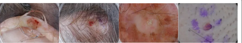
Skin lesion segmentation is a key step in computerized analysis of dermoscopic images. Inaccurate segmenta-tion could adversely impact the subsequent steps of an automated computer-aided skin cancer diagnosis system. However, this task is not trivial due to a number of rea-sons, such as the significant diversity among the lesions; inconsistent pigmentation; presence of various artifacts, e.g., air bubbles and fiducial markers; and low contrast between lesion and the surrounding skin, as can be seen in Fig.1.
In recent years, we have witnessed major advances of convolutional neural networks (CNNs) in many image processing and computer vision tasks, such as object detection [1], image classification [2], and semantic image
*Correspondence:[email protected]
1Electrical Engineering Department, Signal Processing Laboratory (LTS5), École
Polytechnique Fédérale de Lausanne (EPFL), Station 11, 1015 Lausanne, Switzerland
Full list of author information is available at the end of the article
segmentation [3]. A well-known CNN-based
segmenta-tion approach, fully convolusegmenta-tional networks (FCNs) [3], tackles per pixel prediction problems by replacing the fully connected layers with convolutions which kernels can cover the entire input image regions. Doing so, FCNs can process any image size and output pixel-wise labeled prediction map. However, the pooling layers in a down-sampling path cause a loss in the image resolution and make the network fragile to handle the lesion boundary details, e.g., fuzzy boundaries. In addition, the fully con-volutional layers contain a large number of parameters, which produce a computationally expensive network.
Most of the CNN approaches, such as SegNet [4]
and DeconvNet [5], developed for segmentation purposes by using the encoder-decoder structure as the core of their network architecture. Another effective segmenta-tion network is the employment of skip connecsegmenta-tions for the U-Net [6]. The encoder part is responsible for extract-ing the coarse features. It is followed by the decoder, which upsamples the features and is trained to recover the input image resolution at the network output. These
Fig. 1Sample dermoscopic images from the ISBI 2017 Challenge: Skin Lesion Analysis Toward Melanoma Detection. The presence of artifacts such as hairs on the skin and inconsistent pigmentation making accurate skin lesion segmentation difficult
CNN architectures [4,5] use a base network adopted from VGG architecture [7], which is already pre-trained based on millions of images. Having said that, they utilize the deconvolution or unpooling layers to recover fine-grained information from the downsampling layers.
Inspired by the residual networks (ResNets) [2],
recently, a CNN architecture called DenseNet was intro-duced in [8]. The core components of the DenseNet are the dense blocks, where each block performs iterative summation of features from the previous network layers. This characteristic enables DenseNet to be more efficient, since it needs fewer parameters. Moreover, each layer can easily access their preceding layers; therefore, it reuses features of all layers with varying scales.
Even though deep convolutional neural networks have been a significant success for the image pixel-wise seg-mentation, their inefficiency in terms of computational time limits their capability for real-time and practical applications. The motivation for this work is to propose an efficient network architecture for skin lesion segmen-tation, while achieving the state-of-the-art results. Our contributions can be summarized as follows.
1. Our main aim is to perform an efficient segmentation under limited computational
resources, while achieving the state-of-the-art results on skin benchmark datasets.
2. We transform the DenseNets into a fully convolutional network. In particular, our
architecture is built from multiple dense blocks in the encoder part, and we add a decoder part to recover the full input image resolution. This helps the multi-scale feature maps from different layers to be penalized by a loss function.
3. The multiple skip connections are arranged between encoder and decoder. In particular, we link the output of each dense block with its corresponding decoder at each feature resolution. Doing so will enable the network to process high-resolution features from early layers as well as high-semantic features of deeper layers.
4. Since we only upsample the feature maps produced by the preceding dense block, the proposed network uses fewer parameters. This enables the network
achieve the best accuracy within a limited
computational budget. We have conducted extensive experiments on ISBI 2016, ISBI 2017, and PH2 datasets, and we have shown that the proposed approach is superior to the state-of-the-art skin lesion segmentation methods.
The rest of this paper is organized as follows: Section 2 presents the related work. Section 3 describes the pro-posed network architecture in detail. Section 4 conveys and discusses the experimental results. Finally, section5 concludes the paper.
2 Related work
Recently, deep learning has ushered in a new era of com-puter vision and image analysis. It is even more remark-able that the trained models on big dataset seem to trans-fer to many other problems such as detection technology [1, 9, 10] and semantic segmentation [3]. In particular, recent works on applying CNNs to image segmentation demonstrate superior performance over classical methods in terms of accuracy. In particular, convolutional neural networks can be adapted to FCNs [3] and perform seman-tic segmentation by replacing the fully connected layer of a classification network with a convolutional layer. How-ever, due to the resolution loss in the down-sampling steps, the predicted lesion segmentation lacks lesion boundary details. Recently, several alternatives have been presented in the literature to address this shortcoming in FCNs. SegNet [4] and DeconvNet [5] are two examples of these approaches built upon auto-encoder network. In encoder, they both use the convolutional network from VGG16 for image classification. DeconvNet keeps two fully connected layers from VGG16, but SegNet discards them to decrease the number of parameters. Different from FCN in which the segmentation mask is recovered with only one deconvolution layer, the decoder network is composed of multiple deconvolution and unpooling layers both in SegNet and DeconvNet, which identify pixel-wise class labels and predict segmentation masks.
connections help the decoder layers to recover the image details from the encoder part. As a result, a faster con-vergence and a more efficient optimization process are obtained during the training. Farabet et al. [11] pro-posed a segmentation method, where the raw input image is decomposed through a multi-scale convolutional network and produces a set of coarse-to-fine feature maps. Bozorgtabar et al. [12] proposed a skin segmenta-tion method, which integrates fine and coarse predicsegmenta-tion scores of the intermediate network layers. Simon et al. [13] used DenseNets to deal with the problem of semantic seg-mentation, where they achieved state-of-the-art results on urban scene benchmark datasets such as CamVid [14].
In addition, post-processing techniques such as conditional random fields (CRF) have been a popular choice to enforce consistency in the structure of the segmentation out-puts [15]. Zheng et al. [16] proposed an interpretation of dense CRFs as recurrent neural networks (RNN). In their segmentation method, CRF-based probabilistic graphical modeling is integrated with deep learning techniques.
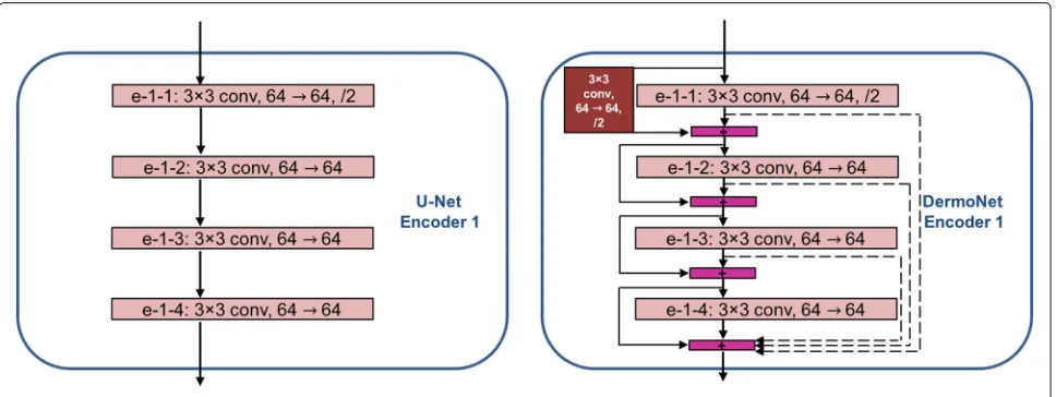
Our proposed DermoNet is based on fully convolutional neural network. Unlike the FCN, in the DermoNet archi-tecture, the outputs of the encoders are linked into the corresponding decoder to recover lost spatial information. The main difference between DermoNet and U-Net is that the encoder in DermoNet consists of four dense blocks with each block having four layers, whereas the encoder of U-Net is a path followed by the typical architecture of a convolutional neural network as can be seen in Fig.2.
3 Method
In this section, we propose a CNN-based architecture to perform lesion segmentation. Our network, DermoNet, consists of an encoder and a decoder; the encoder starts with a block, which performs the convolution on an input image with a kernel size of 7×7 and a stride of 2, and
followed by the max pooling with stride of 2. In Der-moNet, the output feature dimension of each layer within a dense block haskfeature maps, where they are concate-nated to the input. This procedure is repeated four times for each dense block; the output of the dense block is the concatenation of the outputs of the previous layers as in Eq.1.
xl =Fl
xl−1,xl−2,· · ·,x0
(1)
wherexldenotes the output feature of thelth layer.F(·) is a nonlinear function defined as a convolution followed by a rectifier non-linearity (ReLU), and [· · ·] denotes the concatenation operator. By using dense blocks, we enable the network to process high-resolution features from early layers as well as high-semantic features of deeper layers.
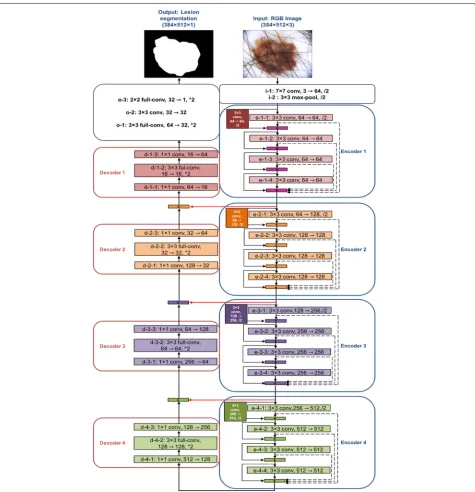
Similar to the encoder, the decoder consists of four blocks, with each block having three layers. Each decoder block is composed of a convolutional layer with a ker-nel of size 1×1, a full-convolution layer with a kernel of size 3×3 followed by an upsampling by a factor 2 and a convolutional layer with a kernel of size 1×1. The net-work ends with three last convolutional layers and two bilinear upsampling steps by a factor of 2 in order to gen-erate a segmented image with the same size as the input. Table1presents the architectural details of the proposed DermoNet.
Figure3illustrates an overview of the proposed archi-tecture; the encoder could be found on the right side of the figure while the decoder is shown on the left side.
Since FCNs perform the image pixel-wise classification, cross-entropy loss is usually used for the segmentation task. However, a skin lesion usually occupies a small por-tion of a skin image. Consequently, the segmentapor-tion network trained with cross-entropy loss function tends to be biased toward the background image rather than lesion itself. Different variants of the cross-entropy loss have
Table 1Architectural details of the proposed DermoNet
Input image, 384×512×3
Encoder Decoder
Input Filter Output Input Filter Output
i-1 7×7, /2 192×256×64 d-4-1 1×1 6×8×128
i-2 3×3, /2 96×128×64 d-4-2 3×3, *2 12×16×128
e-1-1 3×3, /2 48×64×64 d-4-3 1×1 12×16×256
e-1-2 3×3 48×64×64 d-3-1 1×1 12×16×64
e-1-3 3×3 48×64×64 d-3-2 3×3, *2 24×32×64
e-1-4 3×3 48×64×64 d-3-3 1×1 24×32×128
e-2-1 3×3, /2 24×32×128 d-2-1 1×1 24×32×32
e-2-2 3×3 24×32×128 d-2-2 3×3, *2 48×64×32
e-2-3 3×3 24×32×128 d-2-3 1×1 48×64×64
e-2-4 3×3 24×32×128 d-1-1 1×1 48×64×16
e-3-1 3×3, /2 12×16×256 d-1-2 3×3, *2 96×128×16
e-3-2 3×3 12×16×256 d-1-3 1×1 96×128×64
e-3-3 3×3 12×16×256 o-1 3×3, *2 192×256×32
e-3-4 3×3 12×16×256 o-2 3×3 192×256×32
e-4-1 3×3, /2 6×8×512 o-3 2×2, *2 384×512×1
e-4-2 3×3 6×8×512
e-4-3 3×3 6×8×512
e-4-4 3×3 6×8×512
Output image, 384×512×1
Here,/2 and∗2 denote downsampling operator using strided convolution and upsampling using a factor of 2, respectively.
been devised to address this problem, which focus on the class balancing [17]. However, this class balancing strat-egy brings additional computation cost during the training procedure. In this paper, we use a loss function based on Jaccard distance (LJ) [18], which is complementary to the Jaccard index:
LJ =1−
i,j(tijpij)
i,jtij2+
i,jp2ij−
i,j(tijpij)
(2)
wheretijandpijdenote the target and prediction output at image pixeli,j, respectively. The Jaccard index measures the intersection over the union of the labeled segments for each class and reports the average. It takes into account both the false alarms and the missed values for each class. Our experimental results disclose that this loss function is more robust compared to the classical cross-entropy loss function. In addition, it is well suited to the imbalanced classes of the foreground and background, respectively.
4 Results and discussion
The output of DermoNet model is binarized to a lesion and compared with the ground truth provided by clinicians. As the evaluation metrics, Jaccard
coeffi-cient (JC) andDice similarity coefficient(DSC) are used, which measure the spatial overlap between the obtained segmentation mask and the ground truth, respectively. They are defined as follows:
JC = TP+TPFN+FP DSC = 2×TP2×+TPFN+FP where TP, FP, and FN denote the number of true positives, false positives, and false negatives, respectively.
4.1 Datasets
For the experiments, we have used the following three datasets :
ISBI 2017: This dataset [19] contains 2000 training der-moscopic images, while there are 600 test images with the ground truths provided by experts. The images sizes vary
from 771×750 to 6748×4499.
ISBI 2016: This dataset [20] contains dermoscopic
images, where the image sizes vary from 1022×767 to
4288×2848 pixels. There are 900 training images and 379 test images.
PH2: This dataset has been acquired at Dermatology Ser-vice of Hospital Pedro Hispano, Matosinhos, Portugal [21] with Tuebinger Mole Analyzer system. This dataset contains 200 dermoscopic test images with a resolution of 768×560 pixels.
Fig. 3DermoNet is composed of four blocks in the encoder and decoder, respectively. Black arrows show connectivity patterns in the network, while red horizontal arrows represent skip connections between the encoder and decoder
Table 2Datasets summary
Dataset No. of training set
No. of test set
Image sizes
ISBI 2017 2000 600 771×750→6748×4499 ISBI 2016 900 379 1022×767→4288×2848
PH2 – 200 768×560
4.2 Implementation details
We have trained our network using the resized RGB
images of size 384× 512 pixels. For the augmentation,
we flipped the training images horizontally and vertically and did shrinking via cropping. Then, we normalized each image such that the pixel values would be between 0 and 1. The initial weights of our network are sampled from
network is set to 10−4. The maximum number of itera-tion is 5540. The whole architecture is implemented on
the TensorFlow [22]. We used Nvidia Tesla K40 GPU
with 12 GB GDDR5 memory for the training. We apply a threshold value of 0.5 to final pixel-wised score to generate lesion mask.
4.3 Runtime
To verify the effectiveness of the DermoNet in terms of test execution time, we compare it with two related
archi-tectures, namely FCN and U-Net. Table 3 presents the
segmentation execution times per image using a system with Intel Core i7-5820K CPU. Due to the densely con-nected convolutional blocks and having less parameters, the proposed network is found to be faster.
4.4 Results on ISBI 2016 dataset
For the experiments on the ISBI 2016 dataset, for train-ing the models, we used either only the traintrain-ing dataset provided by the ISBI 2016 challenge or the augmented ver-sion of it, in which we include 6500 dermoscopic images from DermoSafe [23] to the ISBI 2016 training dataset, in order to introduce a wider variety of images. These trained models are then evaluated on the ISBI 2016 test dataset. Obtained results on the ISBI 2016 challenge dataset are given in Table 4. In this challenge, the participants are ranked only based on the JC. In addition, we also report the DSC results. The proposed DermoNet improved the segmentation performance both in terms of Jaccard coef-ficient and Dice similarity coefcoef-ficient. As can be seen from the table, in terms of JC, 9.9% and 2.2% absolute performance increase improvement has been achieved with respect to FCN and U-Net, respectively. In terms of DSC, the obtained absolute increase in performance with respect to FCN and U-Net is 7.8% and 1.8%, respec-tively. As can be seen from the last two rows of the table, DermoNet’s performance improves with the use of the additional data provided by DermoSafe. However, even without using the additional DermoSafe data, it stills
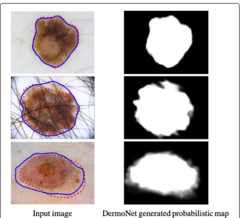
out-performs the state-of-the-art methods. Figure 4 shows
several examples of automatic segmentation results on the ISBI 2016 test set with different cases, such as hairy skin, irregular shape, and low contrast. We observe that the proposed DermoNet is able to separate the skin lesions from these artifacts and is robust to different image acqui-sition conditions.
4.5 Results on PH2 dataset
In these experiments, we have used the trained models
obtained in Section 4.4 and evaluated them on the
Table 3Comparison of average runtime (s) per image
Method FCN U-Net DermoNet
Run time (s) 0.145 0.092 0.081
Table 4Performance comparison between the proposed segmentation and other state-of-the-art methods on ISBI 2016 challenge test set
Method JC (%) DSC (%)
HED [24] 79.3 88.4
SegNet [4] 70.0 82.3
UiT-Seg [25] 80.6 NA
IHPC-CS [20] 79.9 NA
CNN-CRF [20] 79.7 NA
FCN - (JC) trained with DermoSafe data 72.6 81.6
U-Net - (JC) trained with DermoSafe data 80.3 87.6
DermoNet (JC) trained without DermoSafe data 81.1 88.2
DermoNet (JC) trained with DermoSafe data 82.5 89.4
Here, JC denotes Jaccard coefficient
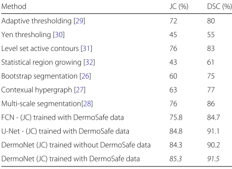
200 skin images from the PH2 dataset. We have also compared the performance of the proposed lesion seg-mentation method with superpixel-based saliency detec-tion approaches [26–28] on the PH2 dataset. Attained results are given in Table5. From the experimental results, it can be observed that DermoNet which is trained using DermoSafe data has outperformed the other skin lesion segmentation methods. Due to dense connectivity in Der-moNet, each layer is connected with all subsequent layers and allows later layers to bypass features and to maintain the high accuracy of the final pixel classification layer in a deeper architecture with fewer parameters. As a result, this brings additional performance gains.
Table 5Performance comparison between the proposed segmentation and other state-of-the-art methods on PH2 dataset
Method JC (%) DSC (%)
Adaptive thresholding [29] 72 80
Yen thresholing [30] 45 55
Level set active contours [31] 76 83
Statistical region growing [32] 43 61
Bootstrap segmentation [26] 60 75
Contexual hypergraph [27] 63 77
Multi-scale segmentation[28] 76 86
FCN - (JC) trained with DermoSafe data 75.8 84.7
U-Net - (JC) trained with DermoSafe data 84.8 91.1
DermoNet (JC) trained without DermoSafe data 84.3 90.2
DermoNet (JC) trained with DermoSafe data 85.3 91.5
Here, JC denotes Jaccard coefficient
4.6 Results on ISBI 2017 dataset
For the experiments on the ISBI 2017 dataset, for train-ing the models, we used either only the traintrain-ing dataset provided by the ISBI 2017 challenge or the augmented ver-sion of it, in which we include 6500 dermoscopic images from DermoSafe [23] to the ISBI 2017 training dataset. These trained models are then evaluated on the ISBI 2017
test dataset. Table 6 compares the performance of
Der-moNet with the state-of-the-art algorithms on ISBI 2017 dataset. Many teams evaluated their segmentation algo-rithms during the ISBI 2017 challenge. Among them, the top two teams used different variations of a fully con-volutional network in their segmentation methods. For
example, Yuan et al. [18] proposed a method based on
deep fully convolutional-deconvolutional neural networks (CDNN) to segment skin lesions in dermoscopic image.
NLP LOGIX [33] used a U-Net architecture followed by
a CRF as post-processing in their segmentation method. Here, we observed that the proposed DermoNet outper-forms the other teams’ approaches.
Table 6Performance comparison between the proposed segmentation and other state-of-the-art methods on ISBI 2017 challenge test set
Method JC (%)
FCN-ensemble[18] 76.5
Modified U-Net[33] 76.2
FCN - (JC) trained with DermoSafe data 66.49
U-Net - (JC) trained with DermoSafe data 75.50
DermoNet (JC) trained without DermoSafe data 77.5
DermoNet (JC) trained with DermoSafe data 78.3
Here, JC denotes Jaccard coefficient
4.7 Effect of loss function
As described in Section 3, due to imbalanced classes,
cross-entropy loss function would not be suitable for the skin lesion segmentation task. Therefore, we used Jaccard distance instead, which enabled the DermoNet’s training to focus more on lesion pixels over background. To also empirically analyze the effect of the loss function, we com-pare the performance of DermoNet using Jaccard distance or cross-entropy on ISBI 2016, 2017 and PH2 dataset. As can be seen from Table 7, using Jaccard distance as the loss function improves the performance significantly compared to using cross-entropy as the loss function.
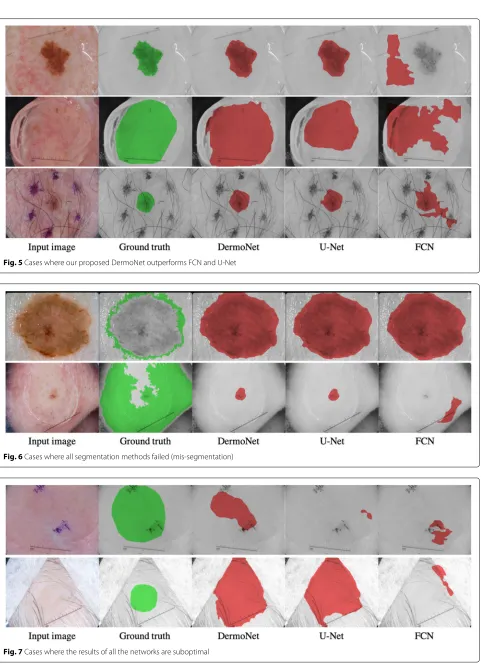
4.8 Qualitative comparison
In this section, we provide qualitative comparison
between DermoNet, FCN, and U-Net. Figure 5 shows
some tricky cases from ISBI 2017 challenge dataset. In this figure, from left to right, we have the original image, ground truth, the output of DermoNet, U-Net, and FCN, respectivly. As can be observed, DermoNet provides bet-ter results compared to FCN and U-Net and is able to sep-arate the skin lesion from artifacts such as ink markings and air bubbles.
Figure6shows cases where the ground truth is wrongly labeled, and it leads to a very low Jaccard coefficients (JC) even though the output of the segmentation is correct. In this figure, from left to right, we have the original image, ground truth, and DermoNet, U-Net, and FCN output.
Finally, Fig. 7 shows some of the challenging cases
among all the ISBI 2017 testing images where all three models (DermoNet, U-Net, and FCN) performed poorly. In these cases, the contrast between lesion and skin is very low.
5 Conclusion and future work
In this paper, we have presented a new fully convo-lutional neural network architecture for automatic skin lesion segmentation. The idea behind DermoNet is shar-ing features across all encoder blocks and takshar-ing benefit of reusing features, while remaining densely connected
Table 7Performance comparison of the proposed segmentation on ISBI 2016 and 2017 and PH2 dataset when using Jaccard coefficient or cross-entropy loss for training
Dataset Method JC (%)
ISBI 2016 DermoNet (CE) 79.16
DermoNet (JC) 82.5
PH2 DermoNet (CE) 72.14
DermoNet (JC) 85.3
ISBI 2017 DermoNet (CE) 75.4
DermoNet (JC) 78.3
Fig. 5Cases where our proposed DermoNet outperforms FCN and U-Net
Fig. 6Cases where all segmentation methods failed (mis-segmentation)
to provide the network with more flexibility in learning new features. The proposed network has fewer parame-ters compared to existing baseline segmentation methods that have an order of magnitude larger memory require-ment. Moreover, it improves state-of-the-art performance on challenging skin datasets, without using neither addi-tional post-processing nor pre-training. We have achieved an average Jaccard coefficient of 82.5% on the ISBI 2016 Skin Lesion Challenge dataset, 85.3% on the PH2 dataset, and 78.3% on ISBI 2017 Skin Lesion Challenge dataset. In our future work, we plan to apply the proposed seg-mentation with some modifications in the network archi-tecture on standard semantic segmentation benchmarks, e.g., MSCOCO, to show the generalization capability of the proposed framework.
Abbreviations
CDNN: Convolutional-deconvolutional neural network; CNN: Convolutional neural networks; CRF: Conditional random fields; DSC: Dice similarity coefficient; FCN: Fully convolutional networks; FN: False negative; FP: False positive; ISBI: International symposium on biomedical imaging; JC: Jaccard coefficient; ReLU: Rectifier non-linearity; ResNet: Residual network; RNN: Recurrent neural networks; TP: True positive
Acknowledgments
The authors would like to thank Mr. Philippe Held, CEO and Founder of DermoSafe, for his support and for giving us access to the DermoSafe’s database of images of pigmented skin lesions, which helped us to achieve the mentioned results.
Funding
This work was supported by the Swiss Commission for Technology and Innovation CTI fund no. 25515.2 PFLS-LS for the project entitled “DermoBrain: advanced computer vision algorithms and features for the early diagnosis of skin cancer.”
Availability of data and materials
The ISBI 2016 [20] datasets analyzed during the current study are available in https://challenge.kitware.com/#phase/566744dccad3a56fac786787. The ISBI 2017 [19] datasets analyzed during the current study are available inhttps:// challenge.kitware.com/#challenge/583f126bcad3a51cc66c8d9a. The PH2 [21] datasets analyzed during the current study are available inhttps://www. dropbox.com/s/k88qukc20ljnbuo/PH2Dataset.rar. The datasets of DermoSafe [23] that are analyzed during the current study are not publicly available due to the protection of patient privacy.
Authors’ contributions
SBS and BB conceived and designed the methods. SBS performed the experiments. PSS, HKE, and JPT supervised the project.
Competing interests
The authors declare that they have no competing interests.
Publisher’s Note
Springer Nature remains neutral with regard to jurisdictional claims in published maps and institutional affiliations.
Author details
1Electrical Engineering Department, Signal Processing Laboratory (LTS5), École
Polytechnique Fédérale de Lausanne (EPFL), Station 11, 1015 Lausanne, Switzerland.2DermoSafe SA, EPFL Innovation Park, Bâtiment D, 1015
Lausanne, Switzerland.3Department of Computer Engineering, 34469 Maslak,
Istanbul, Turkey.
Received: 23 November 2018 Accepted: 15 May 2019
References
1. J. Redmon, S. K. Divvala, R. B. Girshick, A. Farhadi, inIEEE Conference on Computer Vision and Pattern Recognition (CVPR). You only look once: unified, real-time object detection, (Las Vegas, NV, 2016), pp. 779–788 2. K. He, X. Zhang, S. Ren, J. Sun, inProceedings of the IEEE conference on CVPR.
Deep residual learning for image recognition, (2016), pp. 770–778 3. J. Long, E. Shelhamer, T. Darrell, inIEEE Conference on Computer Vision and
Pattern Recognition (CVPR). Fully convolutional networks for semantic segmentation, (Boston, MA, 2015), pp. 3431–3440
4. V. Badrinarayanan, A. Kendall, R. Cipolla, SegNet: a deep convolutional encoder-decoder architecture for image segmentation. IEEE Trans Pattern Anal Mach Intell.39(12), 2481–2495 (2017)
5. H. Noh, S. Hong, B. Han, inProceedings of the IEEE ICCV. Learning deconvolution network for semantic segmentation, (2015), pp. 1520–1528
6. O. Ronneberger, P. Fischer, T. Brox, inProc. Med. Image Comput. Comput.-Assisted Intervention. U-net: convolutional networks for biomedical image segmentation (Springer, 2015), pp. 234–241 7. K. Simonyan, A. Zisserman, inICLR. Very deep convolutional networks for
large-scale image recognition, (2015)
8. G. Huang, Z. Liu, K. Q. Weinberger, in2017 IEEE Conference on Computer Vision and Pattern Recognition (CVPR). Densely connected convolutional networks, (Honolulu, 2017), pp. 2261–2269
9. S. Ren, K. He, R. Girshick, J. Sun, inAdvances in neural information processing systems. Faster R-CNN: towards real-time object detection with region proposal networks, (2015), pp. 91–99
10. C. Yan, H. Xie, J. Chen, Z. Zha, X. Hao, Y. Zhang, Q. Dai, A fast uyghur text detector for complex background images. IEEE Trans. Multimed.20(12), 3389–3398 (2018)
11. C. Farabet, C. Couprie, L. Najman, Y. LeCun, Learning hierarchical features for scene labeling. IEEE Trans. Pattern. Anal. Mach. Intell.35(8), 1915–1929 (2013)
12. B. Bozorgtabar, Z. Ge, R. Chakravorty, M. Abedini, S. Demyanov, R. Garnavi, in2017 IEEE 14th International Symposium on Biomedical Imaging (ISBI 2017). Investigating deep side layers for skin lesion segmentation, (Melbourne, 2017), pp. 256–260
13. S. Jégou, M. Drozdzal, D. Vazquez, A. Romero, Y. Bengio, in2017 IEEE Conference on Computer Vision and Pattern Recognition Workshops (CVPRW). The one hundred layers tiramisu: fully convolutional densenets for semantic segmentation, (Honolulu, 2017), pp. 1175–1183
14. G. J. Brostow, J. Fauqueur, R. Cipolla, Semantic object classes in video: a high-definition ground truth database. Pattern Recogn. Lett.30(2), 88–97 (2009)
15. L. Chen, G. Papandreou, I. Kokkinos, K. Murphy, A. L. Yuille, DeepLab: Semantic Image Segmentation with Deep Convolutional Nets, Atrous Convolution, and Fully Connected CRFs. IEEE Trans Pattern Anal Mach Intell.40(4), 834–848 (2016)
16. S. Zheng, S. Jayasumana, B. Romera-Paredes, V. Vineet, Z. Su, D. Du, C. Huang, P. H. S. Torr, inProceedings of the IEEE ICCV. Conditional random fields as recurrent neural networks, (2015), pp. 1529–1537
17. K. Maninis, J. Pont-Tuset, P. A. Arbeláez, L. J. V. Gool, inInternationalConference on MICCAI. Deep retinal image understanding (Springer, 2016), pp. 140–148 18. Y. Yuan, M. Chao, Y. Lo, inInternational Skin Imaging Collaboration (ISIC)
2017 Challenge at the International Symposium on Biomedical Imaging (ISBI). Automatic skin lesion segmentation with fully
convolutional-deconvolutional networks, (2017).https://arxiv.org/pdf/ 1703.05165.pdf
19. N. C. F. Codella, D. Gutman, M. E. Celebi, B. Helba, M. A. Marchetti, S. W. Dusza, A. Kalloo, K. Liopyris, N. K. Mishra, H. Kittler, A. Halpern, inIEEE 15th International Symposium on Biomedical Imaging (ISBI 2018). Skin lesion analysis toward melanoma detection: A challenge at the 2017 International symposium on biomedical imaging (ISBI), hosted by the international skin imaging collaboration (ISIC), (Washington, 2017), pp. 168–172
20. D. Gutman, N. C. F. Codella, M. E. Celebi, B. Helba, M. A. Marchetti, N. K. Mishra, A. Halpern, Skin lesion analysis toward melanoma detection: a challenge at the ISBI 2016, hosted by the ISIC (2016). arXiv preprint arXiv:1605.01397
21. T. Mendonça, P. M. Ferreira, J. S. Marques, A. R. S. Marçal, J. Rozeira, in
dermoscopic image database for research and benchmarking (IEEE, 2013), pp. 5437–5440
22. M. Abadi, A. Agarwal, P. Barham, E. Brevdo, Z. Chen, C. Citro, G. S. Corrado, A. Davis, J. Dean, M. Devin, S. Ghemawat, I. Goodfellow, A. Harp, G. Irving, M. Isard, Y. Jia, R. Jozefowicz, L. Kaiser, M. Kudlur, J. Levenberg, D. Mané, R. Monga, S. Moore, D. Murray, C. Olah, M. Schuster, J. Shlens, B. Steiner, I. Sutskever, K. Talwar, P. Tucker, V. Vanhoucke, V. Vasudevan, F. Viégas, O. Vinyals, P. Warden, M. Wattenberg, M. Wicke, Y. Yu, X. Zheng, TensorFlow: Large-scale machine learning on heterogeneous systems (2015). software available from tensorflow.org. [Online]. Available:https://www.tensorflow.org/ 23. DermoSafe.https://www.dermosafe.com/en/
24. S. Xie, Z. Tu, inIEEE International Conference on Computer Vision (ICCV). Holistically-nested edge detection, (Santiago, 2015), pp. 1395–1403 25. M. Zortea, S. O. Skrøvseth, T. R. Schopf, H. M. Kirchesch, F. Godtliebsen,
Automatic segmentation of dermoscopic images by iterative classification. J. Biomed. Imaging.2011, 3 (2011)
26. N. Tong, H. Lu, X. Ruan, M. Yang, inProceedings of the IEEE Conference on CVPR. Salient object detection via bootstrap learning, (2015), pp. 1884–1892
27. X. Li, Y. Li, C. Shen, A. Dick, A. V. D. Hengel, inProceedings of the IEEE ICCV. Contextual hypergraph modeling for salient object detection, (2013), pp. 3328–3335
28. B. Bozorgtabar, M. Abedini, R. Garnavi, inProc. Int. Workshop Mach. Learn. Med. Imag.Sparse coding based skin lesion segmentation using dynamic rule-based refinement (Springer, 2016), pp. 254–261
29. M. Silveira, J. C. Nascimento, J. S. Marques, A. R. S. Marcal, T. Mendonca, S. Yamauchi, J. Maeda, J. Rozeira, Comparison of segmentation methods for melanoma diagnosis in dermoscopy images. IEEE J. Sel. Top. Sig. Process. 3(1), 35–45 (2009)
30. M. Sezgin, B. Sankur, Survey over image thresholding techniques and quantitative performance evaluation. J. Electron. Imaging.13(1), 146–168 (2004)
31. C. Li, C. Kao, J. C. Gore, Z. Ding, Minimization of region-scalable fitting energy for image segmentation. IEEE Trans. Image Process.17(10), 1940–1949 (2008)
32. M. Celebi, H. Kingravi, H. Iyatomi, Y. Aslandogan, W. Stoecker, R. Moss, J. Malters, J. Grichnik, A. Marghoob, H. Rabinovitz, S. Menzies, Border detection in dermoscopy images using statistical region merging. Skin Res. Technol.14(3), 347–353 (2008)