A variant extensor indicis muscle and the
branching pattern of the deep radial nerve could
explain hand functionality and clinical symptoms
in the living patient
Myroslava Kumka,
MD, PhD** Department of Anatomy, Canadian Memorial Chiropractic College, Ontario, Canada. Correspondence should be addressed to:
Myroslava Kumka, MD, PhD,
Canadian Memorial Chiropractic College, Department of Anatomy, 6100 Leslie Street, Toronto, ON M2H 3J1, Canada
Tel: (416) 482-2340 ext:175 Email: [email protected] © JCCA 2015
The purpose of this study is to document the topographic anatomy of an extensor indicis (EI) muscle with a double tendon and the associated distribution of the deep branch of the radial nerve (DBRN). Both EI tendons were positioned deep to the tendons of the extensor digitorum as they traversed the dorsal osseofibrous tunnel. They then joined the medial slips of the extensor expansion of the second and third digits. In all other dissected forearms, a tendon of the EI muscle joined the medial slip of the extensor expansion to the index finger. The DBRN provided short branches to the superficial extensor muscles, long branches to the abductor pollicis longus and extensor pollicis brevis muscles, and terminated as the posterior interosseous nerve. Descending deep to the extensor pollicis longus muscle, the posterior interosseous nerve sent branches to the extensor pollicis brevis and EI muscles. Understanding of the topographic anatomy of an EI with a double tendon, and the associated distribution of the DBRN,
Introduction
Anatomical variations of the extensor muscles of the hand, including the extensor indicis (EI), are common, and no standard pattern has been described.1-13 These variations
are often a matter of concern to clinicians as they may lead to a misdiagnosis. For example, a variant EI muscle may give rise to dorsal wrist pain and hence may be diagnosed incorrectly as a ganglion, soft tissue tumor, synovial cyst or tenovaginitis.14-18 For surgeons, the variations of EI
ten-dons are of particular clinical importance, since this is the muscle that is commonly used to perform graft and tendon transfer operations and should be considered during ex-tensor tendon exploration for trauma.5,7,19 Since hand
sur-gery relies mainly on applied anatomy, it is essential to re-visit not only the anatomy of the presented variance of the EI muscle but also the distribution of the deep branch of the radial nerve (DBRN) as it may help to explain clinical symptoms developed by the partial or complete entrap-ment of the main trunk of this nerve or its branches.20-24
To our knowledge, no studies have investigated the cor-relation between the variant EI and the branching pattern of the radial nerve. Even though the distribution of the DBRN is complicated and inconsistent,23 enhancing our
understanding of the relationship between the radial nerve and the variant EI may contribute to accurate diagnosis, effective surgical procedures, and the modeling of neuro-vascular compartments.25 Familiarity with the variations
of extensor muscles and their innervation are crucial for assessment and treatment of pathologic conditions with magnetic resonance imaging, diagnostic sonography, and magnetic resonance neurography.26-30
The rationale of the presented study is to document the existence and topographic anatomy of the EI muscle with a double tendon and the associated distribution of the DBRN. This is done to enhance clinicians’ awareness, so as to provide adequate assessment and treatment of hand
lesions. It may also be useful for modeling of neurovascu-lar and muscuneurovascu-lar compartments of the forearm and hand. The following questions are posed: i) Could the pre-sented variant in the morphology of the EI muscle explain hand functionality and clinical symptoms in the living patient?, ii) What branching pattern of the radial nerve should be expected in the case of an EI with a double ten-don?, iii) What are the clinical implications of the branch-ing pattern of the DBRN?
We present the topographic anatomy of the EI muscle with a double tendon and discuss its functional and clin-ical implications. The distribution of the DBRN within forearms possessing single or double tendons of the EI muscle is investigated and its clinical significance is dis-cussed.
Material and methods
This study utilized 16 embalmed cadavers (32 upper limbs) of both genders and of different ages using ana-tomical macro-and microdissections. An 89-year-old male had a double tendon of the extensor indicis muscle bilaterally. This variant muscle was dissected carefully to expose its origin, course and insertion. All of the fore-arms were dissected to examine the distribution of the radial nerve branches within the posterior forearm to de-termine whether the branching pattern of the radial nerve varies significantly within the forearms possessing single or double tendons of the EI muscle. The specimens were documented and photographed.
Results
In an 89-year–old male cadaver, the EI muscle with a double tendon to the second and third digits was found bi-laterally within the deep layer of the extensor muscle com-partment of the forearm (Figure 1A). This variant muscle originated from the posterior surface of the distal third of
hand lesions.
(JCCA 2015; 59(1):64-71)
k e y w o r d s: extensor indicis, neuropathy, radial nerve, variation, wrist pain
traitement de lésions aux mains.
(JCCA 2015; 59(1):64-71)
the shaft of the ulna, the adjacent interosseous membrane, and from the internal surface of the antebrachial fascia occupying the interface of the superficial-deep forearm extensors (Figure 1B). The lateral part of the muscle belly was shorter and became tendinous proximal to the ex-tensor retinaculum. However, the medial part was long-er, with the musculotendinous junction descending deep to the superior margin of the extensor retinaculum. Both
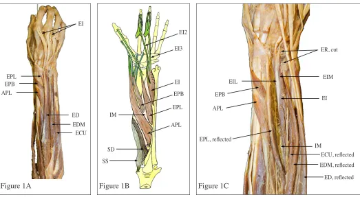
tendons of the EI were positioned medial and deep to the tendons of the extensor digitorum (ED) muscle as they traversed the fourth osseofibrous tunnel under the exten-sor retinaculum (Figure 1C). On the dorsum of the hand, opposite the heads of the second and third metacarpal bones, the two tendons of the EI joined the ulnar sides of the tendons of the ED, enhancing the medial slips of the extensor expansion (EE) for the second and third digits Figure 1
Topography of the extensor indicis (EI) muscle with a double tendon within the muscular layers of the extensor compartment of the forearm.
A – The superficial muscular layer includes ECU, extensor carpi ulnaris; EDM, extensor digiti minimi; ED, extensor digitorum. The deep layer includes EPL, extensor pollicis longus; EPB, extensor pollicis brevis; APL, abductor pollicis longus; EI with a double tendon.
B – Schematic representation illustrates the deep muscular layer. EI2, extensor indicis to the second digit; EI3, extensor indicis to the third digit; SS, supinator superficial part; SD, supinator deep part; APL; EPL; EPB; IM, interosseous membrane.
C – The extensor indicis lateral part (EIL) of the muscle belly becomes tendinous proximal to the extensor retinaculum (ER). The extensor indicis medial part (EIM) is longer with the musculotendinous junction descending deep to the superior margin of the ER. Both tendons of the EI are positioned medial and deep to the tendons of the ED, as they traverse the osseofibrous tunnel under the ER.
ER, cut
EIM
EI
IM
ECU, reflected EDM, reflected ED, reflected EPL, reflected
APL EPB
EIL
Figure 1C
EI2
EI3
EI EPB EPL
APL
SD SS
IM
Figure 1B
EI
ED EDM
ECU APLEPB
EPL
(Figure 2). In all other dissected forearms, a tendon of EI muscle joined the EE to the index finger.
In consideration of the clinical importance of the distri-bution of the DBRN, we dissected 32 upper limbs to de-termine whether the branching pattern of the DBRN var-ied between the forearms possessing single versus double tendons of EI. The radial nerve arose from the posterior cord of the brachial plexus and incorporated the anterior primary rami of C5-T1 spinal nerves. From the axillary fossa, it wound around the posterior aspect of the humerus and pierced the lateral intermuscular septum to enter the anterior compartment of the arm dividing into superficial and deep terminal branches. In the literature, the DBRN is frequently called the posterior interosseous nerve (PIN).31
Figure 2
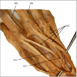
The distal attachments of the EI double tendon. On the dorsum of the hand, opposite the heads of the second metacarpal (M2) and the third metacarpal (M3) bones, the tendons of the EI to the second (EI2), and third (EI3) digits join the ulnar sides of the ED, enhancing the medial slips (MS) of the extensor expansion (EE) of the second and third digits.
MS MS
EE EE EE
EE
M2
M3
ED
EI2 EI3
classify the PIN as the terminal long branch of the DBRN. The DBRN traversed the supinator muscle between the superficial and deep layers, emerging in the posterior compartment deep to the superficial layer of the exten-sor muscles, immediately providing the short branches to the ED, extensor digiti minimi, and extensor carpi ulnaris muscles. Descending superficial to the abductor pollicis longus (APL), the DBRN provided the long branches to the APL and the extensor pollicis brevis (EPB), and ter-minated as the PIN (Figures 3A and 3B). The PIN des-cended deep to the extensor pollicis longus (EPL) muscle on the posterior surface of the interosseous membrane sending two to three short nerve branches to supply the EPB and EI muscles (Figure 3C). Next, the PIN continued within the fourth dorsal osseofibrous tunnel between the tendon of the EPL and the lateral part of the EI terminat-ing within the dorsal fascia and the dorsal surface of the fibrous capsules of the radiocarpal and intercarpal joints. Discussion
It is important understand the anatomy of the variant EI muscle with a double tendon as this variant may explain clinical symptoms of certain hand lesions and should be considered in the differential diagnosis of a swelling, ganglion or other soft tissue tumors on the dorsum of the hand.1,5,9,16
Knowledge of the variant EI muscle with a double tendon is required for precise extensor muscle identifica-tion and confirmaidentifica-tion of a preoperative diagnosis using dynamic sonography or magnetic resonance imaging, as well as for the planning of the best possible surgical treat-ments in this region.28-38
Schmidt et al.39 argued that hand surgery informed by
applied anatomy, and collaboration between anatomists and clinicians are essential to upgrading surgical tech-nique and optimizing patient care.
The present study is congruent with the observations of Ritter et al.40 and Doyle41 with regard to the
an overstretched EI muscle might lead to inflammation within this fascia and an increase in intracompartmental pressure. Moreover, the surgeon should take this attach-ment into consideration when performing any surgical procedure within the extensor forearm region.
Caudwell et al.42 described the musculotendinous
junc-tion of the EI within the confines of the fourth dorsal os-seofibrous tunnel in 75% of the specimens which they examined. Ritter and Inglis,40 confirming the frequency
of the musculotendinosus portion of EI within the fourth
dorsal tunnel, remarked that the contents of the tunnel are extremely tightly confined when the hand and fingers are flexed. That is why an increase in the size of any of the components of the tunnel, including a hypertrophied EI, might produce pain and disability. Similarly, we be-lieve that the presence of two tendons of the EI, espe-cially the musculotendinous junction of its medial part, would increase the volume of the contents of the fourth dorsal osseofibrous tunnel and might cause pain and other clinical symptoms. We observed that in the tunnel itself, Figure 3
The branching pattern of the deep branch of the radial nerve (DBRN) in the forearm with the variant EI muscle. A – Schematic representation illustrates the DBRN which traverses the supinator (S) muscle, and immediately provides the short branches (SB). The long branches include the abductor pollicis longus branch (APLB), the extensor pollicis longus branch (EPLB), and the posterior interosseous nerve (PIN). The PIN provides the abductor pollicis brevis branch (APBB), and the extensor indicis branches (EIB).
B – The DBRN descends superficial to the APL muscle providing the APLB and EPLB, and then as the PIN, descends deep to the EPL muscle.
C – The PIN descends deep to the EPL muscle on the posterior surface of the IM. Being located between the EPL and the lateral part of the EI muscle, the PIN provides the two to three EIB.
PIN
EIB
PIN EPLB
SB DBRN
S APLB APBB
Figure 3A
EIB
EPLB
SB
DBRN S
APLB APLPIN
EPL PIN
Figure 3B
EI medial part EI lateral part
EIB
IM
DBRN PIN EPL, reflected
and deep to the tendons of the ED muscle. Knowledge of this relationship is essential in tendon identification when harvesting for tendon transfer during such procedures as opponensplasty.43
Review of the literature indicates that the EI muscle confers independence to the index finger. Acting alone or together with the ED, it extends the index finger at the metacarpophalangeal and proximal interphalangeal joints and assists with the extension of the hand at the wrist. As a result, the EI muscle enhances tight grip.43, 44 For
this reason, we assume that the double tendon of the EI muscle, joining the EE, may increase the independence not only of the index finger but also of the middle finger, and may further enhance tight grip.
The EE is formed by the four tendinous extensions of the ED muscle on the dorsum of the proximal phalanx of each digit. Each extension is made by three slips, one axial and two collateral slips. The lateral collateral slips are thickened by the tendons of lumbrical and interos-seous muscles and the medial collateral slips by tendons of the interosseous alone.1,31,40,43,45
In the presented case, the two tendons of the EI muscle joined the medial collateral slips of the EE to the mid-dle and index fingers, as reported previously in the liter-ature.40,43,45 We hypothesize that by equalizing the
thick-ness of the medial slips of the EE to the index and middle digits, the EI double tendon assists with the balancing and dissipation of mechanical stresses during the coordinated extension of these digits.
Understanding the anatomical branching pattern of the radial nerve within the posterior forearm is an essential step in recognizing the clinical symptoms of peripheral neuropathy.22,43,46 It is also important in identifying the
level of nerve injury,47 for use during surgical repair using
nerve grafts,48-51 performing nerve blocks,52 neurography,30
and for the modeling of neuromuscular compartments.25
Since total or partial lesions are often encountered in clin-ical settings, we sought to determine the branching pat-tern of the radial nerve in a case of an EI muscle with a double tendon.
According to our observation, the DBRN after emer-ging from the supinator gives off short branches to the superficial extensor muscles. This observation is in agree-ment with the majority of investigations, except that the nerve penetrating the supinator is often referred to as the
ical terminology,32 we classify the PIN as the terminal
long branch of the DBRN.
In our study, the innervation of the deep extensor mus-cles is supplied by the long branches of the DBRN, in-cluding the nerve to the APL, nerve to the EPB, and the PIN. The PIN, descending deep to the EPL, provides two to three short branches to supply the EPB and EI mus-cles. In the literature, all long branches are described as branches of the PIN with morphometric and schematic variances.23,38,49
Thus, we conclude that there are no differences in the branching pattern of the DBRN within forearms pos-sessing either single or double tendons of EI. In both cases, having longer branches to APL and EPL allow for the possibility of isolated neuropathy of these branches. The innervation of the EI and EPB muscles by the short branches from the PIN most likely would be disrupted by the neuropathy of either the main trunk of DBRN or PIN as it descends deep to the EPL muscle.
Conclusions
1. A double tendon of the EI increases the volume of the contents of the fourth dorsal osseofibrous tunnel, which may result in clinical symptoms.
2. By equalizing the thickness of the medial slips of the extensor expansion, the two tendons of the EI may as-sist with the balancing of the mechanical stresses with-in the extensor expansion, contributwith-ing to the coordwith-in- coordin-ation of the extension of the second and third digits. 3. The branching pattern of the DBRN may result in a
predisposition towards isolated neuropathy of the long branches or the posterior interosseous nerve, resulting in specific clinical symptoms.
References
1. Jones BV, Ipswich RN. An anomalous extensor indicis muscle. J Bone Joint Surg Br. 1959;41-B(4):763-765. 2. Schenck RR. Variations of the extensor tendons of the
fingers. Surgical significance. J Bone Joint Surg Am. 1964;46:103-110.
3. Bergman RA, Thompson SA, Afifi AK. Catalog of Human Variation. Baltimore-Munich: Urban & Schwazzenberg, 1984:40-155.
5. GodwinY, Ellis H. Distribution of the extensor tendons on the dorsum of the hand. Clin Anat. 1992;5:394-403. 6. el-Badawi M, Butt M, al-Zuhair A, et al. Extensor tendons
of the fingers: arrangement and variations. Clin Anat. 1995;8(6):391-398.
7. Von Schroeder HP, Botte MJ. Anatomy of the extensor tendons of the fingers: variations and multiplicity. J Hand Surg Am. 1995;20:27-34.
8. Yoshida Y. Anatomical studies on the extensor pollicis et indicis accessorius muscles and the extensor indicis radialis muscle in Japanese. Okajimas Folia Jpn. 1995;71(6):355-363.
9. Tan ST, Smith PJ. Anomalous extensor muscles of the hand: a review. J Hand Surg. 1999;24-A(3):449-455. 10. Shiraishi N, Matsumura G. Anatomical variations of
the extensor pollicis brevis tendon and adductor pollicis longus tendon-relation to tenosynovectomy. Okajimas Folia Anat Jpn. 2005;82:25-29.
11. Ranade AV, Rai R, Prabhu LV, et al. Incidence of extensor digitorum brevis manus muscle. Hand (NY). 2008;3(4):320-323.
12. Zilber S, Oberlin C. Anatomical variations of the extensor tendons to the fingers over the dorsum of the hand: a study of 50 hands and a review of the literature. Plast Reconstr Surg. 2004;113(1):214-221.
13. Li J, Ren ZF. Bilateral extensor indicis brevis: a rare muscular variant. Case report. Rom J Morphol Embryol. 2012;53(1):185-187.
14. Vazquez JM, Linscheid RL. Anomalous extensor muscles simulating dorsal wrist ganglion. Clin Orthop Relat Res. 1972;86:84-86.
15. Reeder CA, Pandeya NK. Extensor indicis proprius syndrome secondary to an anomalous extensor indicis proprius muscle belly. J Am Osteopath Assoc. 1991;91(3):251-253.
16. Baker J, Gonzalez MH. Snapping wrist due to an anomalous extensor indicis proprius: a case report. Hand (NY). 2008;3(4):363-365.
17. Bolla SR, Vollala VR, Bovindala B, et al. Extensor digitorum brevis manus: Its clinical significance and morphology. International J Anatomical Variations. 2008;1:32-34.
18. Hanz KR, Saint-Cyr M, Semmler MJ, et al. Extensor tendon injuries: acute management and secondary reconstruction. Plast Reconstr Surg. 2008;121(3):109e-120e.
19. Celik S, Bilge O, Pinar Y, et al. The anatomical variations of the extensor tendons to the dorsum of the hand. Clin Anat. 2008;21(7):652-659.
20. Otenasek FG. Progressive paralysis of the nervous
interosseus dorsalis. Pathological findings in one case. Bull Johns Hopkins Hosp. 1947;81:163-167.
21. Spinner M. Injuries to the Major Branches of peripheral Nerves of the Forearm. 2nd ed. Philadelphia: W.B. Saunders, 1978:80-157.
22. Hirayama T, Takemitsu Y. Isolated paralysis of the descending branch of the posterior interosseous nerve. Report of a case. J Bone Joint Surg Am. 1988;70(9):1402-1403.
23. Ay S, Apaydin N, Acar H, et al. Anatomic pattern of the terminal branches of posterior interosseous nerve. Clin Anat. 2005;18(4):290-295.
24. Alport AR, Sander HW. Clinical approach to peripheral neuropathy: anatomical localization and diagnostic testing. Continuum (Minneap Minn). 2012;18(1):13-38.
25. Ravichandiran M, Ravichandiran N, Ravichandiran K, et al. Neuromuscular partitioning in the extensor carpi radialis longus and brevis based on intramuscular nerve distribution patterns: A three-dimensional modeling study. Clin Anat. 2012;25(3):366-372.
26. Scott JR, Gobby M, Taggart I. Magnetic resonance imaging of acute tendon injury in the finger. J Hand Surg Br. 1995;20:286-288.
27. Hauger O, Chung CB, Lektrakul N, et al. Pulley system in the fingers: normal anatomy and simulated lesions in cadavers at MR imaging, CT, and US with and without contrast material distention of the tendon sheath. Radiology. 2000;217:201-212.
28. Swen WA, Jacobs JW, Huback PC, et al. Comparison of sonography and magnetic resonance imaging for the diagnosis of partial tears of finger extensor tendons in rheumatoid arthritis. Rheumatology (Oxford). 2000;39(1):55-62.
29. Clavero JA, GolanoP, Farinas O, et al. Extensor mechanism of the fingers: MR Imaging-anatomic correlation. Radio Graphics. 2003;23:593-611.
30. Cudlip SA, Howe FA, Phil D, et al. Magnetic resonance neurography studies of the median nerve before and after carpal tunnel decompression. J Neurosurg. 2002;96:1046-1051.
31. Moore KL, Dalley FD, Agur AMR. Clinically oriented anatomy. 7th ed. Philadelphia: Wolters Kluwer/Lippincott Williams &Wilkins, 2014:750-757; 761-764.
32. Terminologia Anatomica: International anatomical terminology. Federative Committee of Anatomical Terminology (FCAT). Stuttgart: Thieme, 1998:1-292. 33. Soni P, Stern CA, Foreman KB, et al. Advances in extensor
tendon diagnosis and therapy. Plast Reconstr Surg. 2009;123(2):52e-57e.
34. Wu TS, Rosenberg M, VanDillen C, et al. Bedside ultrasound evaluation of tendon injuries. Ann Emerg Med. 2009:54(3):S67-S68.
35. Martinoli C. Musculoskeletal ultrasound: technical guidelines. Insights Imaging. 2010;1:99-144. 36. Li J, Ren ZF. Bilateral extensor indicis brevis: a
rare muscular variant. Rom J Morphol Embryol. 2012;53(1):185-187.
interosseous nerve terminal branches. Clin Orthop Relat Res. 2000;376:242-251.
39. Schmidt HM, Lanz U. Surgical Anatomy of the Hand. 1st ed. New York, NY: Thieme; 2004:1-267.
40. Ritter MA, Inglis AE. The Extensor Indicis Proprius Syndrome. J Bone Joint Surg Am. 1969;51-A(8):1645-1648.
41. Doyle JR. Extensor tendons-acute injuries. In: Green DP, ed. Operative hand surgery. 3rd ed. New York, NY: Churchill Livingstone, 1993:1925-1954.
42. Caudwell EW, Anson BJ, Wright RR. The extensor indicis proprius muscle: a study of 263 consecutive specimens. Q Bull Northwest Univ Med Sch. 1943;17:267-279.
43. Doyle J, Botte MJ. Surgical anatomy of the hand and upper extremity. Philadelphia: Lippincott Williams&Wilkins, 2003:134-135,141-142,221.
44. Williams PL, ed. Gray’s Anatomy. The Anatomical Basis of Medicine and Surgery. 38th ed. Edinburgh: Churchill Livingstone, 1995:852-858.
45. Gonzalez MH, Weinzweig N, Kay T, et al. Anatomy of the extensor tendons to the index finger. J Hand Surg Am. 1996;21(6):988-991.
46. Alport AR, Sander HW. Clinical approach to peripheral neuropathy: anatomical localization and diagnostic testing. Continuum (Minneap Minn). 2012;18(1):13-38.
47. LI Hai, CAI Qi-xun, SHEN Pin-quan, et al. Posterior interosseous nerve entrapment after Monteggia
fracrture-2013;16(3):131-135.
48. Waters PM, Schwartz JT. Posterior interosseus nerve: an anatomic study of potential nerve grafts. J Hand Surg Am. 1993;18(4):743-745.
49. Missankov AA, Sehgal AK, Mennen U. Variations of the posterior interosseous nerve. J Hand Surg Br. 2000;25(3):281-282.
50. Leechavengvongs S, Witoonchart K, Uerpairojkit C. Penetrating injury to the terminal branches of the posterior interosseous nerve with nerve grafting. J Hand Surg Br. 2001;26(6):593-595.
51. Lawton JN, Cameron-Donaldson M, Blazar PE, et al. The anatomic considerations regarding the posterior interosseous nerve at the elbow. J Shoulder Elbow Surg. 2007;16(4):502-507.
52. Abrams RA, Ziets RJ, Lieber RL, et al. Anatomy of the radial nerve motor branches in the forearm. J Hand Surg. 1997;22(2):232-237.
53. Dellon AL, Seif SS. Anatomic dissections relating the posterior interosseous nerve to the carpus, and the etiology of dorsal wrist ganglion pain. J Hand Surg Am. 1978;3(4);326-332.
54. Carr D, Davis P. Distal posterior interossseous nerve syndrome. J Hand Surg. 1985;10(6):873-878.