Abstract— Samples of Bentonite-Clay from Khulays region in S audi Arabia were subjected to mechanical grinding using an agate mortar for 30, 60, and 120 minutes. X-ray diffraction (XRD) patterns of the ground samples exhibited a lack of amorphization. The mean d(001) value and crystallite size were shifted to the lowest values at the first 30 minutes of grinding. FT-IR spectra revealed no decomposition in the structure against grinding for 2h. A reduction in the intensities of water stretching vibration at 3435cm-1(
Al-OH) and at 1637cm -1
(bending-OH) appeared due to the heat produced by grinding which promoted the escape of water molecules. Grinding for 30-60 minutes resulted in a decrease in particles in the µm range and their volume percentage with an increase in particles in the nm range, and aggregation post excessive grinding to 120 minutes. This was translated into a maximum surface area reached at 30 minutes of grinding time. Combined Dynamic Light S cattering (DLS ), surface area, S EM and TEM analyses allowed for the interpretation of this effect in terms of textural modification as a result of change in clay particles morphology. S EM micrographs of the peudo-hexagonal structure of the clay showed that 30-120 minutes of grinding decreased the particle size and increased the dispersion, whilst TEM micrographs revealed that excessive grinding to 120 minutes induced aggregation. Textural properties of the ground clay depended on the time of grinding, where high surface energy of fine particles enhanced aggregation after 30 minute grinding of clay..
Index Term— Aggregation, crystallite size, particle size, surface area.
I. INTRODUCTION
Bentonite-Clays are widely used in several industrial applications, such as foundries, petroleum drilling, civil
T his work is funded by the Deanship of Scientific Research at KSU, research group project No. RGP -VPP-102
M. Al-Qunaibit is an Associate Professor at the Chemistry Department, Science College, King Saud University, Saudi Arabia, P.O. Box. 22452, Riyadh 11495, Saudi Arabia, Phone/Fax 966 -1-4772245.
L. Al Juhaiman is an Associate Professor at the Chemistry Department, Science College, King Saud University, Saudi Arabia, P.O.
Box. 22452, Riyadh 11495, Saudi Arabia, Phone/Fax 966 -1-4772245 [email protected]
engineering, catalysis, environmental remediation and animal feed, due to their high specific surface area, cation exchange capacity and s welling capacity related to the absorption of both water and organic molecules. Clay minerals are natural materials with size dependent properties, and great attention has been paid to the use of auxiliary techniques aimed at modification of clay particle size. Mechanical grinding [1-4], ultrasound and microwave radiation [5, 6] were adopted for shaping up the final textural properties of clays. These processes induce physical and chemical changes in the treated materials that can be exploited either to enh ance known properties, or for new application purposes. Many studies reported on the structural and textural changes upon grinding of kaolinite [7-9], talc [10-12], and montmorillonite [13]. A comparative study between ball mill grinding and ultrasonic treatment showed that sonication gave smaller particles and higher specific surface areas than mechanical grinding which produced significant structural degradation and morphological deformation [14]. Others suffered folding and gliding of layers, and aggregation of the newly formed particles when using an oscillatory mill for kaolinite and pyrophyllite [15]. In addition, mechanical grinding modifies strongly the particle surfaces of clay minerals [8]. Dellisanti et al. [13] found that structural properties of Ca-montmorillonite can be modified by ionized gas treatment and ball milling where ball milling in controlled thermodynamic environment induced a structural destabilization both in the interlayer and in the intralayer with progressive losses of interlayer water.
Fine grinding is carried out in high intensity grinding mills such as oscillating mills, vibration mills, attrition mills, and jet mills. In addition to size reduction, during the fine grinding process, severe mechanical action on the solid surface are known to lead to physical and chemical changes due to the large amount of energy delivered by the grinding mills [16]. Mechanochemical effect is evident in solids when ground with equipment based on impact and shear [17], where energy delivered by the mill is not stored in the particles as thermal energy due to the low thermal conductivity characteristics of most nonmetallic solids, but is applied to the bending and/or breaking of the crystal lattice which results in a loss of crystallinity (amorphization) and formation of active surfaces [17, 18]. Mechanical grinding and microwave radiation [5] were found to produce ordering of saponite layers along the c axis, if the grinding time was large enough (135 min). A M. Al-Qunaibit M., L. Al Juhaiman
simultaneous decrease of the particle size was found with a relationship between layer ordering and the particle size.
The present work reports the influence of grinding time on texture, of the less studied Khulays Bentonite-Clay using eco-friendly manual grinding. For this purpose, changes in crystal structure, particle size distribution, surface area and surface morphology of the mechanically modified clay have been studied.
Scope and Limitations
Mechanically modified clays are used in pharmaceuticals and as catalysts in small amounts, which makes the method adopted in this study appropriate. Different locations in clays usually respond differently to grinding, which makes it difficult to compare this local clay to clays from other sites in the world.
II. MATERIALS AND METHODS Mechanical treatment
Raw Khulays Bentonite-Clay (RB) was collected from the Western Region in Saudi Arabia [19]. RB was mechanically ground using an agate mortar, and samples collected after 30, 60, and 120 min. The samples were labeled B30, B60 and B120. X-ray analysis showed that the dominant components of the clay are: smectite (64.9%), kaolinite (10.6%), geothite (9.5%), hematite (9.4%) and boehmite (5.6%) [20].
Measurements
Phillips Analytical X-ray spectrometer (PW 1710) using CuKα radiation was used for XRD measurements. FTIR spectra were recorded using a Perkin–Elmer FTIR Spectrometer (Spectrum RXI, 4400–440 cm−1). Particle size distribution was measured by a dynamic light scattering technique (DLS) with a Malvern Zeta Sizer 3000HS at 20 °C using a 10 mW He–Ne laser, 633 nm wavelength and 90° fixed scattering angle. Concentration range for all samples was 1% g/mL. The results are given as volume frequency in percent vs. particles size. TriStar II 3020 was used for surface area and pore volume analyses .
Surface Morphology
Surface morphological analyses were carried out using a SEM (JSM-5400 JEOL), working at 20 kV electron
accelerating voltage with a beam current of about 1nA at the specimen level. Samples were gold-coated with a layer about 10 nm thick metal-coating by using a vacuum of 0.13 Pa (10−3 Torr). Transmission electron microscope (TEM) from JEM-2100F with a probe size under 0.50nm working at 80-100 kV was used for the observations. Concentration range for samples was 1% g/mL. The ultrafine fraction (<0.2 μm) was suspended in distilled water and a drop was deposited on holey carbon-covered TEM grids.
III. RESULTS AND DISCUSSION
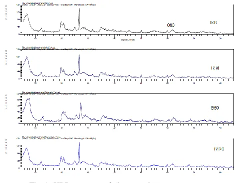
Expected changes in the structure of the ground clay were followed by XRD (Fig.1). Reflections at 001 planes provide information on basal changes, whereas the 060 diffractions refer to in-layers. Line broadening noticed in all patterns may
be ascribed to crystallite size effect, as individual crystals forming the fine clay particles can alter the assumed strain -free environment [21]. The XRD patterns of the ground clay -compared to the raw- showed that the (001) diffractions suffered no decrease in intensity, nor broadness, that usually accompanies amorphization. Hence, the intensity of the 060 diffraction in all samples remained constant, as the (001) diffractions are lost upon grinding much faster than the 060 [22]. The Scherrer crystallite size of the raw clay was 25.4 nm as calculated from FWHM at peak angle 26.5 2.
.
Fig. 1. XRD patterns of clay samples
Grinding shifted both the mean d001 lattice spacing and crystallite size values to a minimum at 30 minutes of grinding (table I).
TABLE I
XRD parameters of clay samples
Sample
Crystallite size
(nm) d(001) nm
RB 25.40 1.30
B30 14.98 1.14
B60 28.25 1.29
B120 25.41 1.19
The slight shift of 001 peak indicates minor structural destabilization of the basal plane, and that the water loss process is not complete [13]. The ground activated particles with high surface energy start reaggregation after 30 minutes of grinding, and the values of the coherent scattering thickness (crystallite size) indicated crystalline degradation [23] accounting for the increase in this value after 30 minutes of grinding.
reported a decrease in the intensity of these bands after grinding the clay in a mortar, which was also observed upon milling of calcium montmorillonite [1, 22] . Broad bands in the O-H stretching region (~3426 cm-1) of the modified clay were assigned to molecular water adsorbed on the amorphous products [13].
FT-IR spectra of the ground clay, compared to the raw, revealed the resistance of the clay structure against decomposition, even after two hours of grinding, (Fig. 2).
5091.7 4000 3000 2000 1500 1000 500 0 -291.7
cm-1 %T
RB
RB30
RB60
RB120
Fig. 2 IR spectra of raw RB (top spectrum) and ground clays.
A general decrease in intensity of (OH) at ~3435cm-1 and (defOH) at ~1637cm-1 for ground samples was observed due to water loss as a result of heat produced during the grinding process, described by Hrachova et al [1].
Dynamic Light Scattering (DLS). It is interesting to see the gradual change in particle size distribution with time using laser light scattering. Fig.3. revealed the size distribution by intensity; the proad peak for RB indicates the heterogeneous size distribution of particles was ( 800-8000 nm). However thirty minutes of grinding was enough to reduce the size of most particles to less than 1000 nm (2% ~2700 nm) but the two large peaks was overlapping. Further grinding to 60 minutes increased the difference in particle size (no peak overlapping) and the same trend was noticed after 120 minutes with the disappearance of the peaks for large particles.
Fig. 3. Particle size distribution versus % of particle volume from DLS for raw bentonite and ground bentonite at different grinding times
BET results
The total surface area of ground samples exhibited a maximum at 30 minutes grinding followed by a decrease (Fig. 4). This finding is in agreement with DLS and XRD results.
Surface Study
Scanning electron micrographs have been recorded to get insight into the particle morphology and texture (Fig. 5). SEM micrograph of (RB) revealed the natural layered structure of bentonite where large aggregates of oriented platelets are present1. Grinding for 30 minutes showed smaller agglomerates of the flaky layered structure of bentonite mixed with smaller particles. Further grinding (B60) increased the percentage of smaller particles while the bulky particles are still seen. Grinding for 120 minutes resulted in a breakdown of the original bulky structure and showed the original pseudo– hexagonal structure of bentonite.
Fig. 5. SEM micrograms for raw bentonite and ground bentonite at different grinding times
Transmission electron microscopy (TEM)
TEM observation of the images showed the original micron sized flaky clay particles with pseudo–hexagonal morphology (RB) (Fig. 6). The clay retained the original morphology after 30 minutes of grinding while the particle size became smaller with submicron particles appearing. Further grinding to 60 minutes decreased the particle size and increased the dispersion. Excessive grinding to 120 minutes induced a reduction of the particle size and revealed the original flaky and plate-like particles of bentonite whereas the black spots seemed to indicate particle aggregation.
Fig. 6. T EM images for raw bentonite and ground bentonite at different grinding times
The results from this study showed that the physical disintegration of the clay minerals by manual milling consisted of many processes: particle size reduction, morphological and structural changes, accompanied by changes of the surface properties. Many aspects should be addressed here. The first process applies to all investigated solids during the milling treatment, refers to the intensive decrease of the initial particle size, associated with morphological changes and the formation of a poly dispersed powder as shown from SEM and TEM results [1-3]. Similar to other studies [13,14, 22, 25], the fracturing and size reduction of the clay mineral particles during this process was accompanied by an increase in the surface area. In the present study there is an agreement in the results from DLS (Fig. 3) after 30 minutes of grinding and BET results (Fig. 4.). This implies that the intensive mechanical treatment of different clay minerals causes the formation of aluminosilicates with similar structural (XRD), morphological and surface area properties.
IV. CONCLUSION
Mortar milling did not have an appreciable influence on the XRD patterns of clay samples, suggesting the absence of delamination. Loss of interlayer water upon grinding was confirmed by reduction in OH-water vibrations intensities in the IR region while retaining the clay structure. The minimum crystallite size and d001 value were observed at 30 min grinding
REFERENCES
[1] J. Hrachová, J. Madejová, P. Billik, P. Komadel, V. Štefan Fajnor., Dry grinding of Ca and octadecyltrimethylammonium montmorillonite J. Col. Inter. Sci., 2007; 316 , 589.
[2] F. Franco, J. Cecila, L. Pérez-Maqueda, J. Pérez-Rodríguez and C. Gomes, Particle-size reduction of dickite by ultrasound treatments: Effect on the structure, shape and particle-size distribution, App. Clay Sci., 2007; 35, 119.
[3] A. Ramadan, A. Esawi and A. Gawad, Effect of ball milling on the structure of Na+-montmorillonite and organo-montmorillonite, Appl. Clay Sci., 2010; 47, 196.
[4] R. Reynolds Jr. and D.L.Bish, Am. Miner., T he Effect of grinding on the structure of a low-defect kaolinite, 2002; 87 1626. [5] R. T rujillano, E. Rico, M. Vicente, M. Herrero, and V. Rives,
Microwave radiation and mechanical grinding as new ways for preparation of saponite-like materials, Appl. Clay Sci., 2010; 48,32.
[6] J. Pe´rez-Rodrı´guez, A. Wiewiora, V. Ramirez-Valle, A. Dura´na and L. Pe´rez-Maqueda, Comparative study of sonication and grinding, Journal of Physics and Chemistry of Solids, J. Phys. Chem. Solids, 2007; 68, 1225.
[7] P. Sanchez-Soto, M. Jimenez de Haro, L. Pérez-Maqueda, I. Varona-Vaira and J. Pérez-Rodríguez, Effect of dry grinding on the structural changes of kaolinite powders, J. Amer. Ceramic Soc., 2000; 83, 1649.
[8] R. Frost, J. Kristof, E. Mako and W. Martens, Modification of the hydroxyl surface of kaolinite through mechanochemical treatment followed by intercalation with potassium acetate, Langmuir, 2002; 18, 6491.
[9] E. Horváth, R. Frost, E. Makò, J. Kristòf and T . Cseh, T hermal treatment of mechanochemically activated kaolinite, T herm. Acta, 2003; 404, 227.
[10] Godet -Morand, A. Chamayou and J. Dodds, T alc grinding in an opposed air jet mill: start -up, product quality and production rate optimisation, Powder T ech., 2002; 128, 306.
[11] G. Christidis, P. Makri and V. Perdikatsis, Influence of grinding on the structure and colour properties of talc, bentonite and calcite white fillers, Clay Miner., 2004; 39/2, 163.
[12] L. Pérez-Maqueda, A. Duran and J. Pérez-Rodríguez, Preparation of submicron talc particles by sonication, Appl. Clay Sci., 2005; 28, 245.
[13] F. Dellisanti and G. Valdrè, Study of structural properties of ion treated and mechanically deformed commercial bentonite, Appl. Clay Sci., 2005; 28, 233.
[14] J. Pérez-Rodríguez, "T ransformation of clay minerals on grinding: A review" in: Applied Study of Cultural Heritage and Clays (Edt. J. Pérez-Rodríguez), Servicio Publicaciones del CSIC, Madrid, Spain, 2003, pp. 425–444.
[15] E. Stepkowska, J. Pérez-Rodríguez, M. Jiménez de Haro, P. Sánchez Soto and C. Maqueda, Effect of grinding and water vapour on the particle size of kaolinite and pyrophyllite, Clay Miner., 2001; 36, 105.
[16] K. Venkataraman and K. Narayanan., Energetics of collision between grinding media in ball mills and mechanochemical effects, Powder T ech., 1998; 96, 190.
[17] M. Al-Wakeel, Egypt, Effect of mechanical treatment on the mineralogical constituents of Abu-T artour phosphate ore, Int. J. Miner. Proc., 2005; 75, 101.
[18] E. Aglietti, J. M.Porto Lopez and E. Pereira, Mechanochemical effects in kaolinite grinding. II. Structural aspects, Int. J. Min. Proc., 1986; 16, 135.
[19] D. Laurent, Atlas of Industrial Minerals, Ministry of Petroleum and Mineral Resources, Kingdom of Saudi Arabia, 1992.
[20] L. Al Juhaiman, W. Mekhamer, and A. Al-Boajan , Effect of poly(vinyl)pyrrolidone on the zeta potential and water loss of raw and Na-rich Saudi bentonite, Surf. Int. Anal., 2010; DOI 10.1002/sia.3669.
[21] B. Culity and S. Stock ,Elem ents of X-Ray Diffraction, Prentice Hall, 2001 p 389.
[22] J. Hrachova, P. Komadel and V. Fajnor, T he effect of mechanical treatment on the structure of montmorillonite, Mat. Letters, 2007; 61, 3361.
[23] P. Sanchez-Soto, A. Wiewióra, M. Avilés, A. Justo, L. Pérez-Maqueda, J. Pérez-Rodríguez and P. Bylina, T alc from Puebla de Lillo, Spain, II Effect of dry grinding on particle size and shape, App. Clay Sci., 1997; 1 (4), 297
[24] I. Bekri-Abbes and E. Srasra, Mater. Sci.,: T he effect of fine dry grinding on the physicochemical properties and textural morphology of T unisian smectite clay, An Indian Journal, 2006; 2(2-3), 46.