www.pharmascholars.com
110
Original Article
CODEN: IJPNL6
EVALUATION OF SOLASODINE FROM THE LEAVES OF
SOLANUM
MAURITIANUM
SCOP. BY HPTLC
Jayakumar K
1and Murugan K
2*1
Department of Botany, SVR NSS College, Kottayam
, Kerala, India
2
Plant Biochemistry and Molecular Biology Lab, Department of Botany, University College,
Trivandrum 695 034, Kerala, India
*Corresponding author e-mail: harimurukan@gmail.com
Received on: 09-11-2015; Revised on: 08-12-2015; Accepted on: 20-12-2015
ABSTRACT
Solanum mauritianum Scop. is an exotic tree Solanum species of South America. High performance thin layer chromatography method was formulated in S. mauritianum to identify the major alkaloid fractions. Optimal extraction of solasodine includes refluxing with 2.5N methanolic hydrochloric acid, precipitation and extraction using non-polar solvent system. Ideal and prominent bands were obtained on HPTLC silica gel plates using chloroform (9.3): methanol (0.7 v/v) solvent system at 0.32±0.06 Rf. and visualized with anisaldehyde-sulphuric acid. The resulted and derivatized plates were scanned at 530 nm for densitometric analysis. The method was found linear (r2 = 0.9978) in a wide range (20-2000 ng/ spot), accurate (88.2-101.4%), precise (% RSD < 2.88), robust (% RSD < 3.48) and specific. The LOD and LOQ of the method was found as 14 and 44 ng/spot, respectively indicating sensitive enough to analyze minute amount of solasodine in multi-component extract. The method was applied for analysis of solasodine content in samples of S. mauritianum.
Keywords: HPTLC, solasodine, steroidal glycoalkaloid, aglycone, Solanum mauritianum.
1. INTRODUCTION
Solasodine are steroidal glycoalkloids, a potential group of plant secondary metabolites. Glycoalkaloids are precursors for the synthesis of steroidal drugs. Most solanaceous species have solasodine and present as aglycone region of glycoalkloids, which are nitrogen analogue of sapogenins. 16-dehydropregnenolone, the potent intermediate in the synthesis of steroidal drugs such as progesterone and cortisone are produced from solasodine (C27 cholestane skeleton). [1] It is reported from many species of Solanum [2]. Solasodine also occur in multiple forms of glycoalkaloids like triosides such as, solasonine and solamargine [2]. Different extraction protocols of solasodine have been reported from fruits, leaves or stems of diverse Solanum
species. Significant importance of solasodine in the pharmaceutical field made way for searching allied compounds of solasodine glycosides from other plant species. Solasodine is a natural lead molecule to synthesize steroidal hormones and drugs which are used for contraceptives, arthritis and behavioural disorders. Initially, solasodine was used as precursor to prepare steroid compounds which display glucocorticoid-like effects. Solasodine shows DNA-damaging activity with remarkable MIC values against Staphylococcus aureus for antibacterial activity [3]. Further, it also has analgesic and antinociceptive activity. Solasodine, the principal aglycone is being isolated using different protocols. It is carried out by two phase system containing
International Journal of Pharmacy
www.pharmascholars.com
111
aqueous mineral acid-organic aqueous solventmixture. In the present work, direct acid hydrolysis of the glycosides is being carried out and aglycone solasodine is obtained via alkali treatment after the hydrolysis of the glycoside solasonine [2].
Solanum constitutes the largest and most complex genus under Solanceae. It is composed of approximately 1500 species, many of which are economically important with cosmopolitan in distribution. However, only a few species of
Solanum are considered to be important for commercial production of solasodine. Steroidal glycoalkaloids aglycones lack chromophore groups in the common operating range of UV spectrophotometry and absorb only at low wave length end of the UV spectrum. This means that they have low UV sensitivity and can be detected and identified by diode array detection/UV only when present in relatively high amounts. Absence of chromophore makes their detection a major challenging problem in the assay of a biological sample [4]. Among various analytical techniques, high performance thin-layer chromatography (HPTLC) in particular appears to be suitable for phytomolecules of varying nature and provides a rational approach in the authentication and quality assessment of crude medicinal herbs and their formulation.
The objectives of present study were to provide a simplified, improved and optimized method for the extraction of aglycone of steroidal alkaloids from S. mauritianum that contains them in glycoside form as well as to develop a rapid, sensitive, accurate and validated HPTLC method which can be applied for the quantitative estimation of solasodine.
2. MATERIAL AND METHODS
2.1 Plant material: Solanum mauritianum leaves
were obtained from Munnar hills of Idukki district, Kerala. Solasodine was used as reference compound. Solasodine, HPLC-grade methanol and chloroform were obtained from Sigma Aldrich. Distilled water was deionized before use. HCl used was of SRL grade.
2.2 Chromatographic Conditions: The following
chromatographic conditions were used to quantify the solasodine:
i. Stationary phase: silica gel precoated TLC plates 60F-254 (20 cm×10 cm)
ii. Mobile phase: chloroform:methanol (9.25:0.75 v/v)
iii. Sample volume: 5 μl
iv. Sample for HPTLC: solosodine from plant sample and standard solution of solasodine
Application mode: Camag Linomat V Development Chamber: Camag Twin Trough Chamber. Plates: Precoated Silica gel plates. Chamber Saturation: 30 min. Development time: 30 min. Development distance: 7 cm. Scanner: Camag Scanner III Detection: Deuterium lamp. Data System: Win cats software.
10 g of fresh leaves of S. mauritianum was extracted with 5 ml of methanol. The material was refluxed in 2.5 N HCl for 2 h at 70°C. The extract was filtered and pH of the filtrate was adjusted to 10 with dilute ammonia solution (12.5% v/v). The precipitate was washed thrice with deionized distilled water, filtered and dried at 60°C. The above crude glycosides, 2-propanol and 37.6% hydrochloric acid in the ratio of 16.4:76:7.6 by weight was optimal in terms of hydrolysis and were placed in a round-bottom flask. Further, the solution was refluxed for 3 h and was then filtered by vacuum pump. The collected precipitate was dissolved in 2-propanol and 20% w/v NaOH solution. A large amount of water was added into the solution to obtain the crystalline solasodine. The dried extract was re-dissolved in methanol and filtered using 0.2 µm syringe filter. The resultant was stored at 4°C for HPTLC quantification.
2.3 Preparation of standard solution: 400 µg/ml
stock solution of standard solasodine was made by dissolving 2 mg of solasodine in 5 ml of methanol. Standard solasodine solutions of 20, 40, 80, 160, 200, 400, 800, 1600, 2000, 4000 ng/spot were spotted repeatedly (6 times) on the plate. Resulted data of peak area vs. solasodine concentration were treated with the linear-least square regression and the regression equation thus formed from the standard curve to quantify solasodine level in the leaf sample. Initially, the plates were pre-washed with methanol. Standard and sample solutions were spotted to the plates as sharp bands by means of Camag Linomat V sample applicator. The spots were dried naturally in the current of air. The mobile phase (20 ml) was poured into a twin trough glass chamber, whole assembly was left to equilibrate for 30 min and the plate was placed in the chamber. The plate was then developed until the solvent front had travelled at a distance of 80 mm above the base of plate. The plate was then removed from chamber and dried in air. Detection and quantification was performed with Camag TLC scanner 3 at 530 nm after spraying the developed plate with anisaldehyde sulphuric acid reagent and heating it on hot plate at 110°C for 5 min.
www.pharmascholars.com
112
2.4 Method validation, accuracy as recovery,
precision, specificity, robustness and sensitivity: The
method employed was validated as per the ICH guidelines and chromatographic HPTLC methods reported by laboratory which are in use for the quality control of herbal drugs [5, 6, 7].
Accuracy was checked in leaf samples by recovery studies in which, pre-analyzed samples were spiked with extra 25, 50, 100, 150 and 200% of standard solasodine and analyzed with the present method. The experiment was repeated, average recovered solasodine was quantified using regression equation, and the % of recovery was calculated.
Precision of the method was evaluated by repeatability, inter and intra-day precisions. Repeatability was studied by spotting 3 varied doses of standard solasodine (100, 200, 400 and 800 ng/ml) for thrice and %RSD of area was calculated. Inter and intra-day precisions were carried by repeating the same experiment in the same day and also in three different days respectively.
Specificity of this protocol was carried by comparing the Rf value with absorption spectra of solasodine in leaf sample and standard. Purity of solasodine peak was assessed by comparing the spectra at three levels i.e. peak start, apex, end position of the band.
Robustness of the protocol was determined at single concentration level i.e., 200 ng/ml in two different ways, i.e. by altering the composition of mobile phase and also changing the detecting wavelength. The % RSD of the method was analyzed to assess the robustness of the method.
Sensitivity was also determined as the limits of detection (LOD) and limits of quantification (LOQ). Decreasing amounts of standard solasodine standard were applied to a plate and chromatographic-densitometric analysis was performed as described above. The dose of sample giving signal to noise ratio of 3 was fixed as the LOD, whereas the concentration of the sample giving signal to noise ratio of 10 was fixed as LOQ.
3. RESULTS AND DISCUSSION
The method describes the utilization of silica gel 60F-254 HPTLC plates as ideal stationary phase and chloroform:methanol (9.3:0.7 v/v) as mobile phase to yield good separation of solasodine (Rf=0.32) standard and the sample. Compact bands were visualized using anisaldehyde-sulphuric acid as spraying reagent and the chromatograms were scanned at 530 nm (Figure 1).
The calibration curve display optimal linear relationship between peak area and concentration in the range of 20-2400 μg/ ml. The regression equation, slope and intercept values were y= 8.8223x + 181.56, 8.833 and 182.55 respectively. The correlation
coefficient was 0.998. The accuracy of the protocol was evaluated by the standard addition method at the concentrations (0, 25, 50, 100, 150 and 200 ng) which showed recovery within the range of 88.2%-101.4% (Table 1).
Repeatability and intermediate precisions were calculated in terms of % RSD. It ranges from 0.84 to 3.42% and the corresponding Rf values were 0.19 to 0.32. Intermediate precision data includes inter and intra-day precisions i.e., 1.1 to 2.4 and 1.48 to 2.88% RSD. Result of precision suggests the protocol may be employed for the routine estimation of solasodine (Table 2 and 3).
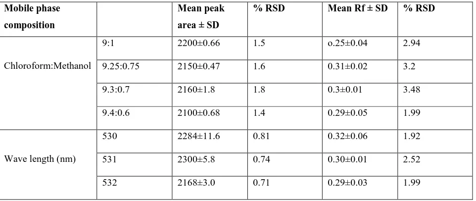
The spectra of standard solasodine and leaf sample track were correlated remarkably and establish the mode of scanning is effective and sensitive. Robustness as in terms of % RSD includes making minor alterations in the composition of mobile phase and wavelength. Mobile phase having compositions; chloroform:methanol (9:1 and 9.4:0.6 v/v) and wavelength 530±2 nm were attempted to analyze any variation (Table 4 and 5).
Optimal mobile phase combinations and wave lengths were 9.3:0.7 chloroform:methanol and 530 nm. Further, the LOD and LOQ by signal to noise ratio was found to be 14 and 44 ng/ml, respectively. Optimization of extraction procedure in terms of variable strength of HCl (1-3 N), volumes of extraction solvents (20-60 ml), temperature (50-100°C), time (30-180 min) and different concentrations of ammonia (5-20% v/v) (Table 5). Maximum solosodine content extraction requires the following parameters such as (a) 2.5 N HCl (b) 40 ml solvent (c) 70°C (d) 2 h and 12.5% v/v ammonia. The present results are comparable with the varied levels of solasodine content of Solanum incanum plants from Sultanate of Oman [8]. Meanwhile, Gangwar et al., (2013)[9] analyzed petroleum ether and alcoholic extract of Solanum xanthocarpum by TLC, HPTLC, IR and NMR obtained a lower values.
4. CONCLUSION
The present developed HPTLC protocol was simple, accurate, precise and cost-effective and can be utilized for the routine analysis of estimation of solosodine. The data was validated in terms of validation, accuracy as recovery, precision, specificity, robustness and sensitivity. Optimal extraction was carried by using 2.5 N methanolic HCl followed by precipitation and extraction using non-polar solvent.
Funding support
www.pharmascholars.com
113
Conflict of Interest
The authors declare no potential conflicts of interest exist.
Figure 1. HPTLC Chromatogram of solasodine at 530 nm using solvent system chloroform:methanol (9.3:0.7 v/v) Rf=0.32.
Table 1. Accuracy of HPLC protocol for solasodine estimation.
% of std. Spiked to the sample
Theoretical value ng/ml
Quantity of drug recovered ng ± SD
% of drug recovered
% RSD
0 119 114.6±0.81 88.2 1.14
25 160 160.2±0.04 94.9 1.21
50 178.6 180±0.38 98.8 1.29
100 201.4 198.5±0.42 101.4 1.78
150 289.8 243±0.33 99.5 0.69
200 329.6 300±0.09 100 0.28
Table 2. Repeatability of HPLC for solasodine estimation. Concentration
ng/spot
Peak area Rf
Mean peak area ±
SD
% RSD Mean Rf ± SD % RSD
100 1688±1.92 1.6 0.19±0.06 0.84
200 2100±35.6 2.1 0.32±0.001 3.42
400 3612±43.9 1.5 0.27±0.04 2.1
www.pharmascholars.com
114
Table 3. Precision of HPTLC in solasodine quantification Concentration
ng/spot
Inter day Intraday
Mean peak area ±
SD
%RSD Mean peak area ±
SD
% RSD
100 1512±2.5 1.9 1596±0.38 1.48
200 2112±0.61 2.4 2246±0.25 1.6
400 3592±0.64 0.51 2467±0.09 2.88
800 4900±0.28 1.1 4916±0.14 1.39
Table 4. Robustness of HPLC in terms of mobile phase and wavelength at 200 ng/spot Mobile phase
composition
Mean peak area ± SD
% RSD Mean Rf ± SD % RSD
Chloroform:Methanol
9:1 2200±0.66 1.5 o.25±0.04 2.94
9.25:0.75 2150±0.47 1.6 0.31±0.02 3.2
9.3:0.7 2160±1.8 1.8 0.3±0.01 3.48
9.4:0.6 2100±0.68 1.4 0.29±0.05 1.99
Wave length (nm)
530 2284±11.6 0.81 0.32±0.06 1.92
531 2300±5.8 0.74 0.30±0.01 2.52
532 2168±3.0 0.71 0.29±0.03 1.99
Table 5. Variable parameters in Solasodine extraction from S.mauritianum
Parameters Variations Mean solasodine ± SD
Strength of Methanolic HCl (N) 1 58.2±0.9
1.5 62.5±0.07
2 69.6±0.01
2.5 73.8±1.4
3 60.2±2.9
Volume of solvent (ml) 20 57.8±0.04
30 63.2±0.18
40 73.9±3.4
50 70.6±1.5
60 68.8±0.67
Temperature (°C) 50 59.6±0.27
60 65.3±3.89
70 71.2±1.8
80 69.6±4.6
www.pharmascholars.com
115
Extraction time (min) 30 52.6±1.41
60 60.7±5.3
90 67.6±8.4
120 75.8±4.6
150 70±3.2
Strength of ammonia (%v/v) 5 59.8±6.85
7.5 67.2±1.2
10 70.5±5.3
12.5 76.4±3.4
15 71.3±2.8
17.5 68.6±6.3
20 52.3±3.3
REFERENCES
1. Manrique-Moreno M, Londono-Londono J, Jemiola-Rzeminska M, Strzalka K, Villena F, Avello M, Suwalsky M. Structural effects of the Solanum steroids solasodine, diosgenin and solanine on human erythrocytes and molecular models of eukaryotic membranes. Biochim Biophys Acta, 2014; 1838(1): 266-277.
2. Patel K, Singh RB, Patel DK. Medicinal significance, pharmacological activities and analytical aspects of solasodine: A concise report of current scientific literature. J Acute Dis, 2013; 2(2): 92-98.
3. Itkin M, Rogachev I, Alkan N, Rosenberg T, Malitsky S. Glycoalkaloid metabolism is required for steroidal alkaloid glycosylation and prevention of phytotoxicity in tomato. Plant Cell, 2011; 23(12): 4507-4525.
4. Solouki M, Hoshyar H, Ramroudi M, Tavassoli A. Comparison and evaluation of steroid alkaloid solasodine on in-vivo and in-vitro cultures of Solanum surattenseBurm L. Afr J Microbiol Res, 2011; 5(23): 3981-3985.
5. Marzouk Z, Cheria J, Bakhrouf A, Guedira K, Boukef K. Influence of vegetation stage on the solasodine content in four natural Solanum sodomeum L. populations of Tunisia. J Food Agric Environ, 2005; 3(2): 243-245.
6. Kamal YT, Singh M, Tamboli ET, Parveen R, Zaidi SM, Ahmad S, Rapid RP-HPLC Method for the quantification of glabridin in crude drug and in polyherbal formulation. J Chromatogr Sci, 2012; 50(9): 779-784.
7. Singh M, Kamal YT, Parveen R, Ahmad S. Development and validation of a stability-indicating HPTLC method for analysis of arjunolic acid in a herbal formulation. J Planar Chromatogr-Mod TLC, 2011; 24(2): 172-175.
8. Sana S, Al Sinani, Elsadig A, Eltayeb, Kamal YT, Sayeed Ahmad. Quantitative estimation of solasodine in
Solanum incanum plants grown in Oman by HPTLC. Int J Pharmacognosy and Phytochem, 2013; 28(1): 1132-1139.