Original Research Article
Role of MRI in Evaluation of Orbital Mass Lesions with
Ultrasonographic and Histopathological Correlation
Rakesh Vijayvargiya
1, Amaresh Kumar Shukla
2*1Associate Professor, 2*P.G. Resident, Department of Radio diagnosis, M.G.M. Medical College, and M.Y. Hospital, Indore, Madhya Pradesh, India.
ABSTRACT
Background: The orbital pathologies now-a-days are rising, which is probably attributed to the increasing awareness to the clinical symptoms, better availability of health care facilities along with newer advanced diagnosis and treatment options. Most of the masses affects the sensory and motor visual pathways. Early diagnosis with institution of early treatment is required to prevent patient sufferings due to vision problems or permanent vision loss.
Aims: The aim of this study was to diagnose, characterize & define the extent of various orbital mass lesions, to differentiate between benign & malignant lesions, to correlate the sensitivity and specificity of MRI findings with Ultrasound and the histopathological diagnosis and to assess its role in the management of patient affected by orbital mass lesions.
Material & Method: In this prospective study, the patients clinically suspicious of orbital mass lesions had undergone orbital B-scan examination using Philips HD7XE & SEIMENS ACCUSON USG machines via high frequency probes, and on revelation of mass in B-Scan, all these patients were then subjected to MRI of the orbit with brain sections for further characterization of the mass lesion. MRI study of 40 patients having orbital mass lesions was conducted from the period of March 2015 to September 2016 using GE 3 tesla, 97 channel, magnetic resonance imaging machine, in the department of Radio diagnosis, Mahatma Gandhi Memorial Medical College & M.Y.H. hospital at KRSNA diagnostic center, Indore, Madhya Pradesh. Subsequent follow up had been done along with biopsy correlation as the reference standard.
Result: In our study 28% of orbital lesions were classified as inflammatory, 15% lesions were benign and the remaining 57%
of orbital lesions were classified as malignant lesions. Among the inflammatory lesions thyroid orbitopathy and pseudo tumor were the most common inflammatory orbital lesions. Cavernous hemangioma was the most common benign orbital mass lesion in our study followed by optic nerve glioma. Retinoblastoma was the most common malignant lesion in our study constituting 25% of the total orbital lesions followed by rhabdomyosarcoma.
Conclusion: The sensitivity and specificity of MRI was higher than USG for detection of malignancy. DWMRI imaging alone may lead to misdiagnosis of lesions as inflammatory lesions also depicts restricted diffusion. Hence, MRI along with DWI is a very valuable noninvasive tool for the Identification, characterization and differentiation of orbital mass lesions.
Keywords: MRI, DWI, USG, Orbital B-Scan, Orbital Mass Lesions.
*Correspondence to:
Dr. Amaresh Kumar Shukla,
P.G. Resident, Department of Radio diagnosis, M.G.M. Medical College, and M.Y. Hospital, Indore, Madhya Pradesh, India.
Article History:
Received: 15-02-2017, Revised: 09-03-2017, Accepted: 19-03-2017
Access this article online
Website:
www.ijmrp.com
Quick Response code
DOI:
10.21276/ijmrp.2017.3.2.032
INTRODUCTION
The orbit and the visual system form the most important sense organ in humans. The orbit is the site of a large number of pathologies of diverse etiologies, and imaging has to be tailored to the symptoms and clinical findings. Multiple disease entities affect the orbit, viz. congenital, inflammatory, infectious, vascular and traumatic and neoplastic. The clinical manifestations and findings are often non-specific, being protean and overlapping.
Of the various pathologies affecting the orbit, orbital masses are an important cause of mortality and morbidity, so it becomes of
immense importance to diagnose, characterize and to know the extent of involvement of the surrounding structures.
space but it has its own limitations in diagnosing retro bulbar pathology, painful orbital lesion and imaging the origin and extension of masses along with its operator dependence, lack of specificity and poor characterization and localization of lesions makes it inferior. Computed tomography has shown its value in characterizing the orbital mass lesions, the results have been variable at times due to various factors. Computed tomography may allow separation of the air, fat, fluid, soft tissue and bone. In many cases, however it is limited by artifacts from bone. Internal chemical makeup of the muscle and inflammatory changes in optic nerve may only be seen as change in size of these structures. So due to higher risk of ionizing radiation exposure and the use of intravenous contrast agents which carry a risk of inducement of acute renal dysfunction and idiosyncratic contrast material reactions along withpoor characterization of soft tissue lesion such as of nerve and muscle in the orbit MDCT is placed inferior to Magnetic resonance imaging.
MRI is a competitive and comprehensive modality for assessing the morphology and characteristics of the orbital lesions. It has advantages of being a multi-planar modality, uses no ionizing radiation, and can be used even in patient with deranged renal function. Technical improvements, such as more powerful gradient systems and phased array coils, as well as the implementation of advanced imaging sequence designs permit high-quality examination of the orbital mass lesions with both T1- and T2-weighted pulse sequences. MR imaging can be used to facilitate clinical management in patients with orbital masses by depicting certain lesions that do not require surgical treatment and suggesting specific surgical approaches for others. MRI imaging along with DWI helps in better characterizing the orbital masses along with their extent of involvement of the surrounding structures and optic nerve pathologies because of absence of radiation risk and superior soft tissue contrast required for better imaging details of orbital structures. Thus we conducted the study on orbital MRI to prove that it helps in better and early diagnosis of orbital mass lesions contributing to the early institution of proper management, which results in better prognostic outcomes.
MATERIAL AND METHODS
In this prospective study, the patients clinically suspicious of orbital mass lesions had undergone orbital B-scan examination using Philips HD7XE & SEIMENS ACCUSON USG machines via high frequency probes, and on revelation of mass in B-Scan, all these patients were then subjected to MRI of the orbit with brain sections for further characterization of the mass lesion.
MRI study of 40 patients having orbital mass lesions was conducted from the period of March 2015 to September 2016 using 3 tesla, 97 channel, magnetic resonance imaging machine, in the department of Radio diagnosis, Mahatma Gandhi Memorial Medical College & M.Y.H. hospital at KRSNA diagnostic center, Indore, Madhya Pradesh.
Subsequently follow up had been done along with biopsy correlation as the reference standard.
RESULTS
Total 40 patients satisfying inclusion criteria were included in this study. The most common age group of patients was 0-9 years (32.5%) with mean age of 26.6 years.
The mean age of patients with benign lesions was 25.5 years and of patients with malignant lesions was 21.25 years.
On MRI, 28% of orbital lesions were classified as inflammatory, 15% lesions were benign and the remaining 57% of orbital lesions were classified as malignant lesions.
Among the inflammatory lesions thyroid orbitopathy and pseudo tumor were the most common inflammatory orbital lesions. We also found a case of orbital cysticercosis.
Cavernous hemangioma was the most common benign orbital mass lesion in our study followed by optic nerve glioma. Single cases of optic nerve meningioma was also found.
Retinoblastoma was the most common malignant lesion in our study constituting 25% of the total orbital lesions followed by rhabdomyosarcoma, orbital lymphoma and choroid melanoma respectively.
USG detected 36 orbital mass lesions however MRI detected 40 orbital mass lesions. 4 lesions were found indeterminate on USG.
Table 1: USG Findings
S.No. TYPE OF LESION No. OF LESIONS % OF LESION
1 INFLAMMATORY 13 36
2 BENIGN 4 11
3 MALIGNANT 19 53
TOTAL 36 100%
Table 2: MRI Findings
S.No. TYPE OF LESION No. OF LESIONS % OF LESION
1 INFLAMMATORY 11 28
2 BENIGN 06 15
3 MALIGNANT 23 57
TOTAL 40 100%
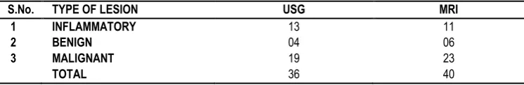
Table 3: No. of Lesions Detected On USG And MRI
S.No. TYPE OF LESION USG MRI
1 INFLAMMATORY 13 11
2 BENIGN 04 06
3 MALIGNANT 19 23
Table 4: Comparision With Follow Up/ Histopathological Findings
S.No. Type of lesion USG MRI FOLLOW UP
01 Inflammatory 13 11 12
02 Benign 04 06 06
03 Malignant 19 23 22
Total 36 40 40
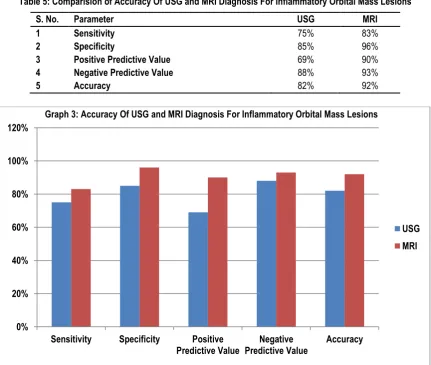
Table 5: Comparision of Accuracy Of USG and MRI Diagnosis For Inflammatory Orbital Mass Lesions
0 5 10 15 20 25
Inflammatory Benign Malignant
Graph 2: Comparision With Follow Up/ Histopathological Findings
USG MRI FOLLOW UP
0% 20% 40% 60% 80% 100% 120%
Sensitivity Specificity Positive
Predictive Value Predictive ValueNegative Accuracy Graph 3: Accuracy Of USG and MRI Diagnosis For Inflammatory Orbital Mass Lesions
USG MRI
S. No. Parameter USG MRI
1 Sensitivity 75% 83%
2 Specificity 85% 96%
3 Positive Predictive Value 69% 90%
4 Negative Predictive Value 88% 93%
Table 6: Comparision of Accuracy of USG and MRI Diagnosis For Benign Orbital Mass Lesions
Table 7: Comparision of Accuracy of USG and MRI Diagnosis For Malignant Orbital Mass Lesions 0%
20% 40% 60% 80% 100% 120%
Sensitivity Specificity Positive
Predictive Value Predictive ValueNegative Accuracy Graph 4: Accuracy of USG and MRI Diagnosis For Benign Orbital Mass Lesions
USG MRI
0% 10% 20% 30% 40% 50% 60% 70% 80% 90% 100%
Sensitivity Specificity Positive
Predictive Value Predictive ValueNegative Accuracy Graph 5: Accuracy of USG and MRI Diagnosis For Malignant Orbital Mass Lesions
USG MRI
S. No. Parameter USG MRI
1 Sensitivity 50% 83%
2 Specificity 97% 97%
3 Positive Predictive Value 69% 83%
4 Negative Predictive Value 88% 97%
5 Accuracy 90% 95%
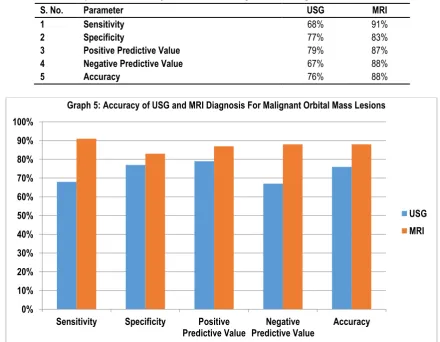
S. No. Parameter USG MRI
1 Sensitivity 68% 91%
2 Specificity 77% 83%
3 Positive Predictive Value 79% 87%
4 Negative Predictive Value 67% 88%
A.
AXIAL T1
B.
AXIAL T2
C.
CORONAL T1
D.
SAGITAL T2
E.
AXIAL DWI
F.
AXIAL ADC
Fig: 1. Axial T1WI (A) T2WI (B) and coronal T1WI (C) shows hypo intense enlarged muscle belly of the extra ocular muscle, sagittal T2WI shows sparing of the tendon of the muscles, DWI (E) and
A.
AXIAL T1
B.
AXIAL T2
C. CORONAL T2 FAT SAT
D. SAGITAL T2
E. AXIAL T2 FAT SAT
F. AXIAL ADC
Fig: 02. Axial T1WI (A) hypo intense, ill-defined solid mass with irregular margins with in the left eye ball, which on T2WI axial (B) and Sagittal (D) appears hyper intense, and on T2WI FAT SAT coronal (C) and
A. AXIAL T1
B. AXIAL T2
C. AXIAL FLAIR
D. AXIAL GRE
Fig: 03. The axial T1 WI(A) shows ill-defined iso intense mass lesion with underlying bone erosions and intracranial extension which is iso to hyper intense on T2WI (B), hypo intense on FLAIR and axial GRE (D) image shows areas of hypo intensities indicating areas of haemorrhage/ necrosis. (RHABDOMYOSARCOMA)
T1WI AXIAL
T2WI AXIAL
T2WI CORONAL
T2WI CORONAL
A.
T2WI AXIAL
B.
T2WI CORONAL
C.
T2WI SAGITAL
D.
DWI
Fig: 05. T2WI axial (A), coronal (B), and sagittal (C) images shows solid, ill-defined mass with irregular margins, in left orbit at super lateral compartment, appearing is intense to hyper intense, and DWI (D) demonstrates
hyper intensity at the lesion, depicting significant restriction of the diffusion. (ORBITAL LYMPHOMA)
A.
AXIAL T1
B.
AXIAL T2
C.
CORONAL T2
D.
AXIAL T2 FAT SAT
Fig: 07. Axial T1WI MR (A) image demonstrates well defined, isointense lesion in the right orbit, which is retro-orbital and intraconal ,the mass appears iso to intermediate intense on T2WI axial (B) and coronal (C) MR images and demonstratesencasement of the optic nerve, and appearsof intermediate intensity on Axial flair image (D).
(OPTIC MENINGIOMA)
A.T1WI AXIAL
B. T1WI CORONAL
A.
T2WI CORONAL
D. T2WI AXIAL
Fig: 08. Axial and coronal T1WI MR (A, B) image demonstrates well defined, isointense lesion in the right orbit, which is retro-orbital and intraconal ,the mass appears hyperintense on T2WI axial (D) and
DISCUSSION
Ultrasonography is usually the first imaging modality to detect an orbital mass lesion. It is widely available, uses no ionizing radiation, is relatively inexpensive and quick to perform and allows dynamic study, 1 but it is inferior in defining origin, extension, bony
involvement and characterizing the orbital masses, along with, it is operator dependent and could not be performed well in painful eye conditions. MRI has many advantages like high contrast resolution, the ability to obtain images in any plane, lack of ionizing radiation makes it a favored modality. Magnetic resonance imaging (MRI) is preferred for diagnosis of orbital masses as it helps in delineating extension and involvement of surrounding structures and grading of orbital masses. Lesion morphology, signal intensity, diffusion restriction pattern on DW MRI is taken into consideration when characterizing masses with MRI, and the data is evaluated together for the differentiation of benign and malignant lesions.
With this background, we attempted in our study to determine the role Of Magnetic Resonance Imaging in the identification and characterization of orbital mass lesions and delineation of extent of mass lesions. Then finally to correlate the MRI findings with sonographic and histopathological findings. The reference standard used in our study, consisted of histopathological confirmation by biopsy or FNAC and clinical follow up.
In the present study the most common age group of patients was 0-9 years (32.5%) with mean age of 26.6 years. The mean age of patients with benign lesions was 25.5 years and of patients with malignant lesions was 21.25 years. Majority of patients were females (23) constituting 57.5% of cases. This finding is in concordance with the study of N. Khandelwal and R.T. Nair et al.2
The most common presenting complaint with which patients presented was proptosis seen in 90% of cases followed by pain seen in 65% of cases. Diminution of vision /loss of vision and diplopia was seen in 45% of cases each. Similar clinical findings was seen in studies by Sarah N. Khan, MD; Ali R. Sepahdari et al.3
On USG thyroid orbitopathy appeared as extraocular intraconal , hypoechoic , ill defined , fusiform enlargement of the extraocular muscles with sparing of the muscle tendon, most common muscle enlargement was seen in medial rectus, and all of them showed vascularity on colour doppler study, depicting active inflammatory process. This was also seen by OP Sharma et al.1 On USG
pseudotumour appeared as extraocular intraconal , hypoechoic , ill-defined enlargement of extraocular muscle without sparing tendons, most commonly involving the lateral rectus, along with evidence hypoechoic, vascular , soft tissue infilterating the surrounding structures seen. This finding was in concordance with the studies of Dr. Hemang D. Chaudhari et al.4 One case (3%) of
cysticercosis was seen, which on USG appeared as extraocular intraconal, hypoechoic with hyperechoic content in it, cystic, well defined lesion with in the extraocular muscle. Similar findings were seen by Neelam Pushker, Mandeep S. Bajaj et al.5
Amongst the inflammatory orbital lesions detected on USG, most common lesions were thyroid orbitopathy and pseudotumor accounting for 17% cases of each. One case (3%) of orbital cysticercosis was also detected. Similar findings were seen by Shields JA, Shields CL, Scartozzi R. et al.6
On ultrasonography two cases of the cavernous haemangioma was diagnosed in our study. These appeared as extraocular
intraconal, retrobulbar, well defined, hypoechoic to hyperechoic, predominantly solid with few cystic areas with in it, in both of them minimal flow of venous spectral pattern was demonstrated on Doppler scanning. Two cases of optic nerve glioma were detected in our study by ultrasonographic examination. These appeared as retrobulbar (extraocular intraconal) well defined, hypoechoic, fusiform soft tissue mass encasing the optic nerve, without evidence of calcification with in it, characteristically defining the optic nerve tumor, more likely optic nerve glioma. Similar findings were seen by Neelam Pushker, Mandeep S. Bajaj et al.5
In our study on USG the most common malignant orbital and intraocular mass lesion detected was retinoblastoma accounting for 27% (10) of all orbital mass lesions in our study, followed by rhabdomyosarcoma (4), choroidal melanoma. One single case each of orbital lymphoma and orbital metastasis was detected. On ultrasonography, 32.5% (13) lesions were diagnosed as inflammatory lesions. There were 10% (4) benign lesions and 53% (19) malignant lesions in our study. Similar findings were also seen by Khandelwal et al.2
On MR Imaging , in Thyroid orbitopathy hypertrophied muscles bellies appeared isointense on T1 weighted images and iso to hyperintensity was seen on T2-weighted images depending on the activity of the disease with sparing of the tendinous insertion of the extraocular muscles, i.e. active Graves’ disease showed more hyperintense muscle on T2WI. There were 6 cases of Thyroid orbitopathy in our study most of them (4) were females of middle age group. Most of them did not show restriction of diffusion while 2 cases of Thyroid Orbitopathy showed restriction of diffusion, appearing bright on DWI. Simillar finding was also seen in study by Nader Roshdy, Maha Shahin and Hanem Kishk et al.7
Pseudotumour on MRI appeared as iso to hypo signal intensity on T1-weighted images and hypo signal intensity on T2-weighted images in affected muscle. Involvement of the tendinous insertion distinguishes it from thyroid associated orbitopathy (TAO) in which the insertion point is spared. Additional inflammations are seen in surrounding tissues. 40% of the pseudotumours on DW MRI showed areas of restricted diffusion that appear bright. This was also seen in study by Nader Roshdy, Maha Shahin and Hanem Kishk et al and M. N. Pakdaman et al.7,8
A case of cysticercosis was seen in our study. The MRI features of orbital cysticercosis was isointense on T1WI and hyperintense with hypointense scolex on T2 weighted images. The affected extraocular muscle (lateral rectus) showed fusiform enlargement of its belly and contained a well-defined, spherical cyst with a nodule attached to its wall. Clinical improvement with praziquintal seen on follow up. This finding was consistent with Sonam Angmo Bodh et al.9
Cavernous haemangioma was the most common benign lesion in our study in the orbit, constituting 7.5% of lesions. MRI appearance of lesion was well defined mostly intraconal , unilateral, solitary lesion causing mass effect, appeared isointense on T1WI and hyperintense on T2-weighted images with one of them showing hypointense pseudocapsule on T2WI. DWI MRI of the lesion showed restriction of the diffusion in two of three cases in our study. Simillar findings were also seen by Tina D. Tailor et al.10
nerve, diffuse involvement of the substance of the nerve differentiates optic nerve glioma from optic nerve sheath meningioma. DWI MRI showed significant restriction of the diffusion in both the cases. Simillar findings were also seen by Tina D. Tailor et al.10
Optic nerve meningioma on MRI appeared as isointense on T1WI and iso to hyperintense on T2WI, however, because the substance of the nerve is spared, a “tram-track” configuration is often observed at axial T2WI MR Images, doughnut sign was demonstrated in coronal T2WI MR Images. On DW MR Images lesion showed significant restriction of diffusion. Similar findings were also observed in study by Tina D. Tailor et al.10
On MRI, malignant lesions of the orbit constituted 57% (23 cases ) of lesions in our study, of which Retinoblastoma is the most common malignant neoplasm (25%). Retinoblastomas were found bilateral in 30% of cases in our study. Similar findings were also seen by Rolando Enrique D et al.11
On MRI Retinoblastoma approximates the signal of grey matter on MR imaging, with the tumor appearing hyperintense to vitreous on T1- weighted images and hypointense to vitreous on T2-weighted images and hypointense on FLAIR imaging, and SWI MR images showed drop out of signals with in it as blooming i.e. hypointensities within the lesion, indicating calcification and hemorrhage with significant restriction of diffusivity seen on DW MRI. Similar findings were also seen by Rolando Enrique D et al.11
Rhabdomyosarcoma was the second most common malignant intraorbital lesion seen in our study, with 12.5% (5) of the cases. On MR imaging, Rhabdomyosarcoma appeared isointense to muscle on T1-weighted images and hyperintense to muscle on T2-weighted images, most of them appearing isointense with cerebral cortex, with decreased diffusivity seen on DWMRI. One (2.5%) of them showed haemorrhage on SW MRI and one case showed intra cranial extension with erosion of underlying cranium bone. Simillar findings were seen by Lama Jurdy et al.12
Orbital lymphoma on MRI appeared characteristically homogeneous and isointense on T1WI and on T2-weighted MR images appeared isointense to hyperintense with white matter, showed significant restriction of diffusivity on DWI, most of the lesions involved mainly superior-lateral quadrant and the orbital structures inside. Simillar findings were seen by K. Haradome, H. Haradome, Y. Usui, et al.13
Choroid melanoma on MRI was depicted as mildly hyperintense on T1-weighted MR images and hypointense to vitreous on T2-weighted MR images, demonstrated significant restriction of diffusivity on DWI, seen arising from the choroid, with evidence of retinal detachment and sub retinal fluid/ blood which is seen hyperintense on T1WI. This finding correlates with the findings of Lemke AJ et al.14
A case of orbital metastasis was detected on MRI in our study, in a known case of breast carcinoma, in which ill defined, solid T1 isointense and T2 hyperintense lesion, showing significant restriction of diffusion with appearing hyperintense on DWI, in superolateral quadrant was seen. Similar findings were also seen by Panagiotis J. Vlachostergios et al.15
Amongst the malignant lesions detected on MRI in our study, retinoblastoma was the most common accounting for 25% of them, followed by rhabdomyosarcoma (15%), orbital lymphoma (7.5%) choroidal melanoma (7.5%) and a single case of orbital breast metastasis was detected. Similar findings were seen by N.
Khandelwal and R. T. Nair et al and Rolando Enrique, D Domingo et al.2,11
Free diffusion usually corresponded to benign lesions while restricted diffusion could be seen in both benign and malignant lesions. Benign lesions showing restricted diffusion were inflammatory mass lesions.On DWI lesions which restricted diffusion appeared hyperintense. In our study 10% inflammatory lesions, 13% benign lesions and 52% malignant lesions demonstrated restriction of diffusion. Similar findings were noted by A.R. Sepahdari, L.S. Politi et al.16
On MRI in our study, 23 (57%) of the cases were diagnosed to have malignant lesions, followed by11 (28%) inflammatory orbital mass lesions and 6(15%) benign orbital mass lesions. Similar findings were also Khandelwal et al and Sa-Ra Ro et al.2,17
On USG in our study we detected 13 inflammatory, 4 benign and 19 malignant lesions, while on MRI inflammatory lesions detected were 11, benign being 6 cases and 23 cases of malignant orbital mass lesions were detected. So it was seen that 10% more lesions were detected on MRI, which were found to be indeterminate on USG.
In our study final diagnosis was made with reference standard taken as medical, surgical and histopathological follow up. The final diagnosis of the malignant orbital lesions came out to be retinoblastoma 10 out of 40 cases, 5 cases of rhabdomyosarcoma seen, 3 cases each of orbital lymphoma and choroidal melanoma and a single case of orbital metastasis, i.e. a total of 22 cases were found to be malignant orbital mass lesions. And follow up study demonstrated 6 out of 40 benign orbital mass lesions including 3 cavernous haemangioma, 2 optic nerve glioma and a single case of optic nerve meningioma. 12 cases were diagnosed inflammatory orbital mass lesions including 6 cases of thyroid orbitopathy, 5 cases of orbital pseudotumours and a single case of orbital cysticercosis was diagnosed. Similar findings were seen by Shields JA, Shields CL, Scartozzi R. et al.6
In our study, inflammatory lesions were 32.5% on USG, 28% on MRI, and 30% on follow up. Benign lesions constituted 10% of the cases on USG, and 15% of cases on both MRI and follow up. Malignant lesions were 48% of the cases on USG, 58% on MRI, and 55% on follow up. Similar findings were seen by Shields JA, Shields CL, Scartozzi R. et al.6
The true positive and false negative for USG to predict inflammatory orbital lesions were 9 and 3. The false positives and true negatives were 4 and 24 respectively. The sensitivity and NPV of USG in predicting inflammatory lesions turned out to be 75% and 88% respectively. The specificity and PPV were 85% and 69% respectively. Similar findings were seen by OP sharma et al.1
The true positive and false negative for USG to predict benign orbital lesions were 3 and 3. The false positives and true negatives were 1 and 33 respectively. The sensitivity and NPV of USG in predicting benign lesion turned out to be 50% and 95% respectively. The specificity and PPV were 97% and 75% respectively. Similar findings were seen by OP sharma et al.1
The true positive and false negative for MRI to predict inflammatory orbital lesions were 10 and 2. The false positives and true negatives were 1 and 27 respectively. The sensitivity and NPV of MRI in predicting malignancy turned out to be 83% and 93% respectively. The specificity and PPV were 96% and 90% respectively. Similar findings were seen by Sa-Ra Ro et al.17
The true positive and false negative for MRI to predict benign orbital lesions were 5 and 1. The false positives and true negatives were 1 and 33 respectively. The sensitivity and NPV of MRI in predicting malignancy turned out to be 83% and 97% respectively. The specificity and PPV were 97% and 83% respectively. Similar findings were seen by Sa-Ra Ro et al.17
The true positive and false negative for MRI to predict malignant orbital lesions were 20 and 2. The false positives and true negatives were 3 and 15 respectively. The sensitivity and NPV of MRI in predicting malignancy turned out to be 91% and 88% respectively. The specificity and PPV were 83% and 87% respectively. Similar findings were seen by Pim de Graaf et al.18
Considering restricted diffusion as a marker for malignancy, we found high sensitivity (95%) and low specificity (50%).The positive predictive value was (70%) and the negative predictive Value was (90%), and accuracy was 75%. Hence nature of diffusion is a sensitive test for the detection of malignant lesions. Similar findings were also seen by A.R. Sepahdari, L.S. Politi et al.16
On comparing the accuracy of USG and MRI in detection of inflammatory orbital mass lesions, the sensitivity, specificity, PPV and NPV of USG was found to be 75%, 85%, 69% and 88% respectively, while the sensitivity, specificity, PPV and NPV of MRI was found to be 83%, 96%, 90% and 93% respectively.
On comparing the accuracy of USG and MRI in detection of benign orbital mass lesions, the sensitivity, specificity, PPV and NPV of USG was found to be 50%, 97%, 75% and 95% respectively, while the sensitivity, specificity, PPV and NPV of MRI was found to be 83%, 97%, 83% and 97% respectively.
On comparing the accuracy of USG and MRI in detection of malignant orbital mass lesions, the sensitivity, specificity, PPV and NPV of USG was found to be 68%, 77%, 79% and 67% respectively, while the sensitivity, specificity, PPV and NPV of MRI was found to be 91%, 83%, 87% and 88% respectively. Similar findings were seen by Rolando Enrique, D Domingo et al.11
The accurate identification of malignant orbital mass lesions is of immense clinical interest because of worse prognosis associated with high grade retinoblastomas, choroidal melanomas as compared to the benign lesions. It also helps in planning of management, as some lesions like lymphomas, melanomas and rhabdomyosarcoma are administered chemotherapy and for some orbital mass radiotherapy and surgical interventions can be done. Accurate diagnosis with extension of the orbital mass lesion helps in planning surgical intervention.
From this study, we found that, the sensitivity, specificity, PPV, NPV and overall accuracy of non-contrast MRI with diffusion weighted imaging in the identification and characterization of orbital lesions was highest as compared to MRI and USG alone. Good correlation existed between the study findings and the findings obtained on follow up.
CONCLUSION
This study aimed at differentiating orbital masses lesions based on the various imaging properties of the lesion and to correlate the
sensitivity and specificity of USG and MRI with histopathological diagnosis & clinical follow-up. We found that the sensitivity and specificity of MRI is higher than USG for detection of orbital mass lesions. And on MRI, DW imaging alone may lead to misdiagnosis of lesions due to inflammatory lesions showing restricted diffusion. Hence, MRI along with DWI is a very valuable noninvasive tool for the Identification, characterization and differentiation of orbital mass lesions.
REFERENCES
1. Sharma OP. Orbital sonography with it’sclinico-surgical correlation. Indian J Radiol Imaging 2005;15(4):537-54.
2. N. khandelwal, R. T. Nair et al. MRI in orbital mass lesion: comparison with US and CT. Journal of Neuroradiology; Vol 29, N° HS 1 - août 2002; pp. 44-47.
3. Khan SN, Sepahdari AR. Orbital masses: CT and MRI of common vascular lesions, benign tumors, and malignancies. Saudi Journal of Ophthalmology. 2012;26(4):373-383. doi:10.1016/j.sjopt.2012.08.001.
4. Chaudhari, H. D. et al. Role of Ultrasonography in evaluation of orbital lesions. Gujarat Medical Journal 2013, 2, 73-[A].
5. Pushker, N., Bajaj, M. S. and Betharia, S. M. Orbital and adnexal cysticercosis. Clinical & Experimental Ophthalmology 2002, 30: 322–333. doi:10.1046/j.1442-9071.2002.00550.x. 6. Shields JA, Shields CL, Scartozzi R. Survey of 1264 patients with orbital tumors and simulating lesions: The 2002 Montgomery lecture, part 1. Ophthalmology 2004;111:997-1008.
7. Roshdy N, Shahin M, Kishk H, El-Khouly S, Mousa A, Elsalekh I. Role of New Magnetic Resonance Imaging Modalities in Diagnosis of Orbital Masses: A Clinicopathologic Correlation. Middle East African Journal of Ophthalmology. 2010;17(2):175-179. doi:10.4103/0974-9233.63077.
8. Pakdaman MN, Sepahdari AR, Elkhamary SM. Orbital inflammatory disease: Pictorial review and differential diagnosis. World Journal of Radiology. 2014;6(4):106-115. doi:10.4329/wjr.v6.i4.106.
9. Bodh SA, Kamal S, Kumar S, Goel R, Nagpal S, Aditya KOrbital Cysticercosis. DJO 2012;23:99-103.
10. Tina D. Tailor, Divakar Gupta, Roberta W. Dalley, C. Dirk Keene, and Yoshimi Anzai. Orbital Neoplasms in Adults: Clinical, Radiologic, and Pathologic Review. RadioGraphics 2013; 33:6, 1739-1758.
11. Domingo, Rolando Enrique D, Lilibeth E Manganip, and Rolando M Castro. “Tumors of the Eye and Ocular Adnexa at the Philippine Eye Research Institute: A 10-Year Review.” Clinical Ophthalmology (Auckland, N.Z.) 9 (2015): 1239–1247. PMC. Web. 29 Sept. 2016.
12. Jurdy L, Merks JHM, Pieters BR, et al. Orbital rhabdomyosarcomas: A review.Saudi Journal of Ophthalmology. 2013;27(3):167-175. doi:10.1016/j.sjopt.2013.06.004.
13. Haradome K, Haradome H, Usui Y et al (2014) Orbital lymphoproliferative disorders (OLPDs): value of MR imaging for differentiating orbital lymphoma from benign OPLDs. AJNR Am J Neuroradiol 35:1976–1982
15. Vlachostergios PJ, Voutsadakis IA, Papandreou CN. Orbital Metastasis of Breast Carcinoma. Breast Cancer: Basic and Clinical Research. 2009;3:91-97.
16. Sepahdari AR, Politi LS, Aakalu VK, Kim HJ, Abdel Razek AAK. Diffusion-weighted imaging of orbital masses: Multi-institutional data support a 2-ADC threshold model to categorize lesions as benign, malignant or indeterminate. American Journal of Neuroradiology. 2014 Jan 1;35(1):170-175.
17. Sa-Ra Ro et al Multiparametric study of orbital mass, Vol-85, issue-2, page 324-336.
18. Pim de Graaf, Frederik Barkhof, Annette C. Moll, Saskia M. Imhof, Dirk L. Knol, Paul van der Valk, and Jonas A. Castelijns. Retinoblastoma: MR Imaging Parameters in Detection of Tumor Extent. Radiology, Apr 2005, Vol. 235:197–207, 10.1148/radiol.2351031301
[
Source of Support: Nil. Conflict of Interest: None Declared.
Copyright: © the author(s) and publisher. IJMRP is an official publication of Ibn Sina Academy of Medieval Medicine & Sciences, registered in 2001 under Indian Trusts Act, 1882. This is an open access article distributed under the terms of the Creative Commons Attribution Non-commercial License, which permits unrestricted non-commercial use, distribution, and reproduction in any medium, provided the original work is properly cited.