R E V I E W
Open Access
What have we learned from brucellosis in the
mouse model?
María-Jesús Grilló
1, José María Blasco
2, Jean Pierre Gorvel
3,4,5, Ignacio Moriyón
6,7and Edgardo Moreno
8,9*Abstract
Brucellosis is a zoonosis caused by Brucella species. Brucellosis research in natural hosts is often precluded by practical, economical and ethical reasons and mice are widely used. However, mice are not natural Brucella hosts and the course of murine brucellosis depends on bacterial strain virulence, dose and inoculation route as well as breed, genetic background, age, sex and physiological statu of mice. Therefore, meaningful experiments require a definition of these variables. Brucella spleen replication profiles are highly reproducible and course in four phases: i), onset or spleen colonization (first 48 h); ii), acute phase, from the third day to the time when bacteria reach maximal numbers; iii), chronic steady phase, where bacterial numbers plateaus; and iv), chronic declining phase, during which brucellae are eliminated. This pattern displays clear physiopathological signs and is sensitive to small virulence variations, making possible to assess attenuation when fully virulent bacteria are used as controls. Similarly, immunity studies using mice with known defects are possible. Mutations affecting INF-γ, TLR9, Myd88, Tγδand TNF-β favor Brucella replication; whereas IL-1β, IL-18, TLR4, TLR5, TLR2, NOD1, NOD2, GM-CSF, IL/17r, Rip2, TRIF, NK or Nramp1 deficiencies have no noticeable effects. Splenomegaly development is also useful: it correlates with IFN-γand IL-12 levels and with Brucella strain virulence. The genetic background is also important: Brucella-resistant mice (C57BL) yield lower splenic bacterial replication and less splenomegaly than susceptible breeds. When inoculum is increased, a saturating dose above which bacterial numbers per organ do not augment, is reached. Unlike many gram-negative bacteria, lethal doses are large (≥108bacteria/mouse) and normally higher than the saturating dose. Persistence is a useful virulence/attenuation index and is used in vaccine (Residual Virulence) quality control. Vaccine candidates are also often tested in mice by determining splenic Brucella numbers after challenging with appropriate virulent brucellae doses at precise post-vaccination times. Since most live or killed Brucella vaccines provide some protection in mice, controls immunized with reference vaccines (S19 or Rev1) are critical. Finally, mice have been successfully used to evaluate brucellosis therapies. It is concluded that, when used properly, the mouse is a valuable brucellosis model.
* Correspondence:[email protected]
8
Programa de Investigación en Enfermedades Tropicales, Escuela de Medicina Veterinaria, Universidad Nacional, Heredia, Costa Rica
9
Instituto Clodomiro Picado, Facultad de Microbiología, Universidad de Costa Rica, San José, Costa Rica
Full list of author information is available at the end of the article
Table of content
Introduction Infection models
TheBrucellastains: replication patterns and related effects
Route of the infection Infective dose The mouse
Resistant and susceptible mouse strains Mutant and knockout mice
Age and sex Transmission Physiopathology
Onset of the infection Acute phase
Acute phase in pregnant mice Chronic steady phase
Chronic declining phase Vaccination
Superinfection and antigen therapy Passive transfer and immunomodulation Antibiotic treatment
Concluding remarks Endnotes
Competing interests Authors’contributions Acknowledgments References
Introduction
The genus Brucella comprises at least eight species named according to their preferred mammal hosts. Bru-cella melitensis, Brucella abortus and Brucella suis are the most economically important species and they pferentially infect goats and sheep, bovines and swine, re-spectively [1]. Livestock is the source of human infections, and brucellosis is a severe disease that affects a considerable number of people in the world [1]. These bacteria cause long lasting chronic infections, mainly col-onizing the reticuloendothelial system and reproductive organs [2,3], replicating in the internal milieu of tropho-blasts, macrophages and dendritic cells [4]. Although able to multiply in life-less media, Brucella organisms are better described as facultative extracellular intracellu-lar parasites [5].
For many years the pathophysiology of brucellosis was investigated in humans and natural hosts [3,6-9]. How-ever, experimentation in ruminants, humans and pri-mates has economical and ethical concerns or is precluded for practical reasons. Consequently, small la-boratory animals are frequently employed as models in brucellosis research. One of the first experimental
models was the chicken embryo [10]. Although this model was useful for evaluating the intracellular multi-plication of Brucella, it does not differentiate virulent from attenuated strains. The rabbit has been used in pro-tocols designed to studyBrucella toxicity and hypersen-sitivity, mainly because of its susceptibility to bacterial endotoxins and toxins [11]. Due to practical reasons related to size, management and cost, the rabbit has never been widely used as a model in brucellosis, although it is used to produce antibodies againstBrucella antigens [12]. Owing to their high susceptibility to Brucella infections and similarities in reproducing human pathology, the guinea pig was extensively used as an experimental animal [13]. These rodents reproduce the pulmonary, hepatic, spleen and genital lesions and the hypersensitivity reactions observed in humans, and match the different phases of the infection caused by Brucella in natural hosts, including abortion [13-15]. Thus, the guinea pig is one of the best models and it is still used for some immunological and vaccine studies [16,17]. However, when large numbers of animals are ne-cessary, guinea pigs become impractical for the same reasons as rabbits. Other laboratory rodents such as rats, hamsters and gerbils have been used sporadically [13].
The mouse (Mus musculus) has been the most widely used brucellosis model. Mice were first used by Holth in 1911 for Brucella vaccine testing. Thereafter, mice were used for the etiological confirmation of samples from infected animals, to test virulence and for the evaluation of the pathological lesions (see [18,19]). The results in mice are not immediately applicable and transferable to humans or to the target animal species. However, the uncovering of a significant phenotype in mice using an appropriate protocol gives useful information. With the arrival of inbred, mutant, knockout and transgenic mice and the understanding of their biology and immunology [20,21], this rodent has become the standard model for brucellosis research.
In this work we have reviewed the models of infection by O-polysaccharide containing (smooth) Brucella spe-cies and strains in mice. Although we have focused on in vivo assays, we occasionally refer to ex vivo studies in cells when they are relevant for understanding the biol-ogy of smooth brucellae in mice. For reasons related to the extent of the document, we have avoided reviewing the infection models induced by the rough brucellae. For a better understanding on the biological behavior of the rough strains in mice, we suggest the work of González et al. [22].
Infection models
sex and physiological status of the mice. Since most studies in mice are devoted to investigate virulence, pathogenicity, immunology and vaccine properties, it is critical to include control groups inoculated with appro-priate referenceBrucella strains [23,24]. In the following sections, we discuss the importance of these factors.
TheBrucellastrains: replication patterns and related effects
The affinity of some Brucella species for a particular mammal host is well-known [1]. It is also notorious that B. melitensis and some biovars of B. suis infect humans more frequently and cause a more severe disease thanB. abortus [1]; though, these infectivity and virulence pat-terns do not always reproduce in mice. For instance, in a comparative study it was found that the order of viru-lence in mice wasB. melitensisH38 (biovar 1)>B. abor-tus 2308 (biovar 1)>B. suis 1330 (biovar 1) [25]. Moreover, there are differences in the pathological be-havior among biovars and strains of some virulent spe-cies, and the virulence demonstrated for their target hosts does not necessarily parallels that observed in mice [25,26]. The appraisal of these issues becomes more complicated when different Brucella strains are simul-taneously inoculated for comparative purposes. In addition to the eight recognized Brucella species and several biotypes (and more in the “waiting list”), a large number of bacterial mutants and constructs have been developed and tested in the murine system. In general, three different categories of Brucella strains may be dis-tinguished in relation to pathogenicity, ability to multiply and persist in mice: virulent, attenuated and avirulent.
Although most organs of the reticuloendothelial sys-tem may become infected (depending upon the bacterial dose), the spleen and liver are the most conspicuously infected organs in mice. In these organs, virulent smooth brucellae (e.g. B. abortus 2308 and 544, B. melitensis 16 M and H38, and B. suis 1330) show a very reprodu-cible pattern that is clearly different from that of the attenuated vaccines and the non-virulent brucellae. In the initial phases, the numbers of CFU/organ are similar in the liver and in the spleen, but the number of CFU/g of organ is lower in liver (one to two logarithms). In sub-sequent phases, the CFU are consistently lower in the liver [27,28] and virulent brucellae are completely cleared from this organ beyond 3–4 weeks post-infection (pi) [29]. This pattern is somewhat different from that observed in spleens (see Physiopathology). In splenecto-mized mice, the liver becomes rapidly colonized [15]. It seems therefore, that the higherBrucella colonization of the spleen during the chronic phases guards the liver from a profound inflammation [15]. Due to this, liver is seldom used for estimating the number of CFU, and the
spleen is the preferred target organ to study Brucella infections in mice.
The enumeration of bacteria in spleen (expressed as the mean ± SD of individual Log10 CFU/spleen or Log10
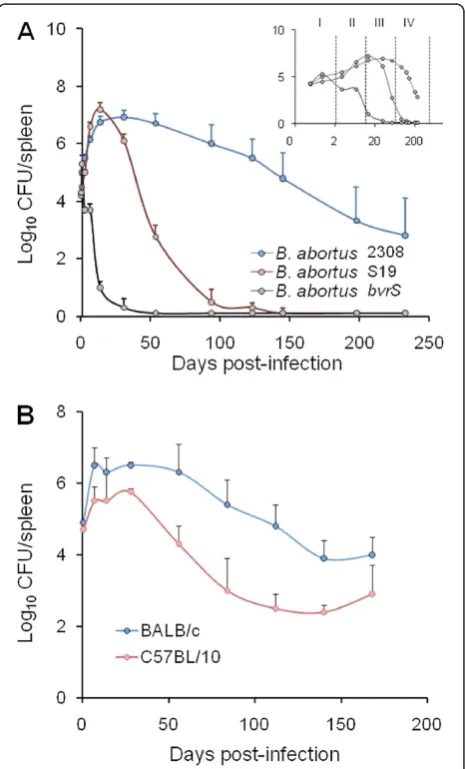
CFU/g of spleen; Figure 1) provides highly reproducible replication profiles. The lapse of the different infection phases (Figure 1) may vary depending upon the inocula-tion protocol. Nevertheless, at the optimal dose of infec-tion (see below) a consistent replicainfec-tion profile for the
Figure 1Replication profiles ofB. abortusin mice spleens.(A)
reference Brucella strains is maintained within a certain range [29]. In the understanding that infection with viru-lentBrucella(at standard doses, see below) is a continu-ous process and that delimitations are not clear cut, this replication profile can be divided into four different phases (Figure 1A): i) the onset of the infection, marked by colonization during the first 48 h pi; ii) the acute phase, extending from the 3rdday to the time when CFU reach their maximum, generally between weeks 2 and 3; iii) the chronic steady phase, that corresponds to the CFU plateau, commonly lasting 8–11 weeks; and iv) the chronic declining phase, at which there is a slow elimin-ation of the bacteria that may last beyond 36 weeks. The span of these phases may vary depending upon the bac-terial dose, route, mouse strain and age [30]. Generally, experiments in mice are not prolonged more than 3– 4 months and, therefore, data on theBrucellapersistence in these animals after this period are scarce. It has been documented that Brucella organisms may still be recov-ered from the spleen and lymph nodes of mice after 6 months of infection [25,31], suggesting that virulent Brucella might remain in mice for life. Although some quantitative variations have been observed, the spleen replication patterns of virulent strains follow similar kin-etics [24,25]. Fully avirulent strains (e.g. B. abortus bvrS mutant; Figure 1A) are unable to multiply or persist, re-gardless of the dose. In contrast, attenuated strains (e.g. S19 and Rev1) can multiply at the levels of the virulent strains at the early phases (Figure 1A) but persist for shorter times, even when inoculated at large doses (e.g. 108 CFU/animal) [31]. The degree to which attenuated bacteria are able to persist is the basis of the recom-mended Residual Virulence quality control of anti-Bru-cellavaccines [16]. It is expressed as the Recovery Time 50 (RT50), i.e. the time (in weeks) at which the bacter-ium is eliminated from the spleens in half of vaccinated mice. Used in this way, the murine model has demon-strated its usefulness to detect batches of poor immuno-genic reference vaccines [32-34].
The course ofBrucellainfections induced by attenuated strains, such as vaccines S19 and Rev1, or non-virulent mutants, such as VirB or BvrS [36,37], is radically shor-tened and modified (Figure 1A). Similarly, the replication profiles in knockout or mutant mice may vary according to the defect displayed by the corresponding mouse strain (Table 1). In the case of vaccine S19 (which shows a con-spicuous Residual Virulence), the replication kinetics in the spleen follows a rapid increase that peaks between weeks 1 and 3, and then decreases steadily at approxi-mately one logarithm per week [23,24]. Nonetheless, this vaccine may still be recovered from spleens 8 to 12 weeks pi. The replication profile of vaccine Rev1 shows some differences with respect to that of S19. In general, Rev1 does not display the rapid increase demonstrated by S19,
declines more slowly, and is still present after 8 to 12 weeks [33]. As stated above, non-virulent strains do not multiply or increase in the initial phases of the infec-tion, and then decline very fast (Figure 1A) being elimi-nated from the spleen and the liver between the 2nd and 7thweek [37,38].
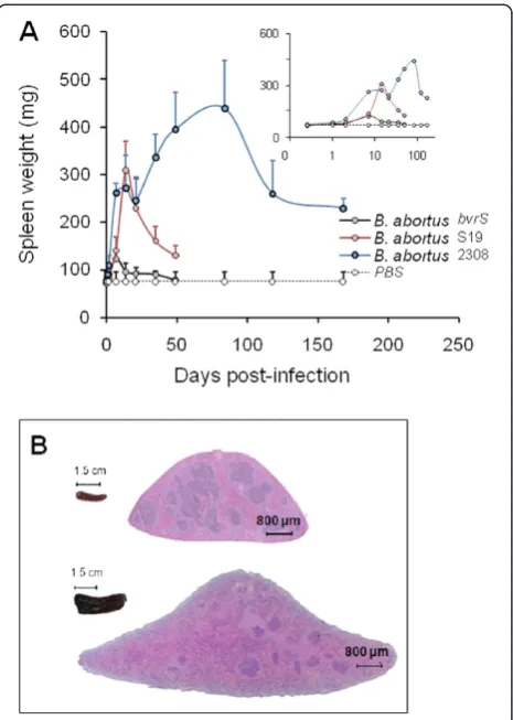
Simultaneously with bacterial replication, there is swel-ling of the spleen (Figure 2) and liver as well as draining through lymphatics from the site of the infection (see Physiopathology). The enlargement of the spleen is char-acterized by a weight peak (e.g.≥ 400 mg) evident from weeks 3 to 16 (Figure 2A). This enlargement is a conse-quence of inflammation [25] and it depends on the Bru-cella dose and virulence [64], as well as on the immune status and genetic background of the mice [50,53,65-67] (see Physiopathology). For example, B. melitensis H38 induces an intense splenic enlargement that generally surpasses that induced by other strains such as B. meli-tensis 16 M, B. suis 1330 or B. abortus 544. Attenuated S19 vaccine induces a characteristic peak of splenomeg-aly that occurs close to 2 weeks after inoculation and that, depending on the dose, may exceed that ofB. abor-tus2308 or 544. However, while in S19 infected mice the weight of the spleen rapidly decreases, the spleen of mice inoculated with virulent Brucella keeps increasing up to the end of the chronic steady phase (Figure 1B). Finally, killed Brucella or non-virulent strains (e.g. B. abortus BvrR/BvrS or VirB mutants) barely cause an increase in spleen size, even when injected in very large quantities (e.g. > 5 × 10⁷/mouse) [68]. This corresponds to a gen-eral phenomenon linked to the rapid removal and killing of non-virulent strains by professional phagocytes.
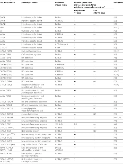
Table 1 Replication and persistence of smooth virulentBrucellain mutant and knockout mice strains
Test mouse strain Phenotypic defect Reference
mouse strain
Brucellaspleen CFU
increase and persistence
relative to mouse reference straina
References
Early before 14 days
Late after 15 days
CBA/H Inbred no specific defect BALB/c # " [39]
CBA/H Inbred no specific defect C57BL/10 " " [39]
CBA/NJ Inbred no specific defect BALB/c ↔ ↔ [40]
DBA2 Inbred no specific defect C57BL/6 " " [41,42]
CD-1 Outbreed Swiss mice BALB/c ↔ ↔ [40]
BALB/c Inbred no specific defect C3H/HeN ↔ ↔ [40]
BALB/c Inbred no specific defect C57BL/6 " " [39]
BALB/c Inbred no specific defect C57BL/10 " " [39]
BALB/c Inbred no specific defect C.CB (Nramp1r) # ↔ [43]
C57BL/10 Inbred no specific defect B10Br ↔ ↔ [39]
C57BL/6 (TLR9) CpG motifs recognition C57BL/6 " " [44,45]
BALB/c (TLR9) CpG motifs recognition BALB/c ↔ ↔ [46]
BALB/c (TLR4) LPS detection BALB/c ↔ ↔ [46]
BALB/c (TLR4) LPS detection BALB/c " ↔ [44]
C3H/HeJ (TLR4) LPS detection C3H/HePas " " [47]
C3H/HeJ (TLR4) LPS detection C3H/HeAu ↔ ↔ [48]
C3H/HeJ (TLR4) LPS detection C3H/Heb ↔ ↔ [41,42]
C3H/HeJ (TLR4) LPS detection C3H/HeN ↔ ↔ [40,49]
C3H/HeJ (TLR4) LPS detection BALB/c ↔ ↔ [40]
C57BL/6 (TLR4) LPS detection C57BL/6 ↔ ↔ [50]
C57BL/6 (TLR2) Lipoproteins detection and peptidoglycan detection
C57BL/6 ↔ ↔ [47,50]
BALB/c (TLR2) Lipoproteins detection and peptidoglycan detection
BALB/c ↔ " [46]
BALB/c (TLR2) Lipoproteins detection and peptidoglycan detection
BALB/c ↔ ↔ [44]
C57BL/6 (TLR2/4) LPS and lipoproteins detection C57BL/6 ↔ ↔ [50]
BALB/c (TLR2/4) LPS and lipoproteins detection BALB/c " ↔ [44]
C57BL/6 (NOD1) muramyl peptides meso-diaminopimelic acid
C57BL/6 ↔ ND [51]
C57BL/6 (NOD2) muramyl dipeptides C57BL/6 ↔ ND [51]
C57BL/6 (Myd88) Low proinflammatory response C57BL/6 " " [44,45,50]
C57BL/6 (TRIF) Low proinflammatory response C57BL/6 ↔ ↔ [44]
C57BL/6 (TRIF) Low proinflammatory response 129 Sv/Ev ND ↔ [44]
C57BL/10 (IRAK-4) Low proinflammatory response C57BL/6 " ↔ [52]
C57BL/6 (Rip2) NOD adaptor protein C57BL/6 ↔ ND [51]
C57BL/6 (gp91phox) Low respiratory burst in phagocytes C57BL/6 ↔ " [53]
C57BL/6 (iNOS) Low respiratory burst in phagocyte C57BL/6 " " [44]
C57BL/6 (iNOS) Low respiratory burst in phagocytes C57BL/6 ↔ " [53]
C57BL/6 (IL-12p40) Early differentiation of Th1 cells C57BL/6 ↔ " [53]
DBA/2j xC57BL/6 (iNOS/IL-12p40)
Early differentiation of Th1 cells and low respiratory burst
DBA/2j
xC57BL/6 ↔ "
[53]
DBA/2 J (ICSBP) Deficient in IL-12p40 and low respiratory burst
C57BL/6 " " [53]
C57BL/6 xDBA/2 J (iNOS/ICSBP)
Deficient in IL-12p40 and low respiratory burst
(like the avirulent VirB or BvrS-BvrR mutants), it is either attenuated or non-virulent. Alternatively, the macrophages may have been activated (e.g. endotoxin in the culture media), increasing their bactericidal abilities [66]. Once the Brucella strain has passed this test, the stored aliquots of the same stock shall be expanded and assayed in mice. In-deed, it may happen that some strains showing the ad-equate profile in macrophages fail to display a virulent
profile in mice and are thus attenuated. This is the case of some mutants that, nevertheless, pass the multiplication test in macrophages [22,71]. One alternative procedure when hesitating about the quality of aBrucella strain of known virulence is to inoculate the bacterium into mice and then recover the organisms from the spleen after 2 to 3 weeks [37,72]. Once selection of the virulent phenotype has been ensured, the isolate has to be handled as described above,
Table 1 Replication and persistence of smooth virulentBrucellain mutant and knockout mice strains(Continued)
C57BL/6 (IRF-2) Deficient in NK cells and dysregulation of IL-12p40
C57BL/6 # ↔ [53]
C57BL/6 (igh6) Affects B cells C57BL/6 ↔ ↔/# [54]
C57BL/6 (igh6) Affects B cells C57BL/6 # # [55]
BALB/c (jh) Affects B cells BALB/c # # [55]
C57BL/6 (rag-1) Affects B and T cells C57BL/6 ↔ ↔ [54]
C57BL/6 (Cd4) Affects CD4 T cells C57BL/6 ↔ # [54]
C57BL/6 (Aβ) Affects CD4 T cells C57BL/6 # ↔ [56]
C57BL/6 (Aβ) Affects CD4 T cells C57BL/6 ↔ ↔ [55]
C57BL/6 (Pfp) Affects Restriction of Tc mediated killing
C57BL/6 " ↔ [57]
C57BL/6 (β2m) Affects CD8 T cells C57BL/6 " " [56]
C57BL/6 (β2m) Affects CD8 T cells C57BL/6 ↔ ↔ [54]
C57BL/6 (β2m) Affects CD8 T cells C57BL/6 " ↔ [57]
C57BL/6 (β2m) Affects CD8 T cells C57BL/6 ↔ # [55]
C57BL/6 (IL12/β2m) Affects CD8 T cells and early differentiation of Th1 cells
C57BL/6 " " [58]
C57BL/6 (nu/nu) Lack thymus derived T cells C57BL/6 # " [59]
BALB/c (ifng) Absence of IFN-γ BALB/c ↔ "Dead [57]
C57BL/6 (ifng) Absence of IFN-γproduction C57BL/6 " " [44,54,58]
C57BL/6 (ifng) Absence of IFN-γproduction C57BL/6 ↔ "Dead [57]
C57BL/6 (ifng) Absence of IFN-γproduction C57BL/6 ↔ " [55]
C57BL/6 (ifng) Absence of IFN-γproduction C57BL/6 " ND [60]
C57BL/6 (IRF-1) CD8 T and NK cells dysregulation of IL-12p40 low respiratory burst
C57BL/6 " "Dead [53]
BALB/c (infar1) More susceptible to viral Infections. Affects NK
BALB/c ↔ ↔ [61]
C57BL/6 (INFαβR) More susceptible to viral Infections. Affects NK
129 Sv/Ev ND # [62]
C57BL/6 (TCRδ) Absence of Tγδcells C57BL/6 " ↔ [60]
C57BL/6 (GM-CSF) Higher lung infection
Defective alveolar macrophages
C57BL/6 ↔ ↔ [60]
C57BL/6 (IL-17Rα) Defective in PMN recruitment and PMN activity
C57BL/6 ↔ ND [60]
C57BL/6 (IL-1β) Low proinflammatory response C57BL/6 ↔ ↔ [50]
C57BL/6 (IL-18) Early activation of NK and Th1 cells
C57BL/6 ↔ ↔ [50]
C57BL/6 (IL-1βx IL-18) Low proinflammatory response and recruitment of NK and Th1 cells
C57BL/6 ↔ ↔ [50]
C57BL/10 (TNF-α) Low proinflammatory response C57BL/10 " ND [63]
Brucellasusceptibility order: CBA/H>CBA/NJ=CD-1=BALB/c=C3H/HeN=C3H/HeJ>C57BL/10=C57BL/6≥B10Br. a
since someBrucellarough variants may arise in the spleens of infected mice [73].
Attenuated and non-virulentBrucella mutants present a particularly difficult problem because stocks of these strains in different laboratories frequently come from serial pas-sages in vitro. This problem is exemplified by the differ-ences in Residual Virulence found for various lots of vaccines [16,32,33]. A drawback of vaccine Rev1 is its ten-dency to dissociate from smooth to rough organisms. This event has a profound negative effect on the immunogenicity and Residual Virulence and efficacy, since rough bacteria are highly attenuated [32,33]. The simultaneous presence of
large and small colonies (evidenced only after 4 days cul-ture) is a common change observed in S19 vaccines stocks that may be related to virulence differences in mice [33]. Similarly, when making mutants by genetic manipulation, a number of passages (frequently in the presence of antibio-tics) is necessary, and these steps bias the selection in favor of bacteria that grow preferentially in vitro and that may introduce further attenuation not associated with the spe-cific genetic defect studied. Therefore, it is always recom-mended to balance these studies with the use of complemented mutant strains, even if this method does not necessarily restore the levels of the original in vivo behavior [37].
The above-described Brucella replication profiles can be expressed either as log10CFU/organ or as log10CFU/g
of organ. However, in some cases the CFU/organ may give statistical significant differences, whereas the normalized CFU/g of organ values may erase this statistical signifi-cance. Expressing the CFU/gram, while correcting for individual variations due to inflammatory responses, elimi-nates information on the absolute bacterial numbers recovered, and then the CFU values are lower. It is thus better to express the CFU per organ. One concern about the latter method is that it assumes that all mice have closely similar spleen weights. If deemed necessary be-cause there are large differences in spleen weights, it is possible to include the individual spleen weights and CFU/ organ in two“dot” graphics. In most cases, however, the overall significance does not change.
Route of the infection
Mice are commonly infected intraperitoneally (i.p.) or intravenously (i.v.) with doses ranging from 104 to 107 CFU/mouse in a volume of 0.2 to 0.05 mL. Both routes infect 100 % of the mice and induce similar levels of infection. However, the i.p. route is preferred: it is technically simpler, admits larger volumes and, therefore, it is less prone to inoculation errors. The i.p. route results in higher bacterial counts and faster colonization of the spleen than other organs [39,65,74]. On the other hand, the i.v. route (commonly in the tail vein) promotes a slightly faster and higher bacterial colonization of the liver in relation to the spleen during the first 10 days, an event that is reversed during the following 2 weeks and then maintained throughout the infection period, up to 120 days or more [29]. Smooth brucellae are quickly phagocytized by leukocytes following i.v. inoculation. The same phenomenon is observed after i.p. infection but with 1 to 3 h delay [14], suggesting that the bacteria promptly reach the blood via the thoracic duct and prob-ably through the peritoneal capillary system as well. No matter if inoculated i.p. or i.v., bacteria are distributed throughout the reticuloendothelial system and placenta within the 1stweek, and depending on the dose, they can
Figure 2Spleen inflammation in mice infected withB. abortus.
also be isolated in testes, joints and salivary glands [25,75,76]. The central nervous system of the adult nor-mal mouse does not seem to be colonized when using these routes, even at high doses [76]. Although strict ex-perimentation concerning the presence of the bacterium in the meninges has not been performed, the behavior of infected mice does not suggest brain infection. It is strik-ing that in contrast to what has been reported in humans, dolphins and the fetuses of ungulates [77-79], there are no reports on neurobrucellosis in other juvenile or adult natural Brucella hosts, such as bovine, caprine or ovine. However, since domestic animals are most often culled upon evidence of the disease, this is an as-pect that has not been studied in all its dimensions. Neu-robrucellosis is quite an interesting syndrome because the hematoencephalic barrier imposes several unique conditions to the invading pathogen.
The subcutaneous (s.c.) inoculation, either in the back zone or in the footpad of mice results in lower levels of infection than the i.p. or i.v. routes [26,74,80,81]. This ef-fect may be due to local recruitment of bacteria at the site of inoculation. The s.c. route in the back zone is recommended for quality control of vaccines [16]. Inocu-lation of Brucella suspensions (e.g. 105 to 106 CFU/ mouse in 0.05 mL) into the footpad causes local inflam-mation; enhanced by the relatively large volume depos-ited in a small area that induces tissue destruction and the subsequent phagocytosis by resident leukocytes. After footpad injection a spreading of theBrucella infec-tion takes place through the lymphatics, favoring the localization of the bacterium in the popliteal lymph node [75,82-84]. At about 1 h pi, brucellae are already detected in blood, spleen and liver, reaching a transient plateau in these organs 6 h later [83,84]. Then, they can extend to other organs [74]. The s.c. infection in the back zone (e.g. 105–109 CFU/mouse) follows a similar course as the footpad inoculation [74]. The s.c. inocula-tion of B. abortus or B. melitensis rarely induces pus. Nevertheless, depending on the bacterial dose and the volume injected, a local granuloma formed by mono-nuclear cells and neutrophils appears in the inoculation site after several days. Vaccine Rev1 can induce encapsu-lated local transient abscesses when inocuencapsu-lated by the s. c. route. However, this only happens at very high doses (≥ 108 CFU) and abscesses are of benign nature being resolved in a few days/weeks [22], in parallel with what it is observed in sheep inoculated by s.c. route with the same strain [85].
The respiratory route of infection (through aerosols or intranasal) has been considered by some authors as the most natural route, and a source of laboratory accidents and of potential bio-terrorism attacks [86,87]. Leaving aside the inherent risks of this procedure, a precise as-sessment of the CFU inoculated is more inexact than
when using other routes. The aerosol method requires estimating the bacterial inocula within a respiratory chamber by sacrificing a group of mice immediately after exposure to the aerosol [86,87]. Moreover, simultaneous infection trough conjunctival, nasal and oral mucosae cannot be excluded during aerosol exposure, with the ensuing problems in interpretation. The intranasal route of infection displays similar problems because mice are very good at sneezing, generating local aerosols. In any case, these routes of inoculation induce an immediate in-fection of the lungs, which is then distributed by blood to the reticuloendothelial system [86-88]. Early in lung infections, Brucella is present and replicates in alveolar macrophages. Then, bacteria are disseminated to the lung-draining mediastinal lymph nodes where they repli-cate in both migratory dendritic cells and migratory al-veolar macrophages. These last phagocytic cells seem to be critical regulators of the early innate immune re-sponse within the lungs [4].
nodes steadily increase during the first 2 weeks, indi-cating the high affinity of Brucella for the reticuloen-dothelial system. A proportion of mice do not show bacteria in the main target organs [91,92], consistent with the idea that Brucella infection through the gastrointestinal route is unfavorable. As an alternative to gavage, large numbers of Brucella have been injected into intestinal loops, favoring the internalization of signifi-cant bacterial numbers by ileal Peyer´s patches dendritic cells [95]. This procedure induces little inflammatory response.
Infective dose
A myriad of bacterial doses have been used in experi-mental murine brucellosis. Indeed, it is essential to deter-mine the dose retrospectively by plating aliquots of the inoculum [33,68]. To know how many live bacteria have been inoculated is critical because, whereas live brucellae predominantly induce a Th1 response [66], dead brucel-lae have a tendency to induce T-independent responses [96]. This precaution is even more relevant when differ-entBrucellastrains are compared.
Brucella hardly induces mortality in mice and, therefore, it is not commonly used as a criterion of virulence. Singer-Brooks [19] performed a comprehensive study on the effects ofBrucelladose in mice. She observed that a larger proportion of mice succumbed after i.p. injections of massive doses of virulent smoothBrucella(>4 × 10⁸CFU/ mouse), displaying obvious clinical signs. On the contrary, doses lower than 107 CFU/mouse did not induce death or relevant clinical symptoms. Nevertheless, these non-lethal doses induced necrotic areas in the liver and spleen enlarge-ment within the first 3 weeks pi. Perusal of the literature shows that these observations have been repeatedly confirmed.
The optimal dose of Brucella infection is defined as the lower number of bacteria that infects the spleen of all mice (between 20–23 grams) at consistent significant levels [97]. This optimal dose varies depending on the bacterial strain and route of infection as well as on the genetic background and physiological status of the mice. The optimal dose has been determined only for classical brucellae [25,35,75,81] as well as for some bacterial constructs [22,98]. When inocu-lated at low doses (<103CFU/mouse),Brucellainduces in-consistent infections in mice, generating wide standard deviations in CFU that complicate statistical interpretations. Doses lower than 103 CFU/mouse do not produce gross anatomical changes, despite the fact that some mice show bacteria in several organs and tissues during the first weeks [19]. In contrast, very highBrucella inocula (> 107 CFU/ mouse) cause saturation of the spleen, to the point that the number of CFU per organ does not increase with respect to the optimal dose. Although these large doses induce a noticeable enlargement of the target organs (e.g. spleen and
liver), distribution of Brucella in the reticuloendothelial system barely changes [29]; however, other organs may be also invaded [76]. If the mouse survives, the reduction of Brucella numbers follows its course albeit with a more protracted elimination time. B. abortus 2308 at i.p. doses between 5 × 10⁸and 109CFU/mouse kill almost 50 % of the mice after 48 h, and 100 % before 1 week [48]. At these large doses, clinical symptoms such tachypnea, lethargy, piloerection, dehydration, and prostration were observed as early as 8 h pi. Since larger doses of killedBrucellaare non-lethal, these symptoms relate to the massive organ invasion by live bacteria [48]. High numbers of attenuatedBrucella (e.g.>5 × 10⁸CFU),such as S19 or Rev1, seldom kill mice, although they may induce some clinical symptoms [22].
A relevant effect observed when inoculating large Brucella doses (e.g. > 5 × 10⁷ CFU/mouse) is that blood and cytokine profiles approach those induced by endo-toxic bacteria like Salmonella[48]. This is an indication that Brucella neither induces an obvious inhibitory ac-tion on immune cells nor hampers the synthesis of proinflammatory cytokines at the onset of the infection, and that there is a threshold over which some molecules carrying pathogen-associated molecular patterns may be available to innate immunity receptors.
The mouse
Mice are highly resistant to brucellosis because they are only killed by very large doses of virulent Brucella [19,48]. However, mice seem more sensitive to brucel-losis than rats, hamsters and rabbits [13,99]. Mice infected with doses of virulent Brucella (e.g. B. abortus 2308) lower than 107 CFU/mouse, hardly show any changes in behavior, or cachexia or wasting syndromes, all signs induced by endotoxic bacteria [19,48]. Never-theless, and depending on the mouse strain,Brucellacan cause long lasting infections that may extend throughout the lifespan, accompanied by characteristic pathological signs.
Resistant and susceptible mouse strains
different levels of splenic colonization one week after in-oculation as well as dissimilar Th1 responses [103]. The spleen and liver CFU during the plateau phase (1 to 10 weeks pi) are commonly about ten-fold lower in the resistant C57BL strains (Figure 1B). In addition, the C57BL mice show less splenomegaly. The central dif-ference between resistant and susceptible mouse strains seems to be the inability of the latter to maintain the production of IFN-γ after the acute phase, a phenomenon that extends throughout the chronic steady phase up to at least the 6th week p.i. [65,103]. As expected, this phenomenon is redundant to other events, such as the recruitment and activa-tion of immune cells [66,103]. A conspicuous differ-ence between the BALB/c and C57BL/6 strains is the lymphocyte/granulocyte proportion in blood, which is close to 80/15 % in the former and to 90/9 % in the latter [20], a phenotype that may be related to Brucella clearance due to the regulatory action that neutrophils can display over macrophages [4].
B. melitensis replicates similarly in macrophages obtained from BALB/c and C57BL mice. Moreover, both strains of mice share the “sensitive” form (Nramp1s) of the Nramp1r (natural resistance-asso-ciated macrophage protein) allele, which codes for a membrane phospho-glycoprotein implicated in the early activation of macrophages [43]. Unexpectedly, during the 1st week after infection, the spleen and liver of “sensitive” BALB/c contain less B. melitensis CFU per organ than mice (e.g. C.CB) harboring the resistant r1r allele form (Table 1). However, the spleen and liver weights in the Nramp1r mice are larger than in the Nramp1s mice, suggesting a more profound inflammation. It is noteworthy that expres-sing the results as CFU/gram of organ reduces the difference between these two strains. The absence of significant role of Nramp1s allele in brucellosis is striking, since this gene is implicated in the resist-ance/susceptibility to other intracellular bacteria [104].
In spite of the quantitative differences, the Brucella replication profiles in the sensitive and resistant mouse strains follow a more or less parallel path [35,39]. This means that the slope of the replication curves is very similar in both strains (Figure 1B). Consequently, any Brucella strain displaying a more negatively pronounced slope must be considered attenuated, no matter whether the bacterium was tested in the resistant or in the sensi-tive mouse strain [22,37]. This parallelism between mouse strains is also maintained when testing attenuated brucellae [35,39]. Therefore, the differences in Brucella replication between the sensitive and resistant mouse strains should be interpreted in quantitative terms rather than in multiplication kinetics.
Mutant and knockout mice
Mice with defects or mutations influencing the innate or/and adaptive immune responses may show significant changes when infected with Brucella (Table 1). For in-stance, athymic nude mice do not clear Brucella after the acute phase [27,59], a time when cell mediated im-mune response has fully developed in immunocompetent mice [103]. During this period, nude mice develop granuloma in the liver and persistent infections of the biliary tract. In spite of this, the infection is not lethal (at least in 3 months), possibly because of immune compen-satory phenomena existing in these mice [59]. In fact, nude mice seem to perform better in eliminating Brucella during the onset and early acute phases of infection. This suggests that the enhanced innate im-mune response displayed by these mutant mice [27,59] is able to partially control the infection, at least during this period. Moreover, nude mice are capable to develop a robust T-independent response against Brucella smooth lipopolysaccharide (LPS) [105]. Since antibodies against LPS are protective in murine brucellosis (Table 2), their generation in nude mice may well exert some protection at later times.
Virulent or attenuatedBrucella extensively replicate in mice deficient in INF-γ production, a cytokine required to develop an adequate Th1 immune response (Table 1). When infected, the INF-γdeficient mice show significant clinical signs, such as cachexia and a severe splenomeg-aly, with macrophages accounting for more than 75 % of the spleen cells; these mice eventually die [118]. Simi-larly, Brucella replicates extensively in knockout mice defective in IFN-γ regulatory factor (IRF)-1 or in mice displaying mutations in the IFN consensus sequence binding protein (ICSBP), which are transcriptional elements regulated by INF-γ[53]. Analogous to what has been observed in the INF-γdeficient mice,Brucella(e.g. >5 × 10⁵CFU/mouse) are lethal for IRF-1 mutant mice. In these animals, not the spleen but the liver is the main target organ [53]. Furthermore, whereas the liver shows significant hepatitis and granuloma formation, the spleen yields CFU numbers similar to those obtained in the
parental immunocompetent mice. Brucella also
replicates better at later times in mice defective in regu-latory cytokine IL-12 (Table 1), involved in maturation of T cells and necessary for the development of Th1 im-mune responses [53].
Table 2 Effect on bacterial counts in mouse spleen (CFU) after passive transfer of antibodies, cells or cytokines at different phases of smooth virulentBrucellainfection
Treatment Administration of treatment in relation to the time of infection
Main effect in infected mice BrucellaCFU
in the spleena
References
Acute before 14 days
Chronic after 15 days
Rabbit
anti-Brucella
2 h, 16 h before or 2 h after
Immune serum from
Brucellainfected mice directed against a variety of different antigens
# # [106]
Murine
anti-Brucella
2 h, 16 h before or 2 h after
Immune serum from
Brucellainfected mice directed against a variety of different antigens
# # [84,98,107]
Anti-LPS 16 h before Immune murine sera againstBrucellaLPS
# # [84]
Anti-O:9 16 h before Immune murine sera againstYersinia enterocoliticaO:9. It protects mice but to a lesser extent than anti-Brucella
# # [84]
Anti-peptido-glycan
16 h before Polyclonal immune sera against peptidoglycan protein complex,
probably contaminated with LPS
# # [84]
Mabs anti-O-chain LPS
4 h before Several antibody isotypes reacting against A, M and C epitopes of theB. abortusO chain of LPS
# # [35,108-110]
Mabs anti-Omps
24 h before Against Omps of molecular weight 10, 16.5, 19, 25–27, 31–34, 36–38 and 89
↔ ↔ [109]
Mab-anti-Omp16
24 h before It induces lower protection than anti- O chain antibodies; IgG2a isotype
# ND [109]
Mab-anti-Omp25
24 h before It induces lower protection than anti-O chain antibodies; IgG2a isotype
# ND [109]
Mab-anti-Omp2b
4 h before Reacts againstB. abortus
Omp2b, which generally is not accessible in smooth bacteria
↔ ND [35]
Mab-anti-Omp31
24 h before It induces lower protection than anti-O chain LPS antibodies; IgG2a isotype
# ND [109]
Spleen cells Same day as infection
Protection was efficiently transferred to naive mice using spleen cells from
mice infected 5 or 12 weeks earlier
ND # [111]
Immune Tcells
2 h after infection
It gave similar protection than CD8+ or CD4+ cells passively transferred. Immune cells from six week infected mice.
Before 4 week there is no protection.
# ND [98,112]
Immune CD4+T cells
2 h after infection
It gave similar protection than CD8+ cells passively transferred. Immune cells from six week infected
Table 2 Effect on bacterial counts in mouse spleen (CFU) after passive transfer of antibodies, cells or cytokines at different phases of smooth virulentBrucellainfection(Continued)
mice. Before 4 week there is no protection
Immune CD8+T cells
2 h after infection
It gave similar protection than CD4+ cells passively transferred. Immune cells from six week infected mice. Before 4 week there is no protection
# ND [107]
Serum anti-Brucellaand T cells
2 h after infection
Enhanced protection over administration of just T cells or Abs alone
# ND [107]
Immune T cell+anti-INF-γ
Anti- INF-γ1day before T cells with challenge
It gave similar protection than passively transferred T cells
↔ ND [112]
Bovine Mø 1 day before infection
Transferred to NK1.1 cell-depleted Rag-1−/− mice
↔ ND [60]
Bovine Mø+γδT cells
1 day before infection
Transferred to NK1.1 cell-depleted Rag-1−/− mice
# ND [60]
Bovine Mø+CD4 T cells
1 day before infection
Transferred to NK1.1 cell-depleted Rag-1−/− mice
↔ ND [60]
INF-γ 1 day before and 2 and 4 day after
It Induces splenomegaly. Mice show enhanced peritoneal and splenic macrophage bactericidal activity
# ND [113]
IL-12 With the infection and every 3 days after
The levels of INF-γ increase during the third week of infection
↔ # [114]
IL-1α 4 h before CSF-1 increases in serum during the first 12 h. Colony forming cells increase in the spleen, mainly Mø and PMNs. Thirty days after treatment, the effect is terminated.
# # [28]
Transfer factor At the sametime No effect in immune enhancement or antibody response
ND ↔ [115]
Indomethacin Daily s. c. for 7 days
Decrease of the cyclooxygenase activity by 80 to 90 % in spleen. Reduction of PGE2
# ND [113]
Poly A:U 2 h before and 2, 4, and 6 days after
Activation of NK cell activity
↔ ND [116]
Poly A:U At the sametime Polyadenylic
acid-polyuridylic acid (poly A: U) is a non-toxic adjuvant that potentiates both humoral and cell-mediated immune responses
# # [27,117]
Cyclosporine Daily for 4 weeks It induces low inflammatory response in spleen and liver. No significant changes in spleen macrophage population
ND " [107]
Corticosteroids 24 h before It has a broad
anti-inflammatory effects " "
[3]
Anti-Ia 24 h before It depletes mostly B cells
and some T cell subpopulation with
“suppressor”activity
Table 2 Effect on bacterial counts in mouse spleen (CFU) after passive transfer of antibodies, cells or cytokines at different phases of smooth virulentBrucellainfection(Continued)
Anti-CD8+ T cells
5 days before and 3 per week
Depletion of CD8+ cells. DTH response was unaffected after treatment. Treatment abolished the IgG antibody response without affecting bacterial numbers.
ND " [111]
Anti-CD8+ T cells
1 day before and every 4 days after
Depletion of CD8+ cells, significant increase of Møs in spleen. No significant effect in the number of CD4+, NK or
γδT cells
ND " [118]
Anti-CD8+ 1 day before and every 3 days after
Depletion of CD8+ lymphocytes involved in cell mediated cytotoxicity of infected cells
ND ↔ [119]
Anti-CD8+ 2 days before and 1,4,7 10 days after
Depletion of CD8+ lymphocytes involved in cell mediated cytotoxicity of infected cells
ND " [56]
Anti-CD4+ 2 days before and 1,4,7 10 days after
Influences the Th1 profile mainly INF-γ. It induces basal levels of IL2 and IL4
ND # [56]
Anti-CD4+ Reduces granulomatous
inflammation, which seems to be mediated mainly by CD4+ T cells
ND ↔ [119]
Anti-CD25+ T cells
3 days before Depletion of CD4+ regulatory T cells. Increase levels of INF-γ in spleen cells
ND # [120]
Anti-NK1.1cells 24 h before Depletion of NK cells and activity ↔ ND [116]
Anti-asialo-GM1 24 h before and 3 day after
Depletion of NK cells and activity ↔ ND [116]
Anti-PMN-RB6 24 h before and3, 6, 9 days after
It depletes neutrophils and a small population of Møs. It does not affect the course of brucellosis. In some cases CFU decrease in numbers after 9 days of treatment
↔/# ND [48,
unpublished results]
Anti-IL-10 1 day before and 4 days after
The levels of INF-γ increase during the first week of infection
# ND [121]
Anti-IL-10 1 day before and 4 days after
Augments the production of INF-γin spleen cells of both, sensitive and resistant mouse strains
# ND [122]
Anti-IL-12 4 h before, or 2 days after, or 7 days after
Decrease in spleen weight and spleen inflammation in relation to infected non-treated mice.
There is granuloma reduction and low levels of INF-γ
" " [123,124]
Anti-IL-4 24 h before and 4 days after
Removal of IL-4 It depresses the Th2 Ab response and indirectly may favor the Th1 response #
ND [122]
Anti-INF-γ 1 day before infection
Reduces splenomegaly " ND [112]
Anti-INF-γ 1 day before and every 5 days after
No significant effect was observed even after administration with IL-12
ND ↔ [114]
Anti-INF-γ 1 day before and 4 days after
It removes secreted INF-γand depressed Th1 response
in murine brucellosis. While two studies [44,47] detected a slightly higher susceptibility in TLR4 knockout mice, others have found that the absence of TLR4 does not in-fluence Brucella replication (Table 1). Brucella LPS sig-nals through TLR4 but very inefficiently [50,125] and the reported susceptibility associated with TLR4 is not as large as that observed for other Gram-negatives [48]. Mutations in the iNOS andgp91phox,which affect several innate immunity pathways, are not lethal and favor the replication of Brucella, mainly at later times (Table 1). Hybrids harboring double mutations in some of these genes (e.g. iNOS/ICSBP) display profound deficiencies that favorBrucellareplication (Table 1).
Defects in the adaptive immune response generate di-vergent phenotypes (Table 1). For instance, mutations such as rag1, which hampers the maturation of some populations of B and T cells, do not have significant in-fluence in Brucella replication. In the case of the igh6 mutation that impedes the development of B lympho-cytes, there are contradictory reports: while some authors detect a decrease ofBrucellaCFU in the spleens of igh6 mutant mice after the 1st week [55], others do not notice significant changes at early times, and barely some decrease at later times [54]. On the contrary, dis-ruption of β2m or perforin (pfp) genes (that impact on the development and function of cytotoxic CD8+ T cells) seems to favor Brucella replication. However, there are reports indicating that theβ2mmutation either does not have any influence [55] or supports the elimination of Brucella later in the infection [54]. Nonetheless, rag1 and igh6 knockouts have problems in eliminating extra-cellular non-virulent Brucella VirB mutants [126], sug-gesting that these genes may play some role [54].
Knockout mice showing other immune defects do not yield clear-cut results (Table 1). For example, mice with defects in IRF-2 (a transcriptional factor regulated by INF-γ),Cd4(with a defect in the CD4+ T subset), or Aβ (MHC-II deficient) seem to eliminateBrucellamore effi-ciently than the parental strains. However, some authors do not report changes in spleen CFU in Aβ deficient
mice [55]. IRF-2 knockout mice also have a conspicuous defect in NK cells, but these cells do not play a relevant role againstBrucella infection [53]. The absence of CD4 +/CD25+ regulatory T cells (involved in the down regu-lation of T cells) in Cd4 and Aβ mutant mice may bal-ance the response towards Th1, thus favoring the elimination ofBrucella[54].
From experiments in mutant and knockout mice a few general conclusions may be drawn. For instance, several factors of the innate immune system that in other bac-terial infections play an essential role, seem to be of minor importance (e.g. iNOS, type-1 INFr, andgp91phox) or irrelevant (e.g. IL-1β, IL-18, TLR4, TLR5, TLR2, NOD1, NOD2, GM-CSF, IL/17r, Rip2, TRIF, NK or Nramp1s) in murine brucellosis. It is also clear that Th1 response via INF-γis crucial for controllingBrucella rep-lication and that any event that negatively influence the generation of this cytokine (e.g.ifng, IRF-1, IL-12KO) se-verely compromise the overall adaptive immune re-sponse against brucellosis. Finally, the absence of some cell populations of the immune system (e.g.igh6, IRF-2, Cd4and Aβ) seems to favor the elimination ofBrucella, a fact that suggests that some regulatory phenomena are induced during infection. All these events are in agree-ment with the evolutionary stealthy strategy that Bru-cella has followed to hide from and modulate the immune system [4,48,125].
Age and sex
In the only study published on the influence of age [30], it was reported that a B. abortus 2308 dose of 4 × 10⁸ CFU/mouse was similarly non-lethal for 2 and 18 month old DBA/2 mice, and that a ten-fold higher dose killed all animals in both age groups. Although such clear-cut differences between these two very high doses of a virulent strain are striking (see Infective Dose, above), these results may indicate that age does not sig-nificantly influences the susceptibility to lethality by B. abortus. However, whereas the bacterial counts in the spleen remained relatively high and stable for at least
Table 2 Effect on bacterial counts in mouse spleen (CFU) after passive transfer of antibodies, cells or cytokines at different phases of smooth virulentBrucellainfection(Continued)
Anti-INF-γ 24 h before and 4 days after
It removes secreted INF-γand depressed Th1 response
" ND [122]
Anti-TNF-α 1 day before and every 4 days after
No significant effect in the number
of PMNs, CD4, CD8, NK,γδT cells or Møs is observed
ND " [118]
Anti-TNF-α 4 h before, or
2 days after, or 7 days after
Decrease in spleen weight and spleen inflammation with respect to the infected non-treated mice. INF-γ is detected at normal levels
" ↔ [60,63,124]
Anti-TCRγδ The same day and 3 days after
Removes Tγδcells if innate immunity. Depletion has similar effect in
IL/17RαKO, INF-γKO and GM-CSF KO mice
" ND [60]
a
(") Significantly higher numbers of CFU in spleen than in controls; (#) significantly lower numbers of CFU in spleen than in controls; (↔) No significant number of
8 weeks in the young adult mice, the numbers decreased in the spleen of older mice after the 5th week. In the same work, it was reported that the anti-Brucella im-mune responses in older mice were less-Th1 specific and showed higher levels of IL-17, and the authors suggested alternative pathways for combating brucellosis in aged mice.
With the exception of theBrucella"resistance" charac-ter of C57BL mice, which seem to be partially dominant with polygenic control in females [41,42], no compara-tive studies have been performed between sexes. The pla-centa becomes infected in pregnant female mice and the testes constitute a site of Brucella colonization in the case of males [74,76,127,128], two facts that are reminis-cent of the events in domestic ungulates and humans with brucellosis [129,130].
Transmission
Brucella horizontal and vertical transmissions are com-mon in natural hosts [131] but rare in mice and humans [132,133]. Although the mammary glands of nursing dams are colonized with Brucella,less than one percent of the mouse pups become infected [132,133]. Similarly, Brucellacolonizes salivary glands, kidneys and testes, but venereal transmission or contagion rarely occurs [25,76]. This is striking since rats (closely related to mice) shed the bacteria in the urine and they are prone to transmit Brucellaby the venereal route [134].
Transmission of virulent B. abortus 544 from the mother to the fetus was demonstrated in mice [25,133], with profuse infection of placentas [127]. Although mice seem to be quite resistant to abortion, this event can be induced at specific time periods. Attenuated B. abortus S19 is also transmitted to the fetus, but it seldom induces abortion [135]. These two events are somewhat reminiscent of what happens in the natural host [136].
Physiopathology
The inoculation of virulent brucellae induces clinical and physiopathological responses in mice that differ from those caused by attenuated or non-virulent Brucella strains [53]. These responses are less severe in Brucella vaccinated or immunostimulated mice and more con-spicuous in pregnant or immunodeficient mice (Table 1). In the following paragraphs, the main events taking place during different infection phases are described (Figure 1A).
Onset of the infection
Optimal doses (e.g. 104 to 5 × 10⁶ CFU/mouse) of viru-lent brucellae by the i.p. or i.v. routes barely induce phy-siopathological symptoms at early stages of infection. The absence of obvious clinical signs correlates with: i) normal blood cell and platelet counts; ii) the lack of a
recruitment of proinflammatory cells at the site of infec-tion; iii) the presence of minimal levels of serum IL-1β, TNF-α, IL-10 and IL-6; iv) very low amounts of MCP-1 and RANTES chemokines [48,137]; and, v) the absence of synthesis and degradation of fibrinogen and coagulo-pathies [48]. IL-10 is not detected in serum and its corre-sponding transcript only appears after 3 days of infection [126]. Although IL-10 may be extracted from murine spleen cells after 1 day of infection [138], the levels of this cytokine are far lower than those induced by other bacteria [139,140]. This suggests that the regulatory role of IL-10 is minor or irrelevant at early times ofBrucella infection. At these early times, INF-γ and IL-12, are barely detected in serum or cell extracts and these cyto-kines become evident only during the next infection phase [63,135,137,138,141]. However, the low levels of INF-γ and IL-12 are not unique to early Brucella infec-tions, since they are also observed at the onset of murine salmonellosis [142]. Although transcripts of CXCL1 and MIP-2 chemokines, and IL-6 can be detected in spleen cells one day after infection [65], their levels are signifi-cantly lower than those induced by other bacteria. Anti-Brucella antibodies and IL-4 are not detected in serum or in spleen cells at the onset of the infection [138,143]. In summary, the proinflammatory response to virulent brucellae is very low and it may have some significance only when compared with that induced by non-virulent strains such as VirB or BvrS [37,126].
A few hours after infection,Brucella is detected inside phagocytic cells in the blood, spleen, liver and bone mar-row of mice [14,15,76]. In the liver, bacteria are detected in sinusoids and within Kupffer´s cells as early as 3 h after i.p. inoculation. During the first 6 h, neutrophils gather around macrophage Kupffer´s cells; thereafter, the number of bacteria decreases and seems to disappear due to engulfment by liver phagocytic resident cells, which become engorged with intracellular brucellae [14,15]. During the early phase of infection, spleen macrophage, neutrophil and colony forming cell num-bers are not significantly different from those of non-infected mice [28,43].
48 h. This is an indication that an early activation of the innate immune system is detrimental forBrucella multi-plication and that, when professional phagocytes are properly activated in vitro or in vivo,they are capable of eliminating the invadingBrucella[48,117].
Acute phase
This phase (from the 3rd day to the 2nd-3rd week) is marked by the rapid increase of bacterial numbers in the target organs, a significant inflammation of the spleen and lymph nodes and the appearance of the first patho-logical lesions in the liver. In addition, there is a develop-ment of type IV delayed type hypersensitivity (DTH), corresponding with the beginning of the Th1 response [65,144].Brucella can be readily isolated from blood and many organs. However, as time passes by, it becomes more difficult to find the bacteria in blood. Organ cell in-filtration becomes significant at the end of this phase, with augmented frequencies of phagocytic cells [14,15,43,65]. During the acute phase the non-gravid uterus, lungs, heart, kidneys, brain and gastrointestinal tract do not show conspicuous pathological signs [145].
The liver is the first organ to show significant histo-pathological changes. Mild perivascular mononuclear infiltrates are observed after the 3rd or 4th day of infec-tion with virulentBrucella, because of the localization of bacteria inside Kupffer´s cells. Thereafter, granulomas become conspicuous, reaching their maximum intensity after the 1stweek of infection (Figure 3). Granulomas are
composed by clusters of macrophages and dendritic cells [146], generally known as epithelioid cells and histio-cytes, several of which demonstrate ingested material as well as Brucella antigens (Figure 3). The presence of plasma cells and lymphocytes becomes evident, but very little or negligible granulocyte infiltration is present in the liver in this phase. At this stage, liver pathologies induced by the virulent B. abortus 2308 and the attenu-ated S19 strain are not significantly different. Mice infected with non-virulent Brucella BvrS/BvrR or VirB mutants do not generate significant pathological responses in the liver or the spleen at any stage of the in-fection [126, and Grilló, Blasco and Moreno, unpub-lished results]. However, some immunodeficient mice, like the IRF-1 mutants, develop more and larger liver granulomas during the acute phase when infected with virulent or attenuatedB. abortus[53].
During the 1stweek of infection with virulentBrucella, the spleen sizes increases (Figure 2A), showing a mild lymphoid depletion in the splenic nodules, moderate macrophage infiltration, few neutrophils and a mild extramedullary hematopoiesis in the red pulp [64]. In spite of this, the overall number of macrophages and neutrophils remains practically unaltered [43]. As expected, higher doses of smooth brucellae (e. g.>107CFU/mouse) induce larger inflammation [19,145]. During the 1stweek of the acute phase, the numbers of macrophages, neutrophils, CD4+, and CD8+ T cells remain grossly within the limits of uninfected spleens (Figure 4) [57,65,118]. After 10 days of
Figure 3Liver pathology and intracellular detection ofBrucellaantigens in macrophages of BALB/c mouse after 10 days of infection
infection, the spleen size increases as the number of CFU augment and the degree of lymphocyte depletion, macro-phage infiltration and extramedullary hematopoiesis dis-playing mitotic cells is prominent. Some macrophages may have intracellularBrucellaantigens.
At the end of the acute phase, the number of macro-phages and neutrophils in the spleen increases slightly (Figure 4A) [43]. While the B-cell areas remain popu-lated and the CD4+ and CD8+ T lymphocytes have decreased in the splenic nodules, the T-cell zones have been displaced by macrophages [112]. The overall number
of B cells and CD4+ and CD8+ T cells in the spleen is slightly higher than in spleens of uninfected mice [57,64,112,118]. Nevertheless, the confined depletion of lymphocytes seems to be relative to the spleen swelling, rather than to a true de-crease in cell content [147].
During the acute phase there are just a few but signifi-cant differences between the spleen cell profiles induced by the virulent B. abortus 2308 in comparison to that generated by the attenuated vaccine S19. One week after inoculation, S19 produces a relatively more severe local lymphoid depletion than strain 2308 [64]. This corre-sponds to a slightly larger spleen size in S19 infected mice (Figure 2A), which also displays more intense neu-trophil infiltration [64]. However, the most significant differences in the pathological signs induced by virulent and attenuated vaccine strains are evidenced at the end of the acute phase and in the next phase. Non-virulent brucellae (e.g. BvrR/BvrS) fail to induce significant spleen changes and hardly any signs of inflammation.
After the 1st week of the acute phase, significant amounts of INF-γ, IL-12, IL-6 and RANTES are present in sera of sus-ceptible mice (Figure 5) [114,135,137,141,148,149]. After the 2ndweek, these cytokines steadily decrease approaching basal levels by the 6th week, already in the next infection phase (Figure 5A). The endogenous IL-12 extracted from spleen cells of infected mice seems to parallel the kinetics in sera, al-though at lower levels [138,148]. Similarly, endogenous
INF-γ(and its transcript) attains maximum levels during the first 2 weeks of the acute phase and is still detected (albeit at sig-nificant lower levels) after 8 weeks, in contrast to endogenous IL-12 [65,138]. The difference between serum and endogen-ous INF-γsuggests that it may still remain as a reservoir pool inside cells of susceptible mice, but not released into circula-tion at later times. In contrast to what happens in the suscep-tible BALB/c mice, the resistant C57BL/10 strain does not display INF-γin serum during the acute phase [114]. More-over, the INF-γkinetics profile depends on theBrucella viru-lence. For example, INF-γ levels decrease faster after inoculation with attenuated S19 than after infection with virulentB. abortus[65,141].
Cultured spleen cells from infected mice are able to generate cytokines after ex vivo challenge with Brucella antigens. In this ex vivo protocol the kinetics of IFN-γ parallels those of GM-CSF and IL-10 production, dis-playing an early rise by the 3rdor 4thday after infection, reaching peak levels between days six and ten, and then declining sharply [65,150,151]. Regardless of whether the assays are performed in sensitive or resistant mouse strains, both IL-12 and IFN-γare produced ex vivo dur-ing secondary stimulation of cultured spleen and CD4+ T cells with Brucella antigens during the 1stweek of in-fection [92,114,122]. However, by the 3rd week of infec-tion, at the beginning of the next phase, there is a decrease in IL-12 receptor-2 expression in spleen cells of
Figure 4Spleen cell population profiles and histopathology
after infection of BALB/c mice withB. abortus2308.(A) Spleen cell populations. The total number of CD4 T cells, CD8 T cells, neutrophils (PMN) and macrophages (Mø) per spleen was
determined by multiplying the percentage of positive cells obtained by differential microscopy observation after cytospin centrifugation and fluorescent flow cytometry analysis by the total leukocyte count. Standard deviation at all points is lower than 10 % of the respective value (adapted from [57]). (B) Spleen histopathology and detection ofBrucellaantigens in the spleen. (a) Normal spleen (arrow points to the central artery). (b-f) Histological sections of spleen nodules during the acute phase of infection with virulentB. abortus2308 (b) Spleen nodule with a clearer area infiltrated by macrophages (arrow points to the central artery). (c) Macrophage and histiocytes (arrow) infiltrating the spleen nodule. (d) Active extramedullary
the susceptible BALB/c mice, corresponding to their in-ability to produce IFN-γ at later times [114,122]. In this mouse strain the IFN-γ levels remain low until the end of the next phase, close to week 10 pi. Then, the spleen cells can be specifically restimulated with Brucella anti-gens to generate this cytokine [65].
During the acute phase, the production of IL-18 (which works synergistically with IL-12 to induce the generation of INF-γ) is depressed in spleen cells of B. abortus infected mice [144]. Therefore, once the infec-tion has been established, the limited secreinfec-tion of IL-18 does not affect the endogenous production of IFN-γ. Simultaneously, small amounts of endogenous IL-10 (and its transcript) reach their maximum during the 1st
week of the acute phase, disappearing from the spleno-cytes during the 2ndweek pi [138]. It may be that the en-dogenous synthesis of IL-10 could, after all, influence the production of IFN-γ and the premature development of the Th1 response.
Although anti-Brucella antibody producing cells are present in the spleen early after infection, relevant levels of anti-Brucella immunoglobulins are detected only after the 2ndweek pi (Figure 5B), with relatively higher levels of IgG3 [30,65,67,138]. DTH to Brucella antigens becomes evident during the acute phase [153], reaching its maximum at 9 days pi (Figure 6). The second event (Figure 6, red line) observed after the 2ndweek may cor-respond to a mixture of type III and IV hypersensitivity reactions. However, macrophages display their maximum unspecific killing activity at 18 days pi, at the end of the acute phase and persist for 4 weeks, albeit, at lower levels. This phenomenon is known as the Mackaness effect [153], described as“an immune response specifically induced but non-specifically expressed”. In addition, cultured spleen cells fromBrucellainfected mice do not proliferate in re-sponse to challenge with killed Brucella or soluble anti-gens (Figure 7) [152]. This suggests the presence of regulatory phenomena at this stage [4]. Only negligible amounts of TNF-α (Figure 5A), IL-4 and MCP-1 are present in the sera ofBrucellainfected mice in the acute phase and thereafter [38,63,135,137,138]. IL-2 and IL-4 are barely detected in spleen cells from infected mice stimu-lated with Brucella antigens during the acute phase [92,122,151]. The quality of Brucellaantigens profoundly
Figure 5Cytokine and antibody serum profiles ofB. abortus
2308 infected mice.(A) Serum cytokine levels in BALB/c mice after infection with virulentB. abortus(INF-γ, TNF-α, RANTES) or attenuated vaccine S19 (IL-6, IL-12) stains (adapted from
[135,137,148,149]). (B) Antibody response of virulentB. abortus2308 infected CD-1 mice (adapted from [138]). Notice that in“A”the absolute units are different for each cytokine, according to the indication (e.g. while TNF-α, INF-γ, RANTES are measured in pg/mL, IL-12 are in pg/mL/5 and IL-6, in Units/mL). For clarity, the SD were not included.
Figure 6DTH inB. abortusS19 infected mice after footpad
injection ofBrucellaprotein extracts.Note the biphasic response between the acute and chronic steady phases. The blue line in the graphic is compatible with type IV hypersensitivity, while the red line is compatible with a mix reaction of type IV and type III
influences the outcome of the immune response. Cultured spleen cells from mice infected with liveBrucelladisplay a Th1 response marked by INF-γ and IL-12 production. However, spleen cells from mice immunized with soluble Brucella antigens generate preferentially a Th2-like re-sponse, with IL-4 and IL-2 production by CD4+ T cells [151]. In addition, there is a higher frequency of precursor IFN-γ-producing CD4+ T cells and a lower frequency of precursor IL-4-producing CD4+ T cells in B. abortus infected mice than in mice immunized withBrucella sol-uble antigens [151].
Acute phase in pregnant mice
Pregnant mice offer a special environment for Brucella replication [145]. Murine brucellosis during pregnancy has been explored mainly throughout the acute phase, because the mouse gestation time has an average of 19 days.B. abortus(i.p. 104 CFU/mouse) induces higher “abortion” rate (death pups on day 18.5, before natural delivery) when administrated on day 4.5 of pregnancy than when injected at later times of the gestation [135,137,154]. The degree of colonization and placental damage depends on the doses and the pregnancy period. After the 7th day of pregnancy, doses lower than 2 × 10⁵ CFU/mouse seldom induce miscarriages or fetal deaths, independently of the infection route. This is an indication of the mouse resistance against Brucella-induced abortion [74,127,132,133]. However, in close parallelism to the events in natural hosts, placentas are
more intensively colonized when mice are challenged during mid pregnancy (days 7–11) than when inoculated during early (e.g. day 3) or late (e.g. day 15) pregnancy [135,137,154]. Placental colonization and abortion are not always linked. In some experiments, virulentB. abor-tus 2308 colonizes the placenta without inducing abor-tions, although it may cause fetal deaths [145]. When mice are infected at day 9 of gestation, the invaded pla-centas have lost weight, look edematous and frequently harbor pale and shrunken autolyzed fetuses 9 days later (day 18 of pregnancy) [145]. Strikingly, mice born alive from infected dams do not demonstrate gross macro-scopic or micromacro-scopic alterations [145], and no differ-ences in bacterial loads between the live and aborted fetus are detected [135]. All these observations suggest the existence of refractory “placental windows” to Bru-cella infection. Attenuated (e.g. S19) and non-virulent Brucella(e.g. VirB mutants) seldom induce abortions, al-though S19 may cause restricted placental infections [38,137].
Brucella replicates within giant trophoblasts located in thedeciduas basalis, 3 days after infection of mice in the 12th day of pregnancy (Figure 8) [135]. Two days later, most bacteria are already found within giant tropho-blasts, and necrotic foci become evident within the spon-giotrophoblastic zone. Inflammatory cells are mainly found along the regressing layer of the endometrium overlying the implanted chorionic vesicle, or free within the newly formed uterine lumen [135]. The occurrence of neutrophils is likely the result of tissue destruction in necrotic areas. Indeed, when bacteria are solely located within giant trophoblasts with no cell destruction, neu-trophilic inflammatory response is not observed. This confirms the absence of granulocyte recruitment by Bru-cellaorganisms at the site of infection [48]. A multifocal necrosis of the spongiotrophoblastic zone of the placenta coalescing in several zones is produced, 7 to 9 days after infection (corresponding to 16 to 18 days of gestation) [135]. In this region, which extends from Reichert’s membrane at the periphery of the disk to the interior, extracellular bacterial colonies are present together with a few giant trophoblasts remaining infected. Throughout the necrotic regions, there is massive bacterial colonization and phagocytosis of Brucella. In some placentas, thrombosis of the uterine vessels in the junctional zone resulted in infarction of the labyrinth zone. All these lesions resemble those observed in the placentas ofBrucellainfected natural hosts [89].
Abortion in mice seems linked to INF-γ increase, RANTES production and to low expression of heme oxygenase-1 in the giant trophoblasts [135,137,154]. Neutralization of INF-γ and RANTES inhibits abortion in mice inoculated withBrucella at day 4.5 of gestation.
Moreover, down-regulation of heme oxygenase-1
Figure 7Spleen cell proliferation (3H-thymidine incorporation)
expression in giant trophoblasts is enhanced by IFN-γ treatment. TNF-αor MCP-1 are not involved inBrucella induced abortion in mice. Non-virulent Brucella VirB mutants do not lower the amounts of heme oxygenase-1 in murine giant trophoblasts and barely induce produc-tion of INF-γ, RANTES.
Chronic steady phase
The chronic steady phase (from the 3rd to the 8th–11th week) is noticeable by high levels of infection, describing a plateau with a maximum and sustained number of CFU in the target organs (Figure 1A). During this phase, bacteremic episodes are transient and the chances to isolate Brucella form blood are scarce. The liver granulomas increase in size, mainly because of the merging of disperse smaller granulomas developed in the acute phase [59]. At this stage,
macrophages containBrucellaantigens, indicating bacterial destruction within phagocytes [59]. Some macrophages fuse and become polykaryons and multinucleated giant cells within the well demarcated liver granulomas. Commonly, these giant cells are located in the granuloma centers and contain from 5 to 20 nuclei (known as Langhans cells). The bone marrow of infected mice also shows granulomas but the lungs, heart, kidneys or gastrointestinal tract do not show significant pathological lesions [88,145]. The appear-ance of granulomas and giant cells in the liver seems to cor-respond to an innate phenomenon not mediated by T cells. Indeed, granulomas and giant cells equally occur in normal and athymic nude mice [59]. The number and size of liver granulomas are larger in mice infected with virulent B. abortus than with attenuated S19. More-over, mice infected with S19 seldom display giant
Figure 8Brucellainvasion of mouse placenta.(A) Model of a mouse placenta with trophoblast giant cells (in ocher) infected withBrucella(in