Neuroprotective Mechanisms of Orientin against Hydrogen
Peroxide-induced Oxidative Damage in SH-SY5Y Cells
Kit Ying Lam
1, Andrew Octavian Sasmita
2, Anna Pick Kiong Ling
2*, Rhun Yian Koh
2,
Kenny Gah Leong
Voon
3, Yee How Say
4, Ying Pei Wong
21School of Postgraduate Studies and Research, International Medical University, Kuala Lumpur, Malaysia.
2Division of Applied Biomedical Sciences and Biotechnology, School of Health Sciences, International Medical University, Kuala
Lumpur, Malaysia.
3Division of Pathology, School of Medicine, International Medical University, Kuala Lumpur, Malaysia. 4Department of Biomedical Science, Faculty of Science, Universiti Tunku Abdul Rahman Perak Campus, Perak, Malaysia.
*Corresponding author: Anna Pick Kiong Ling
Division of Applied Biomedical Sciences and Biotechnology, School of Health Sciences,
International Medical University, Kuala Lumpur, Malaysia.
Tel: +603 - 2731 7222 Fax: +603 - 8656 7229 e-mail: anna_ling@imu.edu.my
INTRODUCTION
Neurodegenerative diseases (ND) are defined as the hereditary, incurable and weaken conditions where there is progressive nervous system dysfunction [1]. As a consequence of inadequate effective treatment, lack of early diagnosis and incurability of ND, it can critically affect the socio-economic status of the country as the patients, patients’ family, as well as the country, have to bear the enormous economic burden of hospital care, medications, physician care and researches. The caregivers also have to tolerate their emotional stress from caregiving and sometimes depression. Therefore, researches should focus on the basic mechanism of ND to have a better early diagnosis for ND to prevent or to treat it.
Until now, there is no big picture for the concrete aetiology of ND; however, some genetic and environmental factors are reported as the possible causes of ND. The mutation in PTEN-induced kinase 1 gene showed association with PARK6 [2], which is a rare familial form of Parkinson’s disease (PD) [3]. Environmental factor takes a role in initiating ND in the specific geographic and social regions, for instance, the Chamorro people from Guam who suffer from the PD- amyotrophic lateral sclerosis (ALS) complex apparently ingested a plant named Cynas circinalis which believed to have toxic compound in it [4].
Mitochondrial dysfunction has found to be one of the possible causes of ND [5]. The mitochondrial dysfunctions such as mitochondria structural abnormalities, mitochondrial fragmentation and/or aggregation, diminished mitochondrial membrane potential, impairment in mitochondrial calcium
HISTORY
Received: 19th Jan 2018
Received in revised form: 25th of Feb 2018 Accepted: 15th of March 2018
ABSTRACT
Natural products or plant derivatives could be used to prevent or treat neurodegenerative diseases (ND). Oxidative stress has been highly implicated in the progression of ND;thus, this leads to the study on the effects of orientin in regulating Nrf2/Keap-1 redox signalling, PI3K/Akt survival and MAPK/ERK apoptosis pathways in the hydrogen peroxide (H2O2)-induced oxidative damage in
SH-SY5Y cells. The cells were treated with half maximum non-toxic dose (½ MNTD) or maximum non-toxic dose (MNTD) of orientin and subsequently with H2O2. Cells were then
subjected to the measurement of nitric oxide (NO), intracellular calcium (Ca2+), mitochondrial
membrane potential (MMP) and annexin V/propidium iodide apoptosis assays. Regulation of the signalling pathways was analysed by Western blotting. Results showed that ½ MNTD of orientin reduced the NO levels. The intracellular Ca2+ level was also reduced by ½ MNTD and MNTD of
orientin. The orientin treatment at ½ MNTD and MNTD restored the loss of MMP. Pre-treatment of orientin reduced the early and late apoptotic cells as well as necrotic cells. Orientin was found to up-regulate the Nrf2/Keap-1 and PI3K/Akt pathways whilst down-regulate the MAPK/ERK pathway. The findings from this study will be useful for the prevention of ND in the near future. KEYWORDS
orientin neuroprotective Nrf2/Keap-1 PI3K/Akt MAPK/ERK
JOURNAL OF BIOCHEMISTRY, MICROBIOLOGY
AND BIOTECHNOLOGY
uptake and decreased mobility of mitochondria have been identified in the major ND [6]. Besides mitochondrial dysfunction, inflammation and apoptosis of neuronal cells also determined to be the causes of ND. Studies show that the activation of cytokines or proinflammatory factors [7, 8] and programmed cell death [9] might worsen neurodegeneration in ND patients. Furthermore, the accumulation of unfolded proteins, endoplasmic reticulum (ER) stress and the defection of the autophagy in neural cells were revealed to cause neurodegeneration and pathogenesis of ND [10-13]. Besides, oxidative stress has been highly implicated in ND.
The reactive oxygen species (ROS) damage have been identified in the particular section in the post-mortem brain tissues of Alzheimer’s disease (AD), PD and ALS patients [14]. The neurodegeneration of the brain is believed to be the result of overproduction or integration of the ROS from the extracellular environment as well as the imbalanced defence mechanism of antioxidants [15]. The massive amount of ROS will then damage DNA and proteins, further initiate inflammation and tissue damage and subsequently apoptosis of the cells [16].
Presently, the existing treatments for ND only treat the symptoms rather than treating the causes [17-19]. As there are no proper effective treatments for the ND, many researches have looked into one of the most abundant sources of active agents, natural products or plants derivatives, which might provide an alternative to prevent or treat ND as they cause fewer side effects than the conventional medications.
This study targets on one of the flavonoid compounds, orientin, which is isolable from natural plants such as passion flower, bamboo leaves and Ocimum sanctum (holy basil). Previous studies reported that orientin at the concentration of less than 20µM was not cytotoxic to SH-SY5Y neuroblastoma cells and the percentage of apoptotic cells was significantly reduced compared to the cells treated with 150 µM hydrogen peroxide (H2O2) alone. This anti-apoptotic effect could have
been attributed to the inhibition of caspases 3/7 and caspase 9 activities [20]. Nevertheless, the basic mechanisms underlying the neuroprotective effect of orientin are yet to be identified. Thus, the possible neuroprotective pathways involved include phosphoinositide 3-kinase (PI3K)/protein kinase B (Akt) survival, nuclear factor erythroid 2-related factor (Nrf)-2/Kelch-like ECH-associated protein (Keap)-1 redox signalling and mitogen-activated protein kinases (MAPK)/extracellular-signal-regulated kinase (ERK) apoptosis pathways were further investigated in this study. The effects of orientin on apoptosis, nitric oxide (NO) level, calcium ion (Ca2+) level and
mitochondrial membrane potential (MMP) in H2O2-induced
apoptotic SH-SY5Y cells were determined as well.
MATERIALS AND METHODS
Cell culture
The SH-SY5Y cells were cultured in DMEM supplemented with 0.1% (v/v) fungizone (GIBCO, UK), penicillin (100 units/mL) streptomycin (100µg/mL) (GIBCO, South America) and 10% (v/v) Foetal Bovine Serum (FBS) (GIBCO, South America). The cultures were incubated at 37ºC in a humidified atmosphere of 5% CO2.
Cell treatments
the positive control (50 µM D-α-tocopherol succinate, vitamin E) were added and incubated for 24 hours in a 37ºC incubator. After 24 hours, H2O2 (Calbiochem, Germany) at 150 µM, which was
the optimal concentration determined by Law et al. [20] was added and further incubated for another 24 hours. After that, the cells were subjected to the measurement of NO level, intracellular calcium level, MMP, cell apoptosis as well as western blot analysis. The complete treatment groups for this study were shown in Table 1.
Table 1. Treatment groups in investigating the neuroprotective mechanism of orientin against hydrogen peroxide-induced oxidative damage in SH-SY5Y cells.
Group Treatments
1 Control (untreated cells) 2 H2O2 (150 μM)
3 Orientin at MNTD (20 μM) + 150 μM H2O2 4 Orientin at ½MNTD (10 μM) + 150 μM H2O2 5 Vitamin E (50 μM) + 150 μM H2O2
Note: H2O2, hydrogen peroxide; MNTD, maximum non-toxic dose
Measurement of nitric oxide level
The supernatants of the treated and untreated cells were collected and assayed by Griess reagent (Sigma Aldrich, USA) at 1:1 ratio in 96-well plates in dark condition. After 30 minutes of incubation at room temperature, optical density values of the mixture were then measured at 540 nm with a microplate reader (Opsys MR, Dynex Technologies, USA) with fresh culture medium as the blank. Subsequently, the amount of nitrite was calculated from a sodium nitrite (John Kollin Chemicals, UK) standard curve constructed from 0 to 250 µM.
Measurement of intracellular calcium level
The intracellular calcium level of treated and untreated cells was measured using Molecular Probes® Fluo-4 NW Calcium Assay Kit (Invitrogen, USA) following the manufacturer’s instruction. The data was further normalised with the total number of cells in each well quantitated by crystal violet staining [21]. The intracellular calcium level in the cells was then expressed as fluorescence reading per cell number.
Measurement of mitochondrial membrane potential MMP was measured using BD™ MitoScreen Flow Cytometry MMP Detection Kit (Biosciences, USA) as per manufacturer’s instruction. Results of mitochondrial membrane potential were presented based on the percentage of green and red fluorescence by a flow cytometer (BD FACSCalibur™, Biosciences, USA), in which the gated cells were presented in dot-plot format.
Flow cytometry analysis of apoptosis
The effects of orientin on apoptosis of SH-SY5Y cells were quantified using annexin V (AV)-FITC apoptosis detection kit (BD Pharmingen, USA) following the manufacturer’s instruction. The data from flow cytometry analysis was presented in dot-plot images and arrangement of quadrants on AV/propidium iodide (PI) dot plots were performed, in which (AV−/PI−), (AV+/PI−), (AV+/PI+) and (AV−/PI+) refer to live cells, cells undergo early apoptosis, cells undergo late apoptosis and necrotic cells, respectively.
Western blot
(SDS) buffer. An ultra-fine needle syringe (BD, USA) was used to damage the cell membrane in order to extract the proteins. The mixture was then heated at 95°C for 3 minutes and followed by centrifugation at 1500 rpm for 5 min. After centrifugation, supernatants were kept at -20°C. The protein concentration of each treatment was determined using Bradford Reagent (BioRad, USA) and measured at 595 nm. The absorbance readings of the treatments were then compared with the standard curve constructed using freshly prepared bovine serum albumin (BSA) (Nacalai tesque, Japan) between 0 to 100 µg/mL.
The extracted proteins were then equalized to the same concentration and stained with the same volume of Laemmli Sample Buffer (BioRad, USA). For the Western Blotting, the Laemmli Sample Buffer-treated proteins were separated by 12% SDS-polyacrylamide gel and transferred onto a 0.45 µM polyvinylidene difluoride (PVDF) membrane (Millipore, USA).
The membrane was incubated with 5% BSA for 1 hour prior to overnight incubation at 4°C with the respective primary antibody, which was nuclear Nrf2, heme oxygenase (HO-1), Keap-1, superoxide dismutase (SOD)-1 (all from Santa Cruz, USA), Phospho-PI3K-p85, PI3K-p85, Phospho-Akt, Akt, Phospho- glycogen synthase kinase (GSK)-3β, GSK-3β, Phospho-phosphatase and tensin homolog (PTEN), PTEN, Phospho-phosphoinositide-dependent protein kinase (PDK)-1, PDK-1, Phospho-p44/42 MAPK, P44/42 MAPK, Phospho- p38 MAPK, P38 MAPK, Phospho-c-Raf, c-Raf and β-actin (all from Cell Signalling Technology, USA).
The blot was then washed and incubated with secondary anti-rabbit antibody conjugated with horseradish peroxidase (Cell Signalling Technology, USA), Nrf2 with secondary anti-mouse antibody and HO-1, Keap-1, SOD-1 with the secondary anti-goat antibody (Santa Cruz, USA) for 1 hour at room temperature. The membrane was then washed thrice with tris-buffered saline/Tween 20 at 5 minutes interval. Proteins bands were visualised and captured by ChemiDoc XRS+ system
(Bio-Rad, USA) after addition of ChemiGlow chemiluminescent substrate solution (Thermo Scientific, USA) to the membranes.
Statistical analysis
All the studies unless otherwise stated were repeated three times and the results were expressed by mean ± standard deviation (SD). Significant differences were examined using Student’s t-test with SPSS 11.0 software. Data with p < 0.05 was considered statistically significant.
RESULTS
Effects on nitric oxide formation
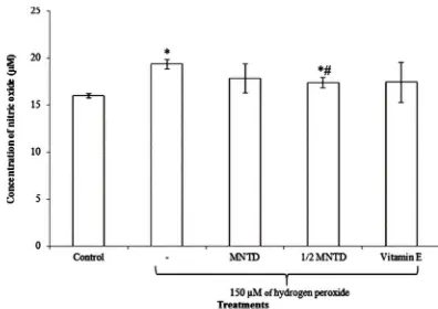
Fig. 1 shows that 150 µM of H2O2 stimulation increased NO level
significantly by 21.0% when compared to the unstimulated control cells. Treatments of orientin at 20 µM (MNTD) and 10 µM (½ MNTD) reduced the production of NO upon stimulated by H2O2, whereby ½ MNTD treatment yielded a significant
reduction in NO concentration in comparison to the H2O2
-stimulated cells alone. The reduction of NO by ½ MNTD was also shown to be more effective and significant than the positive control, Vitamin E.
Fig. 1 Nitric oxide levels in hydrogen peroxide (H2O2)-stimulated SH-SY5Y cells upon 24 hours treatment with orientin at 37°C. Bars indicate the means ± standard deviation. ‘*’ indicates that the treatment was significantly different from the untreated cells using Student’s t-test at p < 0.05. ‘#’ denotes the treatment was significantly different from the H2O2-stimulated cells using Student’s t-test at p < 0.05. MNTD, maximum non-toxic dose.
Effects on intracellular calcium level
The intracellular Ca2+ level of the orientin-treated and untreated
H2O2-stimulated SH-SY5Y cells were stained with Molecular
Probes® Fluo-4 NW dye mix and the fluorescence was analysed by a microplate reader. Fluorescence readings were then normalized by the cell number in each well in which the cell number was assessed by crystal violet staining assay. The intracellular Ca2+ level increased significantly by 82.8 % upon
stimulated by 150 µM of H2O2 when compared to the
unstimulated cells (Fig. 2). The pre-treatment of orientin at MNTD and ½ MNTD alongside the positive control - vitamin E were shown to be capable in significantly decreasing intracellular Ca2+ levels of the H
2O2-stimulated cells by 33.49 %, 28.20 %,
and 40.38 % respectively. Within this intracellular calcium level assay, orientin at MNTD has been revealed to be the most effective treatment group in bringing down the rise in intracellular Ca2+ level by H
2O2, followed by vitamin E and
orientin at ½ MNTD.
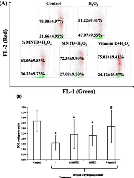
Effects on mitochondrial membrane potential
The red/green fluorescence ratio upon JC-1 staining allows visualization of mitochondrial injury on treated cells as performed by Li et al. [22]. After staining with JC-1, normal healthy cells emitted red fluorescence, but the cells with lower MMP emit green fluorescence. Thus, an increase of green fluorescence denotes a decrease in MMP of SH-SY5Y cells. The flow cytometric analysis of JC-1-stained cells shows increased green fluorescence signals upon oxidative-stressed by 150 μM H2O2 (Fig. 3A). The treatment groups - ½ MNTD and MNTD of
orientin and vitamin E show restoration of the decrease in MMP by increasing the red signals. Fig. 3B displays that the ratio decreased significantly by 56.60 % upon stimulated by H2O2
when compared to the unstimulated cells. MNTD and ½ MNTD of orientin restored the loss of MMP due to H2O2-induced
oxidative damage by 30.66 % and 34.21 %. Treatment of vitamin E recorded the most effective group in restoring the loss of MMP by 1.98 folds, despite not being statistically significant.
Fig. 3 Flow cytometric analysis of orientin effects on mitochondrial membrane potential of SH-SY5Y cells stained by JC-1 fluorochrome. The cells were pre-treated with maximum non-toxic dose (MNTD) and ½ MNTD of orientin for 24 hours, rinsed with phosphate-buffered saline, trypsinised and stained with JC-1 at 37°C for 15 minutes. The stained cells were analyzed for green and red fluorescence by a flow cytometer in which the gated cells were presented in dot-plot format with the Cell Quest Software. (A) Representative images of flow cytometric analysis. (B) The average JC-1 red-to-green fluorescence ratio of hydrogen peroxide (H2O2)-stimulated cells upon 24 hours treatment with orientin at 37°C. Bars indicate the means ± standard deviation. ‘*’ indicates that the treatment was significantly different from the untreated cells using
Effects on apoptosis
In this study, the apoptotic cells were quantitated by AV/PI apoptosis assay, which could be utilized to distinguish the distribution of early and late apoptotic cells. Fig. 4 shows the arrangement of quadrants on AV/PI dot plots which represent live cells (AV−/PI−), cells undergo early apoptosis (AV+/PI−), cells undergo late apoptosis (AV+/PI+) and necrotic cells (AV−/PI).
Fig. 4. Quadrants diagram that represents the different apoptosis stages of cells as shown from the flow cytometric analysis. AV, annexin V; PI, propidium iodide.
In the representative dot plot image in Fig. 5A, stimulation by 150 μM of H2O2 decreased the percentage of healthy cells
(AV-/PI-) along with increased total apoptotic cells. As shown in Fig. 5B, the percentage of early apoptotic cells upon stimulated by H2O2 increased by 1.8 folds and late apoptotic cells increased
by 2.7 folds when compared with the control group. In contrast, pre-treatment with ½ MNTD of orientin reduced 27.36% of early apoptotic cells, 37.22% of late apoptosis cells and 46.37 % necrotic cells whereas the MNTD of orientin reduced 33.21 % of necrotic cells when compared to the H2O2 treatment group.
Orientin of 10 μM (½ MNTD) was shown to be more effective than MNTD to reduce apoptotic cells. However, the differences in the cell populations among the different treatment groups were not statistically significant.
Effects on Nrf2/Keap-1 redox signalling pathway
Fig. 6 shows that H2O2 significantly inhibited the protein
expressions of Nrf2, HO-1 and Keap-1 upon comparison with the negative control. Orientin treatment at MNTD, however, was able to restore the expression of the proteins in SH-SY5Y cells belonging to a generic class of antioxidative-related proteins. This restorative effect was not observed in the ½ MNTD dose of orientin and vitamin E upon H2O2 treatment, which was further
signified by the intensities of said protein bands being identical to the H2O2-treated bands. The expression level of SOD-1 was
observed to constant throughout the treatments.
Fig. 6. Representative blots of the expression of proteins from Nrf2/Keap-1 redox signalling pathway. The SH-SY5Y were pre-treated with maximum non-toxic dose (MNTD) and ½ MNTD of orientin for 24 hours and subsequently stimulated by hydrogen peroxide (H2O2) for 24 hours at 37°C. Proteins were extracted from the treated cell and untreated cells and examined by Western Blotting.
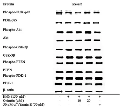
Effect on PI3K/Akt survival pathway
Stimulation by H2O2 moderately reduced the protein level of
phospho-PI3K-p85 but slightly increased the protein levels of phospho-PDK-1 and PDK-1 in SH-SY5Y cells (Fig. 7). Nevertheless, the treatment of orientin at MNTD increased the protein levels of phospho-PI3K-p85, and phospho-Akt when compared to the H2O2-induced cells. Despite the reduction of
their phosphorylated isoforms, it was evident that the total protein expressions of PI3K-p85 and Akt remained unchanged, further signifying that the effects of H2O2 and orientin only
affected these proteins in their activated forms.
The treatment also concurrently decreased the protein levels of PDK-1 in both total and phosphorylated isoforms. Other survival-related proteins we were interested in this study were GSK-3β and PTEN, but the expressions of both proteins in non-phosphorylated and non-phosphorylated forms remained constant throughout all treatments, which suggested that these pathways might belong to downstream protein pathways that remain unaffected in this study.
Fig. 7. Representative blots of the expression of proteins from PI3K/Akt survival pathway. The SH-SY5Y were pre-treated with maximum non-toxic dose (MNTD) and ½ MNTD of orientin for 24 hours and subsequently stimulated by hydrogen peroxide (H2O2) for 24 hours at 37°C. Proteins were extracted from the treated cell and untreated cells and examined by Western Blotting.
Effect on MAPK/ERK apoptosis pathway
As to validate the apoptotic pathway-related proteins, Fig. 8 shows the effect of orientin on MAPK/ERK protein levels in SH-SY5Y cells upon treatments. H2O2-induced oxidative stress was
able to increase the protein levels of phospho-p44/42 MAPK (ERK 1/2), p44/42 MAPK, phospho-p38 MAPK, p38 MAPK, phospho-c-Raf and c-Raf. Upon treatment with orientin and vitamin E, however, no noticeable changes were observed in both non-phosphorylated and phosphorylated isoforms of said proteins. Interestingly, protein expressions of MAP kinase kinase kinase (MAP3K) proteins phospho-c-Raf and c-Raf showed a slight decrease upon orientin pre-treatment at ½ MNTD and MNTD when compared to the H2O2 stimulation group, with a
similar effect shown in the positive control bands as well.
DISCUSSION
In this study, induction of H2O2 increased NO level significantly
which could cause neuronal death. H2O2 has been widely utilized
as an inducer of the nitric oxide signalling pathway, capable of eliciting oxidative stress responses by production of superoxide anions or the more dangerous peroxynitrite, a byproduct of precursor radicals such as NO [23,24]. The product of this stimulation, NO, is a signalling molecule enzymatically produced by three types of nitric oxide synthases (NOS): endothelial NOS, inducible NOS and ʟ-arginine-dependent neuronal NOS [25]. The increase of intracellular Ca2+ level also triggers
calmodulin-dependent protein kinase which concomitantly assists NOS to produce NO. The overproduction of NO or NO-derived reactive nitrogen species can cause inhibition of neuronal respiration resulting in massive glutamate release and subsequent excitotoxicity which may lead to neuronal cell death in inflammation and ND [26-31]. Another pro-apoptotic function that NO was reported to possess is the activation of various caspases required to proceed with apoptosis within SH-SY5Y cells [32]. Furthermore, the process of neuroinflammation and apoptosis of neuronal cells have been determined to be the causes of ND, which provide an avenue to combat ND prior to the event itself by means of plant derivatives such as orientin.
Fig. 1 shows that H2O2 stimulation increased NO level and
treatments of orientin at different doses could reduce the production of NO. Aquilano and colleagues have shown that at normal levels, NO produced via nNOS confers protective effects towards neuroblastoma cells, including SH-SY5Y [33], but at higher levels of NO produced during an event of oxidative stress, the balance of NO and ROS is shifted to fasten apoptosis [34]. Studies showed that the activation of cytokines or proinflammatory factors and programmed cell death had been proven to worsen neurodegeneration in patients [7-9]. Nevertheless, orientin protected the neuronal cells by inhibiting the elevation of intracellular Ca2+ level which would theoretically
lead to the suppression of excessive NO production by NOS and thus potentially protecting neuronal cells from inflammation and cell death.
On a higher degree compared to NO, calcium plays a significant role in all types of cells as most of the activities of cells are regulated by Ca2+ which is the universal second
messenger in cells [35]. In neuronal cells, the production of ROS during oxidative stress precedes the release of cytochrome c from mitochondria and triggers caspase-3 activation. Despite the possible connection between an influx in NO and Ca2+, this does
not seem to be the case as Fig. 1, and Fig. 2 showed an aberrant trend in the decrease between the two when treated with orientin and vitamin E. This might suggest that although Ca2+ influx
might trigger activation of calcium-calmodulin NOS to produce excess NO and its radical derivatives, there are other factors which could lead to the increase of NO, such as presence of stress or pro-inflammatory cytokines [36, 37].
Simultaneously, the influx of ROS-mediated intracellular Ca2+ level activates caspase-2 [38, 39]. The increase in
intracellular Ca2+ level further precedes the release of
cytochrome c which acts on the positive feedback loop to keep on releasing endoplasmic reticulum (ER) Ca2+ though the inositol
trisphosphate receptor (IP3R) [40, 41]. These would eventually
lead to apoptosis of the neuronal cells due to the intracellular Ca2+
The possible mechanism could be that the presence of orientin inhibits the IP3R and prevents the release of ER Ca2+.
This will then lead to the prevention of release of cytochrome c, caspase-2 and -3 and eventually suppress the apoptosis of neuronal cells. The previous study also revealed that the anti-apoptotic effect of orientin could have been attributed to the inactivation of caspases 3/7 and caspase 9 activities based on the caspase assays [20]. Orientin might inhibit the intracellular stress, diminish ROS and Ca2+ levels and restore the MMP. This
led to further inhibition of caspases activity and reduced the neuronal apoptosis. Despite the possible link between the increase in NO and intracellular Ca2+. The study suggested that
the pathological changes related to the AD were affected by glutamate-stimulated mitochondrial dysfunction [42]. It was demonstrated that mitochondrial dysfunction takes place early in the pathogenesis of ND [6]. Hence, the mitochondrial protection seems to be a prospective therapeutic intention. In the present study, orientin showed to moderately restore the MMP of neuronal cells.
Aside from the ample evidence provided by the modulation of NO and intracellular Ca2+ by orientin, various other parameters
were studied to determine the anti-apoptotic effects of orientin, one of which was through the apoptotic assay Under normal conditions, the phosphatidyl serine (PS) of live cells is well-maintained, and AV and PI staining are both negative within the respective steps of staining. AV labels PS on the cell surface and shows positive for AV staining during early apoptosis. As for the latter stages of apoptosis, PS is exposed to the external environment and the cell membrane integrity is lost, PI can now pass through PS and enter the cell to stain the dsDNA and RNA. In order to reduce the false positive events associated with PI staining of RNA, RNase was added during the staining procedures [43]. Reduced number of apoptotic and necrotic cells induced by H2O2 in orientin treatment could be due to the orientin
capability to maintain the PS and cell membrane integrity in order to reduce the cell death. Neuronal cell death has been determined to be one of the causes of ND. Studies showed that apoptosis might worsen neurodegeneration in ND patients [9]. Therefore, the ability of orientin in reducing apoptosis further justifies its neuroprotective effect, and it might be beneficial for ND patients.
Since oxidative stress has been highly implicated in the aetiology of AD and PD [16, 44], direct therapeutic approaches targeting oxidative events are crucial for the AD and PD patients [44, 45]. In this study, Nrf2/Keap-1 redox signalling pathway has been investigated as it is the main defence mechanism used to counteract oxidative stress. The previous study has found that the mRNA and protein expressions of Nrf2 were declined in the motor cortex and spinal cord, while mRNA expression of Keap1 was amplified in the motor cortex and Nrf2 associated antioxidant genes such as catalase and SOD-1 were reduced in ALS patients [46]. Mutations in the Cu-Zn SOD-1 gene are found to be associated with familial ALS patients [47]. Besides, the HO-1 is overexpressed in neurons and astrocytes of PD [48], and AD patients [49]; and this suggests that the patients are experiencing chronic oxidative stress. Thus, the protein expressions of Nrf2, HO-1, Keap-1 and SOD-1 were examined in this study.
protein level was markedly increased upon treatment with orientin at MNTD. HO-1 as an antioxidant enzyme catalyzes the degradation of heme to biliverdin which is subsequently degraded into bilirubin. Both the biliverdin and bilirubin exert antioxidant activities [53, 54]. Orientin was also observed to increase the protein level of Keap-1 in Fig. 6. There was no change in the protein level of SOD-1 which might suggest that the dismutase enzyme is not involved in the H2O2-stimulated
neuroprotection pathway of orientin. These results suggest that orientin has protective effects against oxidative neuronal damage through the up-regulation of Nrf2 and HO-1 proteins with potential implications in utilizing plant derivatives in ameliorating neuroinflammation and neuronal death.
Aside from the restorative Nrf2/Keap-1/HO-1 pathways, molecular mechanisms of oxidative stress-induced neuronal damage seem to be engaged in neuronal apoptosis and thus Western Blot analyses were also carried out to further elucidate the mechanism behind neuronal cell death and potential inhibition of this effect via our compound of interest. Orientin may exert protective effect in the central nervous system by protecting cells against stress-induced injury, or by supporting neurocognitive performance and suppressing the inflammation of neurons through different pathways, for instance, PI3K/Akt pathway [55]. Thus, the protein expressions of PI3K-p85, Akt, PDK-1, GSK-3β and PTEN from this pathway have been studied in the present study. The up-regulation of phospho-PI3K-p85 and phospho-Akt, and down-regulation of phospho PDK and total PDK protein levels by MNTD of orientin displayed the possible pathway that orientin protects neuronal cells. The PDK-1 level unexpectedly decreased during the experiments as most of the studies showed activation of PI3K/Akt by increasing PDK-1 levels [56, 57]. However, low level of PDK-1 has been demonstrated to be advantageous in certain parts of the brain. It is believed that maintaining low level of PDK-1 provides high tolerance to the amyloid-beta which is a metabolite associated to AD development [58]. The presence of orientin, aside from just modulating PDK-1 levels, might also trigger pathways which lead to the phosphorylation of PI3K enzymes and subsequently, phosphorylation of Akt, which will reduce apoptosis and increase neuronal cell survival [59]. The remaining proteins, PTEN and GSK-3β might not be involved in the neuroprotection of orientin as the protein expression did not change with treatments, evident from Fig. 7.
Finally, the MAPK pathway engages an essential role in signalling and phosphorylative activities of cells and subsequently regulates cell growth, cellular differentiation, proliferation, migration, and apoptosis. MAPK does this through activation of ERK through dephosphorylation of Ras, and subsequently activation of Raf which would lead to a cascade of protein expression essential in the event of apoptosis MAPK pathway proteins JNK, p38 and stress-activated protein kinases (SAPK) are commonly related with apoptosis [60], therefore, this study focuses on the proteins p44/42 MAPK (also known as ERK1/2), p38, as well as c-Raf, which belongs to the MAP3K protein family, as a model to demonstrate the viability of our compound of interest in modulating apoptotic responses. The down-regulation of protein expression shows that orientin is capable of inhibiting c-Raf and inadvertently reducing the activation of the downstream MAPK/ERK pathway. Given that c-Raf is a MAP3K and p38 alongside p44/42 are MAPK proteins downstream of MAP3K and MAP2K, it is possible that there are compensatory pathways or crosstalk with PI3K/AKT survival pathways which allow p38 and p44/42 to retain their original levels of expression [61, 62].
Previous study suggested that H2O2 activated the MAPK
apoptosis pathway [63], and the MAPK proteins that were stimulated by oxidative stress would then mediate neuronal cell death [64]. In mediating the event of cell death, the oxidative stress stimulation of HO-1 expression has been revealed to be activated by MAPK [65, 66] in a negative loop feedback manner; and this finding is in line with our finding that the orientin up-regulated the expression of the HO-1. HO-1 itself is an enzyme capable of exerting antioxidative properties, and this serves as a compensatory pathway that SH-SY5Y cells undergo to protect themselves in the event of oxidative stress. A study conducted on osteosarcoma cells have shown evidence which support our hypothesis whereby there is indeed a cross-talk between MAPK/ERK and Nrf2/HO-1 pathways which would lead to a protective effect carried out by the latter [67]. Experimental HO-1 inhibition in dendritic cells via administration therefore, the inhibition of MAPK/ERK pathway by orientin might protect neuronal cells from apoptosis and further activated antioxidant enzyme such as HO-1 to promote survival of the neuronal cells.
In conclusion, orientin is capable of protecting SH-SY5Y cells from H2O2-induced oxidative damage by reducing NO
levels, reducing intracellular Ca2+ levels, restoring the loss of
MMP, decreasing portions of mostly late apoptotic and necrotic cells, up-regulating PI3K/Akt survival and Nrf2/Keap-1 redox signalling pathways while attenuating the activation of MAPK/ERK apoptosis pathway. Based on all these findings, Fig. 9 illustrates the main findings and the probable neuroprotective mechanisms of orientin. While revealing the potential and better targeted effective therapies in the field of neuronal disorders, the results attained in this study will be useful for the prevention of neurodegenerative diseases in the near future as well.
CONFLICT OF INTEREST
The authors declare that there is no conflict of interest regarding the publication of this article.
ACKNOWLEDGEMENTS
The study is funded by the Ministry of Higher Education, Malaysia under the Fundamental Research Grant Scheme (FRGS) [FRGS/1/2013/SKK07/IMU/03/3].
REFERENCES
1. Ross CA, Poirier MA. Protein aggregation and neurodegenerative disease. Nat Med. 2004; 10: S10-17.
2. Valente EM, Abou-Sleiman PM, Caputo V, Muqit MMK, Harvey K, Gispert S, Ali Z, Del Turco D, Bentivoglio AR, Healy DG, Albanese A, Nussbaum R, González-Maldonado R, Deller T, Salvi S, Cortelli P, Gilks WP, Latchman DS, Harvey RJ, Dallapiccola B, Auburger G, Wood NW. Hereditary Early-Onset Parkinson's disease caused by mutations in pink1. Science. 2004; 304(5674):1158-1160.
3. Przedborski S, Vila M, Jackson-Lewis V. Series introduction: neurodegeneration: what is it and where are we? J Clin Invest. 2003; 111(1):3-10.
4. Kurland LT. Amyotrophic lateral sclerosis and Parkinson's disease complex on Guam linked to an environmental neurotoxin. Trends Neurosci. 1988; 11(2):51-54.
5. Chen H, Chan DC. Mitochondrial dynamics–fusion, fission, movement, and mitophagy–in neurodegenerative diseases. Hum Mol Genet. 2009; 18(R2):R169-R176.
6. Lin MT, Beal MF. Mitochondrial dysfunction and oxidative stress in neurodegenerative diseases. Nature. 2006; 443:787-795. 7. Amor S, Puentes F, Baker D, van Der Valk P. Inflammation in
8. Wilms H, Zecca L, Rosenstiel P, Sievers J, Deuschl G and Lucius R. Inflammation in Parkinson's diseases and other neurodegenerative diseases: cause and therapeutic implications. Curr Pharm Des. 2007; 13(18):1925-1928.
9. Vila M, Przedborski S. Targeting programmed cell death in neurodegenerative diseases. Nat Rev Neurosci. 2003; 4:365-375. 10. Hara T, Nakamura K, Matsui M, Yamamoto A, Nakahara Y,
Suzuki-Migishima R, Yokoyama M, Mishima K, Saito I, Okano H, Mizushima N. Suppression of basal autophagy in neural cells causes neurodegenerative disease in mice. Nature, 2006; 441:885-889.
11. Komatsu M, Waguri S, Chiba T, Murata S, Iwata J, Tanida I, Ueno T, Koike M, Uchiyama Y, Kominami E, Tanaka K. Loss of autophagy in the central nervous system causes neurodegeneration in mice. Nature, 2006; 441:880-884.
12. Lindholm D, Wootz H, Korhonen L. ER stress and neurodegenerative diseases. Cell Death Differ. 2006; 13:385- 392. 13. Sherman MY, Goldberg AL. Cellular defenses against unfolded
proteins - a cell biologist thinks about neurodegenerative diseases. Neuron, 2001; 29(1): 5-32.
14. Emerit J, Edeas M, Bricaire F. Neurodegenerative diseases and oxidative stress. Biomed Pharmacother. 2004; 58(1):39-46. 15. Andersen JK. Oxidative stress in neurodegeneration: cause or
consequence? Nat Rev Neurosci. 2004; 5:S18-S25.
16. Bayani U, Ajay VS, Paolo Z, Mahajan RT. Oxidative stress and neurodegenerative diseases: a review of upstream and downstream antioxidant therapeutic options. Curr Neuropharm. 2009; 7(1):65-74.
17. Gomoll BP, Sanders BD, Caserta MT. Psychopharmacologic treatments for Alzheimer disease and related dementias. Psychopharm Rev. 2014; 49(1):9-16.
18. William GO, Lina S, Anthony D. Memantine (Namenda ®) for non-motor features of Parkinson’s disease: A double bind placebo controlled trial. Parkinsonism Relat Disord. 2011; 17(3):156-159. 19. Woolley JD, Khan BK, Murthy NK, Miller BL, Rankin KP. The
diagnostic challenge of psychiatric symptoms in neurodegenerative disease; rates of and risk factors for prior psychiatric diagnosis in patients with early neurodegenerative disease. J Clin Psychiatry. 2011; 72(2):126-133.
20. Law Bnt, Ling APK, Koh RY, Chye SM, Wong YP. Neuroprotective effects of orientin on hydrogen peroxide-induced apoptosis in SH-SY5Y cells. Mol Med Rep. 2013; 9(3):947-954. 21. Kueng W, Silber E, Eppenberger U. Quantification of cells cultured
on 96-well plates. Analyt Biochem. 1989; 182(1):16-19.
22. Li YL, Ma SC, Yang YT, Ye SM, But PP. Antiviral activities of flavonoids and organic acid from Trollius chinensis Bunge. J Ethnopharmacol. 2002; 79(3):365-368.
23. Ahn JH, Song JS. The effect of hydrogen peroxide on inducible nitric oxide synthase expression in murine macrophage Raw264.7 cells. Tuberc Respir Dis, 1999; 47(2):172-183.
24. Thomas SR, Chen K, Keaney Jr JF. Hydrogen peroxide activates endothelial nitric-oxide synthase through coordinated phosphorylation and dephosphorylation via a phosphoinositide 3-kinase-dependent signalling pathway. J Bio Chem. 2001; 277:6017-6024.
25. Alderton WK, Cooper CE, Knowles RG. Nitric oxide synthases: structure, function and inhibition. Biochem J. 2001; 357(3):593-615.
26. Chang CC, Chen SD, Lin TK, Chang WN, Liou CW, Chang AY, Chan SH, Chuang YC. Heat shock protein 70 protects against seizure-induced neuronal cell death in the hippocampus following experimental status epilepticus via inhibition of nuclear factor-κB activation-induced nitric oxide synthase II expression. Neurobiol Dis 2014;62:241-249
27. Choi BM, PaE Ho, Jang SI, Kim YM, Chung HT. Nitric oxide as a pro-apoptotic as well as anti-apoptotic modulator. J Biochem Mol Biol. 2002; 35(1):116-126.
28. Chung HT, Pae HO, Choi BM, Billiar TR, Kim YM. Nitric oxide as a bioregulator of apoptosis. Biochem Biophys Res Commun. 2001; 282(5):1075-1079.
29. Lu Q, Harris V, Rafikov R, Sun X, Kumar S, Black S. Nitric oxide
30. Pacher P, Beckman JS, Liaudet L. Nitric oxide and peroxynitrite in health and disease. Physiol Rev. 2007; 87(1):315-424.
31. Suh S, Hamby A, Gum ET, Shin BS, Won SJ, Sheline CT, Chan PH, Swanson RA. Sequential release of nitric oxide, zinc, and superoxide in hypoglycemic neuronal death. J Cereb Blood Flow Metab. 2008; 28(10):1697-1706.
32. Uehara T, Kikuchi Y, Nomura Y. Mechanism of nitric oxide-induced apoptosis in human neuroblastoma SH-SY5Y cells. J Neurochem. 1999; 72:196-205.
33. Aquilano K, Filomeni G, Baldelli S, Piccirillo S, De Martino A, Rotilio G, Ciriolo MR. Neuronal nitric oxide synthase protects neuroblastoma cells from oxidative stress mediated by garlic derivatives. J Neurochem. 2007; 101:1327-1337.
34. Li CQ, Wogan GN. Nitric oxide as a modulator of apoptosis. Cancer Lett. 2005; 226:1-15.
35. Endo M. Calcium ion as a second messenger with special reference to excitation-contraction coupling. J Pharm Sci. 2006; 100(5):519-524.
36. Li G, Zhao Y, Li Y, Lu J. Up-regulation of neuronal nitric oxide synthase expression by cobalt chloride through a HIF-1α mechanism in neuroblastoma cells. Neuromolecular Med. 2015; 17(4):443-453.
37. Nishiya T, Uehara T, Edamatsu H, Kaziro Y, Itoh H, Nomura Y. Activation of Stat1 and subsequent transcription of inducible nitric oxide synthase gene in C6 glioma cells is independent of interferon-γ-induced MAPK activation that is mediated by p21ras. FEBS Lett. 1997; 408:33-38.
38. Bezprozvanny I, Mattson MP. Neuronal calcium mishandling and the pathogenesis of alzheimer's disease. Trends Neurosci. 2008; 31(9):454-463.
39. Kawahara M, Kuroda Y. Intracellular calcium changes in neuronal cells induced by alzheimer's ß-amyloid protein are blocked by estradiol and cholesterol. Cell Mol Neurobiol. 2001; 21(1):1-13. 40. Boehning D, Patterson RL, Sedaghat L, Glebova NO, Kurosaki T,
Snyder SH. ytochrome c binds to inositol (1, 4, 5) trisphosphate receptors, amplifying calcium-dependent apoptosis. Nat Cell Biol. 2003; 5(12):1051-1061.
41. Sammels E, Parys JB, Missiaen L, Smedt HD, Bultynck G. Intracellular Ca2+ storage in health and disease: a dynamic equilibrium. Cell Calcium. 2010; 47(4):297-314.
42. Swerdlow RH, Burns JM, Khan SM. The Alzheimer's disease mitochondrial cascade hypothesis. J Alzheimers Dis. 2010; 20(Suppl 2):S265-279.
43. Rieger AM, Nelson KL, Konowalchuk JD, Barreda DR. Modified annexin v/propidium iodide apoptosis assay for accurate assessment of cell death. J Vis Exp . 2011; (50): 2597.
44. Bonda DJ, Wang X, Perry G, Nunomura A, Tabaton M, Zhu X, Smith MA. Oxidative stress in Alzheimer's disease: a possibility for prevention. Neuropharm. 2010; 59:290-294.
45. Liochev SI. Reactive oxygen species and the free radical theory of aging. Free Radic Biol Med. 2013; 60:1-4.
46. Sarlette A, Krampfl K, Grothe C, Neuhoff N, Dengler R, Petri S. Nuclear erythroid 2-related factor 2-antioxidative response element signalling pathway in motor cortex and spinal cord in amyotrophic lateral sclerosis. J Neuropatho Exp Neurol. 2008; 67:1055-1062. 47. Rosen DR, Siddique T, Patterson D, Figlewicz DA, Sapp P, Hentati
A, Donaldson D, Goto J, O'Regan JP, Deng HX, Rahmani Z, Krizus A, Mckenna-Yasek D, Cayabyab A, Gaston SM, Berger R, Tanzi RE, Halperin JJ, Herzfeldt B, Van Den Bergh R, Hung W, Bird T, Deng G, Mulder DW, Smyth C, Laing NG, Soriano E, Pericak-Vance MA, Haines J, Rouleau GA, GuseLLA JS, Horvitz HR, Brown RH Jr. Mutations in Cu/Zn superoxide dismutase gene are associated with familial amyotrophic lateral sclerosis. Nature, 1993; 362:59-62.
48. Schipper HM, Liberman A, Stopa EG. Neural heme oxygenase-1 expression in idiopathic Parkinson's disease. Exp Neurol. 1998; 150:60-68.
49. Schipper HM, Cisse S, Stopa EG. Expression of heme oxygenase-1 in the senescent and Alzheimer-diseased brain. Ann Neurol. oxygenase-1995; 37:758-768.
51. Gan L, Johnson JA. Oxidative damage and the Nrf2-ARE pathway in neurodegenerative diseases. Biochimica et Biophysica Acta (BBA) - Mol Basis Dis. 2014; 1842(8):1208-1218.
52. Al-Huseini LM, Yeang HXA, Hamdam JM, Sethu S, Alhumeed N, Wong W, Sathish JG. Heme oxygenase-1 regulates dendritic cell function through modulation of p38 Mapk-creb/atf1 signaling. J Biol Chem. 2014; 289:16442-16451.
53. Dore S, Snyder SH. Neuroprotective action of bilirubin against oxidative stress in primary hippocampal cultures. Ann NY Acad Sci. 1999; 890:167-172.
54. Stocker R, Yamamoto Y, McDonagh AF, Glazer AN, Ames BN. Bilirubin is an antioxidant of possible physiological importance. Science. 1987; 235:1043-1046.
55. Spencer JPE. The interactions of flavonoids within neuronal signalling pathways. Genes Nutr. 2007; 2(3):257-273.
56. Yu ZH, Cai M, Xiang J, Zhang ZN, Zhang JS, Song XL, Zhang W, Bao J, Li WW, Cai DF. PI3K/Akt pathway contributes to neuroprotective effect of Tongxinluo against focal cerebral ischemia and reperfusion injury in rats. J. Ethnopharmacol. 2016; 181:8-19.
57. Zhong Y, Huang Y, Cao JH, Lu X, Feng MJ, Shen G, Shen AG, Yu XW. Increase in phosphorylation of PDK1 and cell survival after acute spinal cord injury. J Neuro Sci. 2012; 320(1-2):38-44. 58. Newington JT, Rappon T, Albers S, Wong DY, Rylett RJ,
Cumming RC. Overexpression of pyruvate dehydrogenase kinase 1 and lactate dehydrogenase A in nerve cells confers resistance to amyloid β and other toxins by decreasing mitochondrial respiration and reactive oxygen species production. J Biol Chem. 2012; 287(44):37245-37258.
59. Brunet A, Datta SR, Greenberg ME. Transcriptiondependent and -independent control of neuronal survival by the PI3K–Akt signalling pathway. Curr Opin Neurobiol. 2001; 11(3):297-305. 60. Schroeter H, Boyd C, Spencer JPE, Williams RJ, Cadenas E,
Rice-Evans C. MAPK signalling in neurodegeneration: influences of flavonoids and of nitric oxide. Neurobiol Aging, 2002; 23(5):861-880.
61. Chera S, Ghila L, Wenger Y, Galliot B. Injury-induced activation of the MAPK/CREB pathway triggers apoptosis-induced compensatory proliferation in hydra head regeneration. Dev Growth Differ, 2011; 53(2):186-201.
62. Du J, Tong A, Wang F, Cui Y, Li C, Zhang Y, Yan Z. The roles of PI3K/AKT/mTOR and MAPK/ERK signaling pathways in human phechromocytomas. Int J Endo. 2016: 5286972.
63. Chen L, Liu L, Yin J, Luo Y, Huang S. Hydrogen peroxide-induced neuronal apoptosis is associated with inhibition of protein phosphatase 2A and 5, leading to activation of MAPK pathway. Int J Biochem Cell Biol. 2009; 41:1284-1295.
64. Rincon M, Flavell RA, Davis RA. The JNK and p38 MAP kinase signalling pathways in T cell-mediated immune responses. Free Radic Biol Med. 2000; 28:1328-1337.
65. Aggeli IK, Gaitanaki C, Beis I. Involvement of JNKs and p38-MAPK/MSK1 pathways in H2O2-induced upregulation of heme oxygenase-1 mRNA in H9c2 cells. Cell Signal. 2006; 18:1801-1812.
66. Benvenisti-Zarom L, Chem-Roetling J, Regan RF. Inhibition of the ERK/MAP kinase pathway attenuates heme oxygenase-1 expression and heme-mediated neuronal injury. Neurosci Lett. 2006; 398:230-234.