Original Research Article
A study on relationship of age at first coitus and number of conceptions
with the development of uterine cervical dysplasia in women
undergoing VIA screening at community health centre,
Muradnagar, U.P., India
Sunita Vashist
1, Narendra Singh
2*
INTRODUCTION
Cervical cancer is the commonest cancer causing death among women in developing countries.1 It is usually a very slow growing cancer that may not have symptoms for a long time but can be detected in early stage with regular screening methods.86% of all deaths due to cervical cancer are in developing, low and middle income countries.2-4 Cancer is leading cause of death in
economically developed countries and the second leading cause of death in developing countries.5
There are several suggested reasons for increasing incidence of cervical cancer cases and mortality associated with it. The long interval between initial infection and disease indicates that there are other factors involved such as sexual habits, local hygiene, reproductive factors, nutritional deficiencies, genetic
ABSTRACT
Background: Cancer of cervix is a common cancer that affects Indian women physically psychologically, socially and financially. The disease affects not just the women but also her family and society. Development of cervical dysplasia is highly associated with age of first coitus and number of conception of women. This study was designed to study the correlation between age of first coitus and number of conceptions in women with development of uterine cervical dysplasia.
Methods: This study was done at the Community Health Centre (CHC), Muradnagar. It was a cross-sectional study done by using VIA (visual inspection using acetic acid) technique on uterine cervix on 1250 women aged above 30 years of age attending gynaecology OPD of CHC, Muradnagar, Ghaziabad, U.P. Purposive sampling was used to enrol all the women who were attending the gynaecology OPD at CHC and were coming in the eligibility criteria.
Results: Out of 1250 women 14 were found to be VIA positive, out of which 4 cases came out to be positive for dysplasia after doing biopsy under colposcopy.
Conclusions: Development of cervical dysplasia is highly correlated with early age at first coitus and more number of conceptions in women. As cervical dysplasia is highly associated with early age at first coitus and more conceptions, so more screening for cervical dysplasia required in women having first coitus at early age and having more conceptions.
Keywords: Cervical dysplasia, VIA screening, Early age at first coitus, Number of conceptions Department ofCommunity Medicine, Santosh Medical College and Hospital, Ghaziabad, U.P., India
Received: 15 January 2018
Revised: 02 February 2018
Accepted: 03 February 2018
*Correspondence:
Dr. Narendra Singh,
E-mail: [email protected]
Copyright: © the author(s), publisher and licensee Medip Academy. This is an open-access article distributed under the terms of the Creative Commons Attribution Non-Commercial License, which permits unrestricted non-commercial use, distribution, and reproduction in any medium, provided the original work is properly cited.
susceptibility, use of hormonal contraceptives and high parity. Specific religious practices also modify the risk of developing cancer in women following HPV infection.6
In the absence of a nationwide screening program, there are disparities in screening, treatment, and also survival. An analysis of population-based surveys indicates that coverage of cervical cancer screening in developing countries is 19% compared to 63% in developed countries and ranges from 1% in Bangladesh to 73% in Brazil.7 However the older and the poorer women who are most likely to develop cervical cancer are least likely to undergo screening. Opportunistic screening in various regions of India varied from 6.95 in Kerala to 0.006% and 0.002% in the western state of Maharashtra and Tamil Nadu respectively.8-10 Most of the cervical cancer cases (85%) presented in advanced and late stages.11 Cervical cancer diagnosis and treatment in the advanced stages makes it a costly exercise, with poor prognosis resulting in poor compliance.12
Following are brief descriptions of the methods used for screening / diagnostic test, their sensitivity, specificity and characteristic for cancer cervix.13,14
Exfoliative cervico-vaginal cytology
Also known as Pap smear screening technique or conventional cytology screening has been used as far as 1927 when Papanicolau was introduced. In this technique Pap smear collection is obtained from the cervix and the endo-cervical canal by the use of Ayre’s spatula and cyto brush. Samples taken are then smeared on a slide which is then fixed with cytology fixative. Sensitivity of this test is moderate between 44-78, percent, specificity is high 91-96%, and epidemiological data suggests that it is unlikely to detect 60 percent of the cervical cancer cases. Requires adequate healthcare infrastructure, lab based, stringent training and quality control.
HPV DNA testing
These nucleic acid hybridization methods, involving use of Real time PCR, are aimed at detecting high risk HPV types 16, 18, 31, 33, 35, 39, 45, 51, 52, 56, 59 and 68 HPV DNA. All these tests have high sensitivity (66-100%) and moderate specificity (61-96%). They are lab based, have high output, objective during interpretation of results, highly reproducible. However, currently the high cost and the availability are the main hindering factors in their usage in low resource settings.
Visual screening
This type of screening involves direct inspection of the cervix without taking of samples and although it is less tedious in terms of preparation and inspection it is considered to be less accurate in identifying precancerous changes. However with the use of acetic acid (vinegar) the precancerous cells temporarily turn white therefore is
making it easy to identify them. In VILI iodine-based solution is used which turns normal cervical cells brown and abnormal cells yellow or unstained making them clearly visible its sensitivity being moderate to high 78-98% specificity low 73-93%, VIA (visual inspection with acetic acid is suitable for its cost effect since it requires low resources.13
A review study observed cervical cancer is relatively neglected disease in terms of advocacy and indicated cytology, human papilloma virus (HPV) testing, visual inspection of cervix using acetic acid (VIA) and visual inspection of cervix using Lugol’s iodine (VILI) are known to be accurate and effective method to detect cervical cancer and could contribute to the reduction of disease in low resource setting.
Visual inspection with 3-5% acetic acid (VIA)13
VIA also known as direct visual inspection (DVI), the acetic acid test (AAT) or cervicoscopy, involves examination of the cervix with naked eye; using a bright light source, after one to two minutes of 3-5% diluted acetic acid application using a cotton swab or a spray. Detection of well-defined aceto-white areas close to the squamo columnar junction (SCJ) indicates a positive test. Although aceto-whitening can occur in immature squamous metaplasia and in inflamed and regenerating cervical epithelium, aceto-whitening associated with Cervical–intra-epithelial neoplasia (CIN) is well demarcated, intensely opaque and localized to the transformation zone. Aceto-whitening is thought to be due to a reversible coagulation of intracellular proteins following acetic acid application. Its sensitivity is moderate 67-79%, sensitivity low 49-86%, requires low technology, low cost, linkage to immediate treatment possible, and is suitable for low –resource settings.
With the literature knowledge for cervical dysplasia that we could gather, our aim was to do a study on cervical dysplasia in the rural and urban areas of district Ghaziabad in order to add further knowledge regarding this crucial disease of such a paramount importance. Also our objective was to find correlation between age at first coitus and number of conceptions with the development of cervical dysplasia, regarding importance of cervical cancer and its early detection through screening amongst all of the women attending CHC (community health centre) Muradnagar, Ghaziabad, UP, India and channelizing our study population towards early diagnosis, treatment and follow-up of cervical cancer, beside health education.
METHODS
women with cross-sectional sectional study design. All of the women coming under eligibility criteria aged above 30 years, attending Gynaecology OPD of CHC, Muradnagar, Ghaziabad was the study population. Purposive sampling was used to enrol all the women patients, attending the gynaecology OPD at CHC and coming in the eligibility criteria.
Criteria for selection
Inclusion criteria
All of the women aged above 30 years, attending Gynaecology OPD of CHC, Muradnagar, Ghaziabad with all type of gynaecological complaints.
Exclusion criteria
Exclusion criteria were all women aged below 30 years; all women with history of hysterectomy done; all women who had undergone cervical cancer treatment in past; all currently pregnant women; women who were not willing to give consent for the study.
Information of individual and family was collected by pre- designed, pre-tested and structured schedule. Reproductive health related complaints were noted followed by clinical examination and cervical cancer screening. VIA (Visual inspection under acetic acid) of cervix was done, women which came out to be VIA positive were send for colposcopical guided biopsy to see dysplastic changes. The data collected was entered in MS Excel.
Method of doing VIA
(VIA) Visual examination was done with 4% acetic acid. Once the per speculum examination was done, 4% acetic acid was applied on the cervix with the help of cotton swab and observed after 1 minute. The respective changes in the colour and texture of the cervix were
observed carefully. Any areas turning aceto -white over the cervix were identified as a positive sign of presence of cellular abnormalities. All the positive cases with VIA were sent for colposcopy aided cervical biopsy.
Ethical consideration
The study was initiated after getting clearance from the Ethical Board Committee of the Santosh University. The objective, the purpose and the method of the study were explained to all the participants in their local easy to understand language. A written consent, in the local language was obtained from all the participants who were eligible for the study.
Data analysis
The data collected was entered in MS Excel. After the master chart was prepared it was coded as categorical variables. The data was analysed using SPSS version 17. (Statistical Package of Social Sciences) appropriate statistical test for the analysis of categorical variables was applied. Results were produced in form of frequencies and percentages. Chi-square and Fisher’s exact test was applied for the relevant variables. A ‘p’ value obtained as less than 0.05 was considered as statistically significant.
RESULTS
A total of 1250 women in the age group between 30 to 70 years who visited the CHC, Muradnagar OPD for gynaecological complaints were enrolled in the study and out of them 14 women were found to be VIA positive (i.e. positive for abnormal cells in the cervical epithelium under visual inspection with 4% acetic acid). Out of the 14 VIA positive cases, 4 cases came out to be positive for cervical dysplastic changes under colposcopy guided biopsy. Considering colposcopy guided biopsy as our gold standard test for confirmation of cervical cancer we thereby detected a total of four cervical cancer cases.
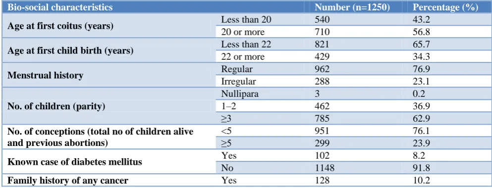
Table 1: Distribution of the study population according to their bio-social characteristics.
Bio-social characteristics Number (n=1250) Percentage (%)
Age at first coitus (years) Less than 20 540 43.2
20 or more 710 56.8
Age at first child birth (years) Less than 22 821 65.7
22 or more 429 34.3
Menstrual history Regular 962 76.9
Irregular 288 23.1
No. of children (parity)
Nullipara 3 0.2
1–2 462 36.9
≥3 785 62.9
No. of conceptions (total no of children alive and previous abortions)
<5 951 76.1
≥5 299 23.9
Known case of diabetes mellitus Yes 102 8.2
No 1148 91.8
Table 2: Association of the age at first coitus of the participants with their cervical biopsy specimen finding.
Age at first coitus of the participants Biopsy finding Total
(n=1250) (%)
Positive (%) Negative (%)
<20 years 4 (0.7) 536 (99.3) 540 (43.2)
≥20 years 0 710 (100) 710 (56.8)
Total 4 (0.3) 1246 (99.7) 1250 (100)
Fisher's exact test value=5.276, p=0.022.
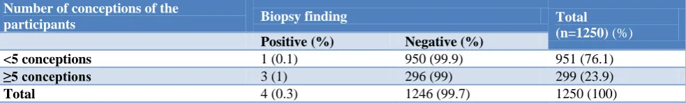
Table 3: Association of the number of conceptions of the participants with their cervical biopsy specimen finding.
Number of conceptions of the
participants Biopsy finding Total
(n=1250) (%)
Positive (%) Negative (%)
<5 conceptions 1 (0.1) 950 (99.9) 951 (76.1)
≥5 conceptions 3 (1) 296 (99) 299 (23.9)
Total 4 (0.3) 1246 (99.7) 1250 (100)
Fisher's exact test value=5.753, p=0.016.
More than half of the women (56.8%) reported age at first coitus as 20 or more and most of them (65.7%) had their first child birth at the age of less than 22. Majority (76.9%) reported to have regular menstrual cycles and most (62.9%) of them had three or more children. Majority (76.1%) reported to have less than or equal to 5 conceptions; which included live births and abortions.
Nearly hundred cases (8.2%) of the 1250 participants were known case of diabetes and similarly 128 participants (10.2%) reported positive history for family cancer.
All the four biopsy positive cases were found amongst participants whose age of first coitus was less than 20 years and there is a statistically significant difference in the age of first coitus of the participants and cervical cancer cases.
One biopsy positive case was found amongst participants who had less than 5 conceptions (including total live births and abortions) and three cases were found positive amongst the participants who had 5 or more conceptions and this difference in number of conceptions and biopsy positive cases was found to be statistically significant.
Our study concluded that development of cervical dysplasia is highly correlated with first coitus at early age and more conceptions.
DISCUSSION
The present study showed that (Table 2) all the four biopsy positive cases were found amongst participants whose age of first coitus was less than 20 years and there is a statistically significant difference in the age of first coitus of the participants and cervical cancer cases. Rotkin in his study on a comparison review of key epidemiological studies in cervical cancer related to current researches for transmissible agents found that 11
studies were confirming an excess of cervical cancer cases with early age of marriage and early age of coitus, compared to controls. A mean of 40% of patients who were first married before ages of 20 or 21 calculated from all these studies, with individual excess ranging from 20 to 90 percent.15
Similarly Franceschi et al in their multicentre case-control study in Chennai, Southern India revealed that ORs and corresponding 95% CIs for Age at first intercourse <15 years (OR vs. ≥21 years= 2.2) compared to ≥21 years doubles the risk for development of cervical cancer.16
Our study showed that (Table 3) one biopsy positive case was found amongst participants who had less than 5 conceptions (including total live births and abortions) and three cases were found positive amongst the participants who had 5 or more conceptions and this difference in number of conceptions and biopsy positive cases was found to be statistically significant. In line with our finding Franceschi et al in their multicentre case-control study in Chennai, Southern India revealed that women who reported 7 or more births had a higher risk than those with 1 or 2 births (OR=5.7) for development of cervical cancer.16 They also revealed that the OR for age at first pregnancy <17 years (vs. ≥19= 2.2) was lowered to 1.7 (non-significant) after adjustment for number of pregnancies. Similar finding was observed in our study as well.
Funding: No funding sources Conflict of interest: None declared
Ethical approval: The study was approved by the Institutional Ethics Committee
REFERENCES
1. Dennny L. Cancer Cancer: Prevention and
2. Satiga A. Cervical Cancer in India. South Asia Centre for Chronic Disease. Accessed on 16 February 2017.
3. Arbyn M, Castellsaque X, de Sanjosé S, Bruni L, Saraiya M, Bray F, et al. Worldwide burden of cervical cancer. Ann Oncol. 2011;22(12):2675-86. 4. Yesle BB, Kumar AV, Kerkureet A., Sunny L.
Population based survival from cancers of breast, cervix and ovary in women in Mumbai. Asian Pac J Cancer Prevention. 2004;5:308-15.
5. World Health Organization. The Global Burden of Disease: 2004 Update. Geneva: World Health Organization: 2008.
6. Seema P, Paul B, Boffetta P. Meta-analysis of social inequality and the risk of cervical cancer. Int J Cancer. 2003;105(5):687-91.
7. Gakidou E, Stella N, Ziad O. Coverage of cervical cancer screening in 57 countries: low average levels and large inequalities. PloS Med. 2008;5(6):132. 8. Aswathy S, Quereshi MA, Kurian B, LeelamoniK.
Cervical cancer screening: current knowledge and practice among women in a rural population of Kerala, India. Indian J Med Reas. 2012;136:205-10. 9. Sankaranarayanan R, Nene BM, Shastri SS, Jayant
K, Muwonge R, Budukh AM, et al. HPV screening for cervical cancer in rural India. N Engl J Med. 2009;360:1385-94.
10. Sankaranarayanan R, Esmy PO, Ramkumar R, Muwonge R, Swaminathan R, Shanthakumari S, et al. Effect of visual screening on cervical cancer incidence and mortality in Tamil Nadu: a cluster-randomised trial. Lancet. 2007;370:398-406. 11. Dutta S, Biswas N, Mukheriee G. Evaluation of
sociodemographic factors for non-compliance to
treatment in locally advanced cases of cancer cervix in a rural medical college hospital in India. Indian J Palliat Care. 2013;19(3):158-65.
12. Sreedevi A, Javed R, Dinesh A. Epidemiology of cervical cancer with special focus on India. Int J Women’s Health. 2015;7:405-13.
13. Denny L, Quinn M, Sankaranarayanan R. Screening for cervical cancer in developing countries. Vaccine. 2006;24:71-7.
14. Hakama M, Miller AB, Day NE. Screening for Cancer of the Uterine Cervix.from the IARC Working Group on Cervical Cancer Screening and the UICC Project Group on the Evaluation of Screening Programmes for Cancer. WHO (Geneva), IARC (Lyon), and UICC (Geneva) IARC Sci Publ. 1986;76:1–315.
15. Rotkin ID. A comparison review of key
epidemiological studies in cervical cancer related to current researches for transmissible agents. Cancer Research. 1973;33(6):1353-67.
16. Franceschi S, Rajkumar T, Vaccarella S, Gajalakshmi V, Sharmila A, Snijders PJ, et al. Human papillomavirus and risk factors for cervical cancer in Chennai, India: a case-control study. Int J Cancer. 2003;107:127–33.