
Synthesis of Silver Nanoparticles Using Silybum Marianum
Seed Extract
R. Mohammadinejad
1*,
Sh. Pourseyedi
2,
A. Baghizadeh
3,
Sh. Ranjbar
1,
G. A. Mansoori
4*1- Department of Agricultural Biotechnology, College of Sciences and Modern Technologies,
Graduate University of Advanced Technology, Kerman, I. R. Iran
2- Department of Biotechnology, Shahid Bahonar University of Kerman, Kerman, I. R. Iran
3- Department of Biotechnology, Institute of Science and High Technology and Environmental
Sciences, Graduate University of Advanced Technology, Kerman, I. R. Iran
4- Department of Bioengineering, University of Illinois at Chicago, Chicago, USA
(*) Corresponding authors: [email protected], [email protected]
(Received:20 Oct. 2013 and Accepted: 19 Nov. 2013)
Abstract:
Green synthesis of nanoparticles, as fundamental building blocks of nanotechnology, has recently attracted considerable attention. Silver nanoparticles have unique physiochemical, biological and environmental properties which make them useful in a wide range of applications. In the present paper we report our research results on biosynthesis of silver nanoparticles from silver precursor using milk-thistle plant (Silybum marianum) seed extract. The resulting synthesized Ag nanoparticles (AgNPs) were characterized with UV– visible spectroscopy, X-ray diffraction (XRD) and transmission electron microscopy (TEM). Our measurements showed that S. marianum seed extract can mediate facile and eco-friendly biosynthesis of colloidal spherical silver nanoparticles (AgNPs) of size range 1–25 nm. The colloidal AgNPs were formed at room temperature and were observed to be highly stable even after 6 months.
Keywords: milk-thistle plant, Silver nanoparticles, Silybum marianum, stable colloids, UV–vis spectroscopy, XRD, TEM
Int. J. Nanosci. Nanotechnol., Vol. 9, No. 4, Dec. 2013, pp. 221-226
1. INTRODUCTION
Advancement of nanotechnology is based on the
availability of well-defined and well-characterized
molecules, macromolecules, nanostructures, and supramolecules as its fundamental molecular building blocks. These are small particles of 1-100
nm with high specific surface area. Nanostructure
materials show unique physical, chemical, biological and environmental properties, including catalytic activity, optical, electronic and magnetic properties, which have increased their applications in research, industry, agriculture, environment and medicine (Khataee and Mansoori 2011; Mansoori
2005; Mansoori et al. 2007, 2008, 2012).
Facile green silver nanoparticles (AgNPs) are
blossoming field of research and have high potential
as commercialized nanomaterials (Chaloupka et al. 2010; Vahabi et al. 2011; Mansoori 2013). As an effective antimicrobial agent, facile green AgNPs have the potential for large-scale applications in the formulation of dental resin composites (Kassaee et al. 2008), bone cement (Alt et al. 2004), water
and silver nanocomposites or nanohybrids have other interesting characteristics which will further enable them to be used in catalysts, biosensors, conductive inks, electronic devices and solar cells (Tsuji et al. 2012; Wijnhoven et al. 2009). They can be produced economically and in large / industrial scale (Vahabi et al. 2011; Mansoori 2013). Several techniques to manufacture AgNPs are proposed. Generally, AgNPs are prepared by a variety of biological, chemical and physical methods, but majority of these techniques are either expensive and/or environmentally hazardous. In addition the synthesized nanoparticles by most methods may be unstable and tend to agglomerate quickly and become useless unless capping agents are applied for their stabilization (Sintubin et al. 2012). Chemicals used for synthesis and stabilization of nanoparticles could be also toxic.
The need for clean and reliable synthetic protocols for nanomaterials synthesis leads to the developing interest in benign / green biological approaches (Vahabi et al. 2011; Zaki et al. 2011; Mansoori 2013). In recent years many live bio-organisms such as bacteria, fungi, algae, plants and extracts or metabolites from them have been mediated for synthesis of AgNPs. The reduction of Ag+ to Ag0 occurs by combinations of biomolecules such as proteins, polysaccharides,
and flavonoids (Park et al. 2011; Vahabi et al. 2011; Mansoori 2013). Certain biological synthesis of metal and their alloy nanoparticles is
nontoxic, eco-friendly and a low-cost technology for the large-scale (industrial) production of well-characterized nanoparticles (Vahabi et al. 2011; Mansoori 2013). However, exploration of the plant systems as another potential nature nanofactory has heightened interest in the biosynthesis of nanoparticles.
In this article, we report biosynthesis of stable colloidal AgNPs using Milk-thistle plant (Silybum marianum) seed extract. Milk-thistle plant, as shown in Figure 1, is ecofriendly and an important medicinal crop. Seeds of Milk-thistle plant, as
shown in Figure 1, contain silymarin flavonolignans
and 25% (w/w) oil.
Silymarin is a strong antioxidant and it is the commonly used herbal product for chronic liver disease and prevention of cancer (Dewick 2002; Karkanis et al. 2011). Milk-thistle plant (Silybum marianum) seed extract is used for production of AgNPs through the Keto-enol Tautomerization as shown by Figure 2.
The Ag nanoparticles we produced by the S. marianum were very stable in the solution, even six months after their synthesis.
In what follows we report the materials and methods we used to produce silver nanoparticles. Reagents, biosynthesis details, and the characterization methods we used are presented. Then, we report the results of our biosynthesis and characterization tests. Finally, conclusions and discussion followed by our bibliography are reported.
Figure 1: A. Milk-thistle plant (Silybum marianum), B. Its flower, C. Its dried flower. D. Its seeds, E. Its seeds extract (silymarin flavonolignans). In this figure pictures A, C. and D are public-domain-pictures. B is from
2. MATERIALS AND METHODS
2.1. Reagents
Silver nitrate (AgNO3) was purchased from Merck, Germany. Seeds of S. marianum were obtained from, Pakanbazr, Isfahan, Iran.
2.2. Biosynthesis of AgNPs
For seed extract preparation 5 g dry seeds of S. marianum were washed several times with deionized (DI) water to remove dust. Seeds were added to 100 mL boiling DI water. After boiling for 20 min, 3 mL of seed extract was added to 47 mL of 10-3 M AgNO
3 solution for AgNPs synthesis at room temperature.
2.3. Characterization of AgNPs
2.3.1. UV–vis spectroscopy
The biosynthesis of AgNPs was monitored periodically using a UV–vis spectrophotometer (Cary 50, Australia) at different times at room temperature. These measurements operated at a resolution of 1 nm and wavelength range between 300 and 600 nm.
2.3.2. X-ray diffraction
The formation and quality of compounds were gained by XRD technique. For this purpose, biosynthesized AgNPs colloid was centrifuged
(at 18,000 rpm; 25◦C) for 20 min’s, washed with
DI water and re-centrifuged in four cycles. Then
purified AgNPs were dried and subjected to XRD
experiment. AgNPs were then coated on silicon wafer and X-ray diffraction was performed on an X-ray diffractometer (X’Pert Pro MPD) operated at 40 kV and 40 mA. The scanning was done in the
region of 2θ from 20◦ to 80◦.
2.3.3. Transmission electron microscopy
Transmission Electron Microscopy (TEM) was performed on Philips CM-10 model (HT 100KV) for determining the morphology of AgNPs. The sample was sonicated for 15 min. A drop of this solution was loaded on carbon-coated copper grids, and allowed to evaporate.
3. RESULTS AND DISCUSSION
3.1. UV–vis absorbance studies
Reduction of the Ag+ to Ag0 during exposure to the S. marianum seed extract was followed by color change of the solution from colorless to yellow. These color changes aroused out of the excitation of surface plasmon vibrations with the AgNPs (Mulvaney, 1996). The UV–vis spectra produced are shown in Figure 3.
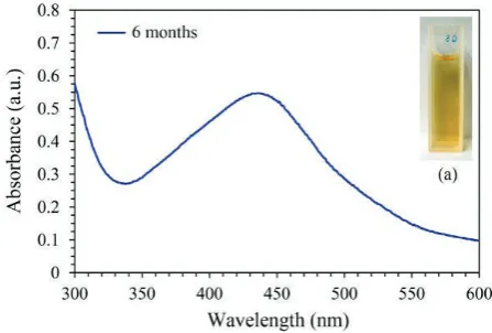
It is observed that the maximum absorbance of Ag nanoparticles occurs at 425 nm, indicating that AgNPs were produced. It was also observed that reduction of silver ions into nanoparticles started after 3 hours of reaction and completed after almost 24 hours. Figure 4 shows the UV–vis absorption spectra of silver synthesized nanoparticles after storage for 6 months to test the stability of the AgNPs.
Figure 3: UV–vis spectra showing absorption recorded as a function of 10−3 M aqueous solution
of silver nitrate with S. marianum seed extract as a function of time. (a) Color of S. marianum seed extract before adding silver nitrate (b) Color of S. marianum seed extract after adding silver
nitrate at 24 h.
As it can be seen, the absorption peaks of the AgNPs
shift only slightly, without a significant change in
the color. This indicates that the as-prepared AgNPs are stable over a long period (Liu et al. 2009).
3.2. XRD analysis
The formations of the nano-crystalline Ag particles
were further confirmed by the XRD analysisdepicted
in Figure 5.
Figure 4: UV–vis spectra of biosynthesized silver nanoparticles by S. marianum seed extract after
six months.
Figure 5: XRD pattern of silver nanoparticles synthesized by treating AgNO3 solution with
Silybum marianum seed extract.
Intense peaks were observed at 2θ values of 38.098o, 44.154o, 64.674o, and 77.544o, corresponding to
(111), (200), (220) and (311) Bragg’s reflection
based on the face-centered-cubic (fcc) crystal structure of AgNPs. The broadening of Bragg’s peaks indicates the formation of nanoparticles. The XRD pattern thus clearly shows that the AgNPs formed by the reduction of Ag+ ions by S. marianum seed extract are crystalline in nature. No additional peak appeared in XRD pattern, indicating a high purity of biosynthesized AgNPs.
3.3. TEM analysis
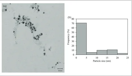
Transmission electron microscopy (TEM) was used to determine the morphology (size and shape) of nanoparticles. The TEM images of the prepared AgNPs at 50 nm scales are shown in the Figure 6a. TEM images show that they have spherical shape. Particle size distribution histogram determined from TEM is shown in Figure 6b. Ag nanoparticles range in size from 1 to 25nm.
4. CONCLUSION
many months without any obvious sedimentation. This was achieved without the use of external stabilizing or capping agents.
We conclude that S. marianum seed extract as a bioreductant and capping agent and also as an easily available plant source plays an important role in the synthesis of highly stable AgNPs. Structural analysis by X-ray diffraction pattern strongly indicated a high purity of biosynthesized AgNPs. This pristine method is facile, cost effective, clean and greener for the synthesis of AgNPs and therefore is applicable for a variety of purposes. Moreover, it is easy to scale-up the production of Ag nanoparticle to industrial scale using this method.
REFERENCES
1. Alt, V., Bechert, T., Steinrücke, P., Wagener, M., Seidel, P., Dingeldein, E., Domann, E., Schnettler, R. An in vitro assessment of the antibacterial properties and cytotoxicity of nanoparticulate
silver bone cement. Biomaterials. Vol. 25, (2004), pp. 4383-4391.
2. Azeredo, H. Nanocomposites for food packaging applications. Food. Res. Int., Vol. 42, (2009), pp. 1240-1253.
3. Chaloupka, K., Malam, Y., Seifalian, A. M. Nanosilver as a new generation of nanoproduct in biomedical applications. Trends. Biotechnol. Vol. 28, (2010), pp. 580-588.
4. de Mel, A., Chaloupka, K., Malam, Y., Darbyshire, A., Cousins, B., Seifalian, A. M. A silver nanocomposite biomaterial for blood-contacting implants. J. Biomed. Mater. Res. A. Part A. Vol. 100A, No. 9, (2012), pp. 2348–2357.
5. Dewick, P. M. Medicinal Natural Products A Biosynthetic Approach. 2nd Edition, John Wiley & Sons, Ltd. (2002)
6. Jain, P., Pradeep, T. Potential of silver nanoparticle-coated polyurethane foam as an antibacterial water filter. Biotechnol. Bioeng., Vol. 90, (2005), pp. 59-63. 7. Karkanis, A., Bilalis, D., Efthimiadou, A. Cultivation
of milk thistle (Silybum marianum L. Gaertn.), a
medicinal weed. Ind. Crop. Prod., Vol. 34, (2011), pp. 825-830.
8. Kassaee, M., Akhavan, A., Sheikh, N., Sodagar, A. . Antibacterial effects of a new dental acrylic resin containing silver nanoparticles. J. Appl. Polym. Sci., Vol. 110, (2008), pp. 1699-1703.
9. Khataee, A. and Mansoori, G.A. Nanostructured titanium Dioxide Materials. Word Scientific, Hackensack, USA (2011).
10. Kokura, S., Handa, O., Takagi, T., Ishikawa, T., Naito, Y., Yoshikawa, T. Silver nanoparticles as a safe preservative for use in cosmetics. Nanomed-Nanotechnol., Vol. 6, (2010), pp. 570-574.
11. Liu, Y., Chen, S., Zhong, L., Wu, G. Preparation of high-stable silver nanoparticle dispersion by using sodium alginate as a stabilizer under gamma radiation. Radiat. Phys. Chem., Vol. 78, (2009), pp. 251-255.
12. Mansoori, G. A. Principles of nanotechnology: molecular-based study of condensed matter in small systems. Word Scientific, Hackensack, USA (2005). 13. Mansoori, G. A., George, T. F., Assoufid, L., Zhang,
G. Molecular building blocks for nanotechnology: From diamondoids to nanoscale materials and applications. Springer. New York, NY (2007).
14. Mansoori, G. A. Synthesis of Nanoparticles by Fungi, U.S. Patent “US 8,394,421 B2”, (2013).
15. Mansoori G. A., Bastami T. R., Ahmadpour, A., Eshaghi, Z. . Environmental application of nanotechnology. Annual Review of Nano Research, Vol. 2, Chapter 10, Cao, G. and Brinker, C.J. (Editors). Word Scientific, Hackensack, USA (2008).
16. Mansoori G.A., De Araujo PLB, De Araujo ES Diamondoid molecules: with applications in biomedicine, materials science, nanotechnology and petroleum science. Word Scientific, Hackensack, USA (2012).
17. Mulvaney, P. Surface plasmon spectroscopy of nanosized metal particles. Langmuir., Vol. 12, (1996), pp. 788-800.
18. Park, Y., Hong, Y., Weyers, A., Kim, Y., Linhardt, R. Polysaccharides and phytochemicals: a natural reservoir for the green synthesis of gold and silver nanoparticles. IET Nanobiotechnol., Vol. 5, (2011), pp. 69-78.
19. Sharma, V. K., Yngard, R. A., Lin, Y. . Silver nanoparticles: green synthesis and their antimicrobial activities. Adv. Colloid Interfac., Vol. 145, (2009), pp. 83-96.
20. Sintubin, L., Verstraete, W., Boon, N. . Biologically produced nanosilver: Current state and future perspectives. Biotechnol. Bioeng., Vol. 109, No. 10, (2012), pp. 2422–2436.
21. Tsuji, M., Gomi, S., Maeda, Y., Matsunaga, M., Hikino, S., Uto, K., Tsuji, T., Kawazumi, H. . Rapid Transformation from Spherical Nanoparticles, Nanorods, Cubes, or Bipyramids to Triangular Prisms of Silver with PVP, Citrate, and H2O2. Langmuir., Vol. 28, (2012), pp. 8845-8861.
22. Vahabi, K., Mansoori, G.A. and Karimi, S. . Biosynthesis of Silver Nanoparticles by Fungus Trichoderma Reesei (A Route for Large-Scale Production of AgNPs). Insciences J., Vol. 1, No. 1, (2011), pp. 65-79; doi:10.5640/insc.010165 ISSN 1664-171X.
23. Wijnhoven, S.W.P., Peijnenburg, W.J.G.M., Herberts, C.A., Hagens, W.I., Oomen, A.G., Heugens, E.H.W., Roszek, B., Bisschops, J., Gosens, I., Van De Meent, D. Nano-silver-a review of available data and knowledge gaps in human and environmental risk assessment. Nanotoxicology., Vol. 3, (2009), pp. 109-138.


![Production and quality of milk thistle (Silybum marianum[L ] Gaertn ) cultivated in cultural conditions of warm agri climatic macroregion](data:image/gif;base64,R0lGODlhAQABAIAAAP///wAAACH5BAEAAAAALAAAAAABAAEAAAICRAEAOw==)