Endovaginal magnetic resonance imaging of stage 1A/1B cervical cancer with A T2- and diffusion-weighted magnetic resonance technique: Effect of lesion size and previous cone biopsy on tumor detectability
Authors: Elizabeth Charles-Edwards, Veronica Morgan, Ayoma D. Attygalle, Sharon L. Giles, Thomas E. Ind, Michael Davis, John Shepherd, Norman McWhinney, Nandita M. deSouza
Introduction
Frequently, the diagnosis of cervical cancer is made at an early stage particularly as tumors detected at screening are of smaller volume than those identified in women who present with vaginal bleeding [1]. Often, the cone or large loop excision of the transformation zone (LLETZ) biopsy on which the diagnosis is made and which leads to routine use of T2-W magnetic resonance imaging for assessing disease extent within the cervix [2], removes a large amount of the disease. The determination of residual disease is crucial in treatment planning; fertility-sparing procedures demand a precise knowledge of the site and extent of any residual disease in order to ensure a curative and optimal surgical strategy. However, even with the use of an
endovaginal coil to improve spatial resolution [3], image contrast between tumor and surrounding granulation tissue following cone biopsy can be insufficient [4] to
delineate the extent of residual disease within the cervix, particularly when disease involves the margins of the cone. This has lead to investigation into alternative contrast mechanisms for identifying tumor.
The use of intrinsic contrast mechanisms such as diffusion-weighted imaging is proving useful in a variety of extra-cranial cancer related
pathologies [5], [6], [7], [8] and [9]. Diffusion-weighted (DW)-MRI is sensitive to the motion of water within tissues of the order of 10–20 μm [10]. Application of a series of diffusion sensitizing gradients allows calculation of an apparent diffusion
coefficient (ADC) for each pixel in the image which is then represented as a map. Tumors generally appear as areas of relatively restricted diffusion, compared to non-tumor regions and histopathological correlations have indicated that this is largely a function of cell density, with limited extracellular space limiting water diffusivity [11]. In the cervix, pilot data has documented values for ADC within tumor and
Materials and methods Patients
This was a prospective, single-institution study with approval from the local research ethics committee. Written informed consent was obtained from each subject.
Patients unsuitable for MR imaging because of metallic implants or claustrophobia were excluded. No account was taken of stage of menstrual cycle. Over a 3-year period (July 2007 to July 2010) 113 women with previously diagnosed cervical cancer were recruited prospectively to this study by gynaecologists in the clinic and subsequently had MR imaging performed on a 1.5-T MRI scanner (Intera; Philips, Best, the Netherlands) using a 37-mm ring coil [13] designed specifically to image the cervix. Of these, 2 were subsequently diagnosed with endometrial cancer, 22 women were upstaged and required chemotherapy and/or radiotherapy rather than a primary surgical approach, 3 patients were found not to have invasive cancer on histological review and 1 patient became pregnant soon after diagnosis and required conservative management. These 28 women were excluded from the study. Of the 85 women (aged, 23–74; median, 34 years), 58 had cone or LLETZ biopsy
performed prior to imaging (median, 35 days; 11–125 days; interquartile range, 25), the remaining 27 patients having had their cancer previously confirmed by less invasive methods of biopsy (e.g. punch biopsy, curettage). After MR imaging 24 were treated conservatively (extended cone biopsy/LLETZ/colposcopic follow-up), 30 had trachelectomy and 31 had radical hysterectomy (2 of whom had neoadjuvant chemotherapy).
Imaging methods
Images were obtained on a 1.5-T Philips Intera using a 37 mm ring design solenoidal receiver coil that has been previously described. The coil was inserted following digital vaginal examination and positioned around the cervix. Air in the vagina introduced during coil insertion was aspirated via a 4 mm diameter tube (Ryles; Pennine Healthcare, London, England) to reduce susceptibility-based artifacts occurring at the air–tissue interface. Each patient was examined supine and coil immobilization achieved using an externally sited clamp whose stand was placed under the patients’ knees. Hyoscine butyl bromide 20 mg was administered routinely intramuscularly to reduce artefact from bowel motion.
T2-W images (repetition time ms/echo time ms, 4500/80; acquisition matrix, 256; field of view, 11 cm; section thickness, 3 mm; no intersection gap) were obtained in three orthogonal planes to the cervix [sagittal, coronal and transverse] with a
view, 20 cm) employing b-values of 0, 100, 300, 500, and 800 s/mm2 were obtained in the coronal plane to match the T2-W images. Twelve 4-mm-thick sections
provided coverage of the cervix (acquisition time 1 min 24 s, in-plane resolution 2.1 mm, voxel size 17.6 mm3). Isotropic ADC maps were generated with the system software using all b-values and an average value for the three directions of diffusion sensitization. Following endovaginal imaging, the internal receiver coil was removed and axial images through the abdomen and pelvis obtained for assessment of lymph node status which did not form part of this study.
Image analysis
All images were evaluated prospectively at the time of reporting using all available clinical information by a single gynaecological radiologist with > 10 years experience of endovaginal MRI. Images were scored in a binary fashion as positive or negative for the presence of tumor based on the combined information from T2-W and DW images viewed together. Images were deemed to be positive for tumor if there was a mass lesion (definite tumor) or nodularity (high index of suspicion) on T2-W imaging with corresponding diffusion restriction on the ADC map. Images were deemed to be negative for tumor if there was no mass (definitely negative) or nodularity (low index of suspicion) on T2-W imaging with no corresponding diffusion restriction on the ADC map. In patients scored positive for invasive disease, tumor volume was measured by drawing a region of interest around the tumor on the T2-W images on every slice containing the lesion, and multiplying the summed area on all slices by slice thickness. Maximum radiological dimensions of the tumor visualised in
superior–inferior, anterior–posterior and right–left directions also were noted on T2-W images.
On the ADC maps, using visual correlation with anatomic information on the
corresponding T2-W image, a slice with the largest tumor area with adjacent cervical epithelium visible was selected, and regions of interest encompassing the entire tumor drawn around the lesion and normal cervical epithelium. In those without visible cervical epithelium, the slice with the largest tumor dimensions was used.
Histopathological analysis
Trachelectomy and hysterectomy sectioning was carried out in the standard manner for our institution. Section thickness was 4 mm. The cervix was sectioned
sagittal plane, while lateral sections related to coronal MR images. For conisation samples, sections were sliced serially in a sagittal/parasagittal plane starting at the 3 or 9 o'clock end. The lateral end-slices were often embedded whole, as they were too small to slice serially in the coronal plane.
Statistical analysis
Statistical analysis used SPSS for Windows v 18.0 and tested for normality using a Shapiro-Wilk test. Mann-Whitney U was used for comparisons of non-normally distributed data. Sensitivity, specificity, positive and negative predictive values of T2-W combined with DT2-W-MRI in distinguishing tumor was established for patients without versus those with previous cone biopsy/LLETZ. Area under receiver operating characteristic (ROC) curve (Az) was used to determine the cut-off lesion volume measured on MRI for best accuracy in identifying the presence of residual invasive disease and cut-off lesion size (maximum dimension on histopathology in all positive cases) for best accuracy in identifying disease on MRI in patients without and with previous cone biopsy/LLETZ. Maximum MR tumor dimension measured on T2-W images was correlated with maximum tumor dimension on histology using Spearman's rho.
In patients with definite radiological and histological evidence of tumor, ADC values from tumor regions of interest (ROIs) were compared with those of adjacent cervical epithelium using a paired t-test after log transformation of non-normally distributed data. ADC values of tumor and normal epithelium were compared between the two groups using a Mann-Whitney U test. A p value of less than .05 was used to indicate significance.
Results
The procedure was tolerated well in all subjects and in no case was termination of the examination requested by any patient. There was no significant difference in age between the 27 patients without (median 36 years, lower quartile [LQ] 28 years, upper quartile [UQ] 45 years) and 58 patients with prior cone biopsy/LLETZ (median 33 years, LQ 29 years, UQ 39 years, p = 0.18).
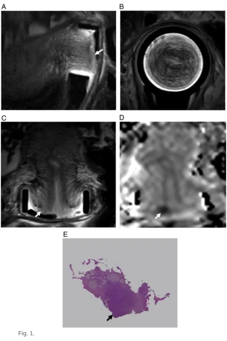
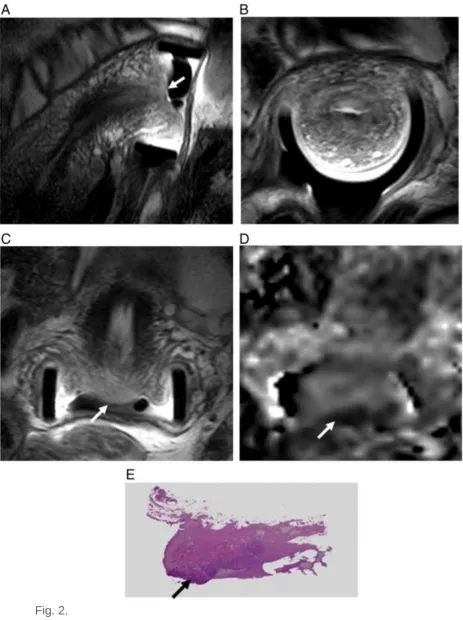
In the 27 patients who had not undergone cone biopsy/LLETZ prior to imaging, 26 were classed as definite disease (Fig. 1) and 1 as low suspicion of disease. In 58 patients with cone biopsy/LLETZ prior to imaging 24 were classed as definite (n = 15 Fig. 2) or a high suspicion (n = 9) of residual invasive disease and scored positive for tumor on imaging. Thirty-four were classed as either no disease (n = 5) or low suspicion of disease (n = 29) and were scored negative radiologically.
Cervical cancer in a 28-year-old female with no cone biopsy or LLETZ prior to MRI: sagittal (A), axial (B) and coronal (C) T2-W images using an endovaginal coil show a 200 mm3 (8 mm maximum dimension) lesion (arrows) on the right anterior cervical lip which, on the coronal ADC map (D) in the same slice position as (C), shows markedly restricted diffusion (arrow). A LLETZ biopsy following MRI (E) confirmed a 9 mm tumor nodule (arrow).
Fig. 2.
Cervical cancer in a 38-year-old female with cone biopsy or LLETZ prior to MRI: sagittal (A), axial (B) and coronal (C) T2-W images using an endovaginal coil show a concave LLETZ defect with an irregular 150 mm3 (17 mm
coronal ADC map (D) in the same slice position as (C), shows markedly restricted diffusion (arrow). A hysterectomy following MRI (E) confirmed a 30 mm tumor plaque (arrow).
Figure options Table 1.
Sensitivity, specificity, positive and negative predictive values for identifying invasive cervical carcinoma using T2-W and ADC maps in patients without or with previous cone biopsies (TP = true positive, TN = true negative, FP = false positive, FN = false negative, PPV = positive predictive value, NPV = negative predictive value).
TP TN FP FN Sensitivity %
Specificity %
PPV %
NPV % Cone
(n = 58)
16 30 8 4 80.0 78.9 66.7 88.2
No cone (n = 27)
26 1 0 0 100 100 100 100
Table options Median tumor volume on the T2-W images in all 50 cases scored positive on
imaging was 900 mm3 (LQ 200 mm3, UQ 4200 mm3), maximal lesion dimension 15.4 mm (LQ 8.6 mm, UQ 25.8 mm). All cases without prior cone biopsy/LLETZ were correctly scored; the smallest tumor volume was 200 mm3 and the smallest untreated maximum dimension on histology was 8 mm. In those with prior cone biopsy/LLETZ a cut-off volume of 83 mm3 (approximately 150 T2-W voxels) detected tumor with 80.0% sensitivity and 94.7% specificity (Az = 0.86; Fig. 3).
Fig. 3.
ROC curve of MRI-derived tumor volume in patients who had cone biopsy/LLETZ prior to MRI gives an area under the curve (Az) of 0.86.
Figure options Of 46 patients with residual disease on histology, 2 had received neoadjuvant
maximal dimension of tumors on histology in the remaining 44 (25 without and 19 with prior cone biopsy/LLETZ) was 20 mm (LQ 10.8 mm, UQ 30.5 mm). An
independent samples t-test demonstrated a significant difference in maximum tumor dimension on pathology in those without (mean ± standard deviation
25.5 ± 13.8 mm) and with (mean ± standard deviation 14.9 ± 9.9 mm) previous cone biopsy/LLETZ (p = 0.007). In patients with prior cone biopsy/LLETZ, a cut-off value of 3 mm maximal histological tumor dimension was detected on MR with 100% sensitivity, 75% specificity; at 5.3 mm, 100% sensitivity and specificity was achieved (Az = 1). The maximum dimension on histopathology in those with prior cone
biopsy/LLETZ was significantly larger in those with a true positive result (7.5–35 mm) than those with a false negative result (1–3 mm) (p = 0.003), although the time
interval between prior cone biopsy/LLETZ and imaging was not statistically significant between these groups (p = 0.42).
Maximal tumor dimension in the 48 MRI positive patients managed surgically without neoadjuvant chemotherapy showed significant positive correlation with histological dimensions (rs = 0.78, p < 0.001) which remained so when the groups were
considered separately (no previous cone biopsy/LLETZ n = 25, rs = 0.79, p < 0.001; with previous cone biopsy/LLETZ n = 23, r = 0.73, p < 0.001).
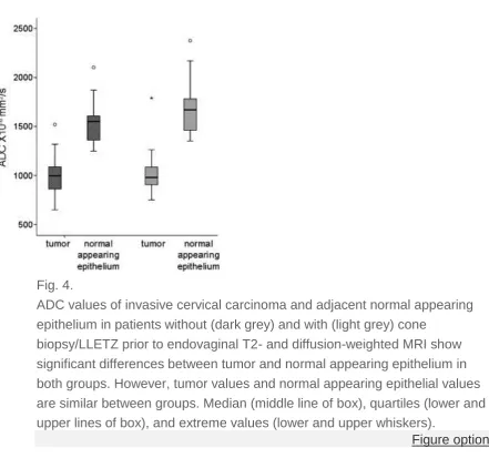
Of the 50 patients with positive radiological diagnosis, 42 had histological evidence of tumor and it was possible to identify tumor and normal epithelium on the T2-W images in 41. ADCs of invasive cervical carcinoma (median 988, LQ 869, UQ
1101 × 10−6 mm2/s) and paired normal epithelial tissue (median 1564, LQ 1430, UQ 1694 × 10−6 mm2/s) were significantly different (p < 0.001) but tumor or cervical epithelial ADCs did not differ significantly between patients who had not (n = 26 and 25, respectively) and those who had (n = 16) prior cone biopsy/LLETZ (p = 0.48 and 0.15 respectively, Fig. 4). Of the patients with prior cone biopsy, ADCs of tissue identified as tumor in the 8 false positive patients (median 1210, LQ 1135, UQ
Fig. 4.
ADC values of invasive cervical carcinoma and adjacent normal appearing epithelium in patients without (dark grey) and with (light grey) cone
biopsy/LLETZ prior to endovaginal T2- and diffusion-weighted MRI show significant differences between tumor and normal appearing epithelium in both groups. However, tumor values and normal appearing epithelial values are similar between groups. Median (middle line of box), quartiles (lower and upper lines of box), and extreme values (lower and upper whiskers).
Figure options
Discussion
This study establishes the sensitivity and specificity of detecting cervical tumor using T2-W and diffusion-weighted MRI with an endovaginal receiver coil in patients
alternative of scanning of a large number of colposcopically abnormal women without definite invasive disease is not practicable.
The sensitivity and specificity with which the radiological assessment could correctly detect tumors with a cut-off size of 5 mm on subsequent histological specimens in patients with prior cone biopsy/LLETZ, indicates that this technique is a useful diagnostic technique prior to fertility-sparing surgery where measurement of the maximal tumor dimensions, particularly the estimation of extent within the endocervical canal, is crucial for surgical planning [15]. Correlation of maximum tumor dimension as visualised on T2-W MRI and that subsequently determined on histology was good regardless of prior cone biopsy/LLETZ despite the fact that some tumor configurations (e.g. a thin, flat plaque) may be associated with a large
maximum tumor dimension but small tumor volume and hence more difficult to visualize radiologically. Because of the varying orientations of the histological
sections and the MRI scans, a maximal dimension from each technique was used for comparison. In future, a volume-based MRI acquisition technique may enable more accurate orientation of the MRI abnormality with the histology.
The ADCs of tumor remained significantly different to that of adjacent normal epithelium independent of the existence of a prior cone biopsy. This extends the finding from previous work where we have shown that the ADC values of cervical tumor are significantly lower than surrounding normal tissue [12]. Interestingly there was no significant difference in normal epithelium in patients with previous cone biopsy compared to those without, despite the likelihood that such a procedure would disrupt the normal epithelium. With a median time between cone biopsy and MR scanning of ~ 30 days it is feasible that much of the acute reaction to injury may have subsided by the time the scan was performed.
The success of a T2- plus diffusion-weighted diagnostic approach is necessarily limited by the resolution achievable with our current diffusion-weighted sequence (2.1 mm in-plane, 17.6 mm3 voxel size). Although the maximum dimensions on histology of the false negative tumors were 3 mm, shrinkage is likely during
histological fixation and has been estimated at 15% for cervical tissue [16], so that these tumors may have occupied less than 3 voxels on a diffusion-weighted image and resulting ADC values increased through partial volume effects. It may be possible to reduce the false negative rate in patients with previous cone biopsy by improvements in spatial resolution of the technique whilst maintaining an appropriate signal-to-noise ratio. This may be afforded by scanning patients at a higher field strength, increasing the sequence scan time or using fewer b-values whilst increasing the number of signal averages.
contributed to significantly higher ADCs in tissue identified as tumor in the false positive group compared to the true positives in the post cone biopsy cohort. There were no false positives or negatives associated with patients who had not undergone a previous cone biopsy/LLETZ, which shows superiority to quoted values for
standard external array techniques [17].
A limitation to this study is that, whilst prospective, a single experienced operator reported all images immediately after data acquisition and prior to any surgery, and therefore the study did not benefit from intraobserver data. Our previous
study [12]indicated that the inclusion of ADC maps resulted in good intraobserver agreement. In future a multicentre prospective study should establish whether such an observation holds for multiple observers in a larger group of patients.
In investigating the effects of a cone biopsy/LLETZ prior to MR imaging we did not differentiate between the two methods despite a potential difference in the type of trauma associated with the two techniques [18] and [19]. As an initial investigation into the impact of significant tissue removal prior to an MR examination it was thought more appropriate to combine the two types of biopsy in the analysis. Investigating the potentially different impacts of the two approaches on the
subsequent accuracy of MR diagnoses will form the basis of further investigation. Such future work and the improvement of the spatial resolution of the diffusion-weighted MR sequence has the potential to further improve the management of women diagnosed with cervical cancer who wish to be considered for conservative fertility-sparing surgery.
In summary, a combination of T2-W with diffusion-weighted imaging using an endovaginal technique is invaluable for detecting small cervical cancers, prior to fertility-sparing procedures although sensitivity and specificity are lower following a previous cone biopsy/LLETZ procedure. However, the size of tumors detected even post cone/LLETZ is of the order where fertility-sparing surgery remains a major management option. This procedure remains to be evaluated in multicentre trials, but offers enormous potential in the pre-operative management of this group of patients.
Conflict of interest statement
None of the authors have a financial interest in the consequences of the findings reported here and none of the authors have a conflict of interest represented by their involvement in the work reported.
Acknowledgments
References
1. N.M. deSouza, W.P. Soutter, G.A. McIndoe, D.J. Gilderdale, T. Krausz. Stage I cervical cancer: tumor volume by magnetic resonance imaging of screen-detected versus symptomatic lesions
J Natl Cancer Inst, 89 (17) (Sep. 3 1997), pp. 1314–1315
2. D.A. Milliken, J.H. Shepherd. Fertility preserving surgery for carcinoma of the cervix
Curr Opin Oncol, 20 (5) (Sep. 2008), pp. 575–580
3. N.M. deSouza, R. Dina, G.A. McIndoe, W.P. Soutter. Cervical cancer: value of an endovaginal coil magnetic resonance imaging technique in detecting small volume disease and assessing parametrial extension
Gynecol Oncol, 102 (2006), pp. 80–85
4. N.M. deSouza, M. Whittle, A.D. Williams, M. Sohail, T. Krausz, D.J. Gilderdale, G.A. McIndoe, W.P. Soutter. Magnetic resonance imaging of the primary site in stage I cervical carcinoma: a comparison of endovaginal coil with
external phased array coil techniques at 0.5 T
J Magn Reson Imaging, 12 (6) (Dec. 2000), pp. 1020–1026
5. H.C. Thoeny, F. De Keyzer. Extracranial applications of diffusion-weighted magnetic resonance imaging
Eur Radiol, 17 (6) (Jun. 2007), pp. 1385–1393
6. Whittaker CS, Coady A, Culver L, Rustin G, Padwick M, Padhani AR. Diffusion-weighted MR imaging of female pelvic tumors: a pictorial review. Radiographics. 2009 May-Jun;29(3):759-74; discussion 774-8.
7. T. Namimoto, K. Awai, T. Nakaura, Y. Yanaga, T. Hirai, Y. Yamashita. Role of diffusion-weighted imaging in the diagnosis of gynecological diseases
Eur Radiol, 19 (3) (Mar. 2009), pp. 745–760
8. A.A. Abdel Razek, G. Gaballa, A. Denewer, I. Tawakol. Diffusion weighted MR imaging of the breast
Acad Radiol, 17 (3) (Mar. 2010), pp. 382–386
9. N.M. deSouza, S.A. Reinsberg, E.D. Scurr, J.M. Brewster, G.S. Payne. Magnetic resonance imaging in prostate cancer: the value of apparent diffusion coefficients for identifying malignant nodules
10. E.M. Charles-Edwards, N.M. DeSouza. Diffusion-weighted magnetic resonance imaging and its application to cancer
Cancer Imaging, 6 (Sep. 13 2006), pp. 135–143
11. T. Sugahara, Y. Korogi, M. Kochi, I. Ikushima, Y. Shigematu, T. Hirai, T. Okuda, L. Liang, Y. Ge, Y. Komohara, Y. Ushio, M. Takahashi. Usefulness of
diffusion-weighted MRI with echo-planar technique in the evaluation of cellularity in gliomas
J Magn Reson Imaging, 9 (1) (Jan. 1999), pp. 53–60
12. E.M. Charles-Edwards, C. Messiou, V.A. Morgan, S.S. De Silva, N.A.
McWhinney, M. Katesmark, A.D. Attygalle, N.M. DeSouza. Diffusion-weighted imaging in cervical cancer with an endovaginal technique: potential value for improving tumor detection in stage Ia and Ib1 disease
Radiology, 249 (2) (Nov. 2008), pp. 541–550
13. N.M. deSouza, D. Scoones, T. Krausz, D.J. Gilderdale, W.P. Soutter. High-resolution MR imaging of stage I cervical neoplasia with a dedicated transvaginal coil: MR features and correlation of imaging and pathologic findings
AJR Am J Roentgenol, 166 (1996), pp. 553–559
14. D. Fischerova, D. Cibula, H. Stenhova, H. Vondrichova, P. Calda, M. Zikan, P. Freitag, J. Slama, P. Dundr, J. Belacek. Transrectal ultrasound and magnetic resonance imaging in staging of early cervical cancer
Int J Gynecol Cancer, 18 (4) (Jul-Aug. 2008), pp. 766–772
15. A. Sahdev, S.A. Sohaib, A.E. Wenaden, J.H. Shepherd, R.H. Reznek. The performance of magnetic resonance imaging in early cervical carcinoma: a long-term experience
Int J Gynecol Cancer, 17 (3) (May-Jun. 2007), pp. 629–636
16. H. Boonstra, J.W. Oosterhuis, A.M. Oosterhuis, G.J. Fleuren. Cervical tissue shrinkage by formaldehyde fixation, paraffin wax embedding, section cutting and mounting
Virchows Arch A Pathol Anat Histopathol, 402 (2) (1983), pp. 195–201
ultrasonography and magnetic resonance imaging for assessment of presence, size and extent of invasive cervical cancer
Ultrasound Obstet Gynecol, 34 (3) (Sep. 2009), pp. 335–344
18. O.A. Oyesanya, C. Amerasinghe, E.A. Manning. A comparison between loop diathermy conization and cold-knife conization for management of cervical dysplasia associated with unsatisfactory colposcopy
Gynecol Oncol, 50 (1) (Jul. 1993), pp. 84–88
19. P. Mathevet, D. Dargent, M. Roy, G. Beau. A randomized prospective study comparing three techniques of conization: cold knife, laser, and LEEP