ORIGINAL ARTICLE
DISEASE EXPRESSION AND HLA TYPES IN EARLY AND LATE
ONSET DISEASE OF MALAYSIAN PATIENTS WITH SYSTEMIC
LUPUS ERYTHEMATOSUS
Azizah Mohd Radzi, Kuak Soo Huan, Normaznah Yahaya, Ainol Shahera*, Norella Kong**, Rahim Mohd Noah***
Biotechnology Unit, Institute for Medical Research, Kuala Lumpur *Hospital Penang, Penang
**HUKM, Cheras, Kuala Lumpur
***Department of Bio-medicine, Faculty of Allied Science, UKM, Kuala Lumpur
Age has been suggested to modify systemic lupus erythematosus expression. In this study we have attempted to study 13 patients with late onset (40 years and above) and 90 with early onset disease (below 40 years) to determine whether age-related differences in disease expression exist and whether the genetic make-up influences the age of disease onset. We found that patients with late onset disease initially presented with pericarditis (31% vs 3%, P<0.005) and a lower incidence of malar rash (31% vs 57%, p<0.05). During the disease course, there was a lower incidence of mucocutaneous symptoms especially malar rash (p<0.005) and psychosis (p<0.05) in the late onset group. Serological parameters were similar in both groups. There was a prevalence of HLA-DQA1*0103 in Chinese patients with late onset disease (pcorr=0.004). These findings suggest that a subgroup of late onset patients may experience milder disease and that the risk conferred by the HLA-DQA1*0103 may be significant among these patients.
Key words : Systemic lupus erythematosus, disease onset, autoantibodies, autoimmunity, HLA, clinical, serological
Introduction
Systemic lupus erythematosus (SLE) is a chronic autoimmune disorder which may affect multiple organ systems. It occurs predominantly in young women with a peak incidence from the second to fourth decades of life (1). The onset of SLE later in life is uncommon and form 6.1 to 18% of the lupus population (2-4). Elderly patients with the disease have been known to have an insidious onset (4-9) and follow a more benign course (2,5,6,9,10).
studied the clinical, serological manifestations and the HLA types of patients with late and early onset SLE.
Materials and Methods
Patients
The study cohort consisted of 103 patients with SLE attending the SLE Clinic of the National University Hospital of Malaysia, Kuala Lumpur, between 1997-1998. All patients met the American College of Rheumatology (ACR) revised criteria for the classification of SLE (13). Patients were divided into two groups with regard to their age at disease onset; early (less than 40 years of age) and late (40 years and above) onset. One hundred and twenty-five unrelated healthy individuals matched for age and sex served as the control group for HLA-typing. Clinical and laboratory manifestations
A retrospective chart review provided data on the demography, physical findings and laboratory
investigations of these patients. The present physical assessment was carried out according to a pre-established questionnaire by an internist or a rheumatologist. Sera for serological studies and anti-coagulated blood for HLA typing was collected. The present age was defined as the age when the patient entered into the study; age at onset of the disease was defined as the initial manifestation clearly attributable to SLE; age at diagnosis was defined as the age when the patient fulfilled 4 or more of the 1982 revised ACR criteria for the classification of SLE. Disease duration was also recorded. The clinical manifestations assessed at the onset and during the disease course were: fever ; mucocutaneous involvement: malar rash, discoid rash, alopecia, photosensitivity and oral ulcers; arthritis; pericarditis and/or pleuritis; lymphadenopathy; neurological involvement: psychosis and seizure; renal involvement; haematological disorders: autoimmune hemolytic anaemia, thrombocytopenia of less than 100,000/ mm3, and other clinical features, including
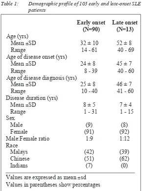
Early onset Late onset
(N=90) (N=13)
Age (yrs)
Mean
±
SD
32 ± 10
52 ± 8
Range
14 - 61
40 - 69
Age of disease onset (yrs)
Mean
±
SD
24 ± 8
45 ± 7
Range
8 - 39
40 - 60
Age of disease diagnosis (yrs)
Mean
±
SD
25 ± 8
46 ± 7
Range
10 - 40
41 - 60
Disease duration (yrs)
Mean
±
SD
8 ± 5
7 ± 4
Range
1 - 31
1 - 15
Sex
Male
(9)
(8)
Female
(91)
(92)
Male:Female ratio
1:9
1:12
Race
Malays
(42)
(39)
Chinese
(51)
(62)
Indians
(7)
(0)
Values are expressed as mean
±
sd
Values in parentheses show percentages
Raynaud’s phenomenon and thrombosis.
The laboratory assessment included measurements of antinuclear antibodies (ANA) as detected by indirect immunofluorescence using mouse liver as substrate (IMMCO Diagnostics, USA); anti-ds DNA antibodies, antibodies to the extractable nuclear antigens (Sm, U1RNP, SSA (Ro), and SSB (La)) and anti-cardiolipin antibodies (IgG ACA and IgM ACA) assayed by ELISA (IMMCO, USA) as well as complement levels using turbidimetry technique (Behring Marburg, Germany). The information collected from the questionnaire was transferred to a computerized database program and further analysed using SPSS.
HLA typing
Genomic DNA was purified from peripheral blood leucocytes using the salting-out method (14) . DNA typing for “broad” DR groups (DR1,2,3,4,5,6,7,8,9,10) were determined by PCR while DQA1, DQB1 and DPB1 genotyping was performed by a modified PCR-RFLP (15,16) . Genomic DNA was amplified by PCR with 2.5 units
of the Taq DNA polymerase (Fermentas AB, Lithuania), 200mM dNTPs, 2.5mM Magnesium chloride, 0.25mM primers in a 20ml reaction buffer. This mixture was subjected to 35 cycles of 1 min at 96ºC, 1 min at 55ºC and 2min at 72ºC in an automated thermocycler (Perkin Elmer Cetus Inc) for the DQB1 gene. As for the DQA1 and DPB1 gene, amplification was done for 30 cycles of 1 min at 94ºC, 1 min at 62ºC and 2 min at 72ºC. After amplification, aliquots of the amplicon were digested by restriction endonucleases (FokI, ApaI, HaeII, SfaNI and BssHII, HphI, BglI, SacI, AcyI and HpaII for DQB1, ApaLI, HphI, BsaJI, FokI, MboII and MnlI for DQA1 and Bsp12861, FokI, DdeI, BsaJI, BssHII, Sau96I, RsaI, EcoNI and AvaII for DPB1). Electrophoresis was then performed and the bands visualised by ethidium bromide staining.
Statistical analyses
Statistical analyses were carried out using conventional Chi square test and Fischer’s exact test for comparing qualitative differences , uncorrected for multiple comparisons. The HLA antigen
Manifestations
At onset
Disease course
Early-onset Late-onset
Early-onset Late-onset
(%)
(%)
(%)
(%)
Fever
59
54
66
62
Mucocutaneous
74
54
93
62
**Malar rash
57
31
*74
31
**Discoid lesion
5
0
15
0
Photosensitivity
44
31
58
31
Alopecia 50
39
64
39
Arthritis
29
46
42
54
Pleuritis
8
15
17
23
Pericarditis
3
31
**11
31
Renal involvement
44
54
65
62
Neurologic14
15
18
23
involvement
Seizures 30
10
8
Psychosis
6
8
16
8
*Thrombocytopenia
15
8
23
15
Hemolytic anemia
12
23
19
39
Raynaud’s
6
0
15
8
phenomenon
Thrombosis
3
0
8
8
Lymphadenopathy
13
15
19
31
P value from chi-square and Fischer’s exact tests:
*<0.05,
**<0.005
frequencies in patients and controls were done using a 2x2 table (EPI-INFO statistical programme, CDC, Atlanta, GA). A p value of <0.05 was considered to be significant. Significant p values were corrected for the number of antigens/alleles tested. The non-parametric Mann Whitney U test was used to compare age differences between groups. The data are presented as mean ± standard deviation .
Results
Patient population
The study consisted of 103 patients with SLE (Table 1) comprising of 93 females and 10 males with a female:male ratio of 9:1. They consisted of the different races; 43 (42%) Malays, 53 (51%) Chinese and 7 (7%) Indians . In 10 (10%) patients, the onset of disease occurred at or above the age of 40 years (late onset) while in 93 (90%), disease onset was below 40 years (early onset). The female:male ratio was 9:1 and 12:1 in the early and late onset groups respectively. Chinese constituted the majority of patients in both groups (51% and 62%) while there were no Indians in the late onset group. The mean age at onset and disease diagnosis for both groups are shown in Table 1. No significant difference was found between the groups with regard to disease duration, sex and racial distribution.
Main clinical and serological manifestations The frequeny of SLE manifestations at disease onset and in the course of the disease for the two groups are listed in Table 2. The late onset group predominantly with pericarditis as compared to the artly onset group (31% vs 3%, p<0.005)). Malar rash
(31% vs 57%) occurred at a lower incidence in the late onset group (p<0.05). Arthritis (46% vs 29%), renal involvement (54% vs 44%) and hemolytic anaemia (23% vs 12%) were also common presentations of this group although these manifestations were not significantly more prevalent than the early onset group. In our study cohort, no patients with late onset disease presented with discoid lesion, seizures, Raynauds phenomenon and thrombosis. The other presentations occurred at similar frequencies in both groups.
After analysing clinical manifestations during the course of the disease, we found that mucocutaneous symptoms, especially malar rash was a less common presentation of patients with late onset disease (31% vs 74%, p<0.005). Psychosis was also another uncommon presentation of this group (8 % vs 16%, P<0.05). There were no patients from the late onset group with discoid lesion. Although found to be non-significant, there was a lower incidence of photosensitivity, oral ulcers, alopecia but a higher incidence of arthritis, pericarditis, hemolytic anaemia and lymphadenopathy in patients in the late onset group. Table 3 summarizes the findings of serological parameters of both groups. There was no significant difference in the serological status or parameter in both groups.
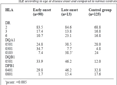
HLA antigens
When the values were corrected for the number of HLA alleles tested, a significant difference was found between DQA1*0103 (p corr=0.004) and late onset disease (Table 4). As for DPB1*0801, p “unocrrected” was 0.046 but did not
Parameter
Early-onset
Late-onset
(%)
(%)
ANA
95
92
Anti ds DNA
66
69
Anti SSA(Ro)
32
54
Anti SSB(La)
47
54
Anti U1RNP
36
31
Anti Sm
17
8
IgG ACA
62
77
IgM ACA
6
8
Low C3
9
23
Low C4
15
15
ANA: antinuclear antibodies
ACA: anticardiolipin antibodies
remain significant after correction.
Discussion
SLE is an autoimmune disease which may affect multiple organs. It is highly heterogenous in its clinical expression. The hallmark of this disease is the production of autoantibodies against tissue components leading to the deposition of antigen-antibody complexes in various organs finally resulting in organ failure. Various studies have attempted to identify subgroups of these patients based on their age at disease onset and have documented variability in clinical and laboratory manifestations between early and late onset disease (2,9,17-22). To analyse the clinical and laboratory manifestations of SLE in relation to the age of onset, we studied patients with late onset disease (40 years and above) and early onset disease (below 40 years). However, accurate comparison between studies are difficult to make because division of patients into groups of late or early onset have been arbitrary and inconsistent. While a study divided patients into juvenile (0-18 years) and adult onset (>18 years) (19) another grouped patients into 3 age groups below 16 years, 17 to 49 years, and those above 50 years (20), while others grouped them into those below and above 50 years (8,11,21,22). Furthermore, both Catoggio et al (9) and Ballou et al(2) divided their patients into those below and above 55 years.
The female to male ratio of our patients were somewhat similar in both groups, and this was in agreement with that reported by others (6,10,23). Ballou et al(2) found it to be less apparent in the older patients (3:1) than in the younger age group (8:1), which was supported by other findings (1,24,25,27) . However a significantly lower female: male ratio (4:1) in the early onset group (20) was supported by King et al (26).
Other studies have reported more male patients among elderly subjects with SLE (2,24) while Shaikh & Wang (21), Catoggio et al (9) and our current study found that all late onset patients above the age of 55 were females. We did not find any significant difference between the two groups of patients in terms of age of disease duration, sex or race. Costallat et al (20) also found no difference among the three groups of patients studied in terms of mean follow-up, disease duration or age. Koh & Boey (18) did not find sex differences with regard to age at onset and diagnosis, in contrast to a study which reported more men with older age of diagnosis compared to women (23).
Several investigators have reported on age-related differences in clinical and serological manifestations in SLE. Ballou et al (2) found renal disease, central nervous system involvement, cutaneous involvement and hemocytopenia and anti-ds DNA antibodies occurred with similar frequency in both groups, while hypocomplementemia was
HLA
Early onset
Late onset
Control group
(n=90)
(n=13)
(n=125)
DR
2
83.5
84.6
68.8
3
17.4
13.8
16.8
6
10.7
23.1
16.8
DQA1
0501
24.8
38.5
28.0
0301
34.7
7.7
4.8
0103
7.4
38.5
*24
DQB1
0501
33.9
46.2
12.0
DPB1
0401
29.8
46.2
32.8
0801
1.7
15.4
17.6
*
pcorr: <0.005
more common in the younger patients. While a decreased incidence of renal involvement in the older age was observed (10), others found skin involvement was less frequent in the older age group (6,28). Costallat et al (20) noted alopecia as an early manifestation as well as seizures, nephrotic syndrome, gastro-intestinal involvment, higher numbers of positive LE cells and anti-DNA antibodies in the younger age group while elderly patients presented more frequently with pericarditis and milder disease. Cervera et al (17) found malar rash, photosensitivity, arthritis and nephropathy to be less common in the older onset group with serositis and pulmonary involvement as the most common feature in this age group which was in agreement with that found by us and several others (6,8,9). Catoggio et al (9) noted that the initial presentations of the elderly onset group were predominantly cutaneous, neuropsychiatric and pulmonary, and less frequently with arthritis compared to the younger patients. Shaikh & Wang (21) observed rash and arthritis to be the commonest initial presentation, and nephritis to be a less common initial presenting manifestation in the older onset group.
Our findings of a high incidence of pericarditis and arthritis at disease onset was supported by Costallat et al (20). In addition, we also found renal involvement and haemolytic anaemia to be common initial presentations of the older onset group. During the disease course, the lower incidence of mucocutaneous symptoms especially malar rash in the late onset group (p<0.005) was also supported by the findings of Costallat et al(20).
It has been suggested that anti-SSA (Ro) and anti-SSB (La) antibodies were more prevalent in older patients and may serve as a useful aid in establishing the diagnosis of SLE in this group of patients (2,8,23). With regard to serological parameters, we did not find any significant difference in their incidence between the two groups. Others found a higher incidence of anti-SSA (Ro) antibodies in the late onset group (9,18) . Cervera et
al (17) , however, noted that the incidence of anti-SSB (La) had a tendency to decrease in the older onset group. Others observed a significantly increased incidence of hypocomplementemia and anti-ds DNA antibodies (21) in late onset disease. While a higher incidence of positive LE cells in younger patients (29) were observed, others did not find any differences in prevalence of ANA in the two groups (19).
The association of HLA antigens and the age of onset has been studied by several investigators (12,30,31). In this study, HLA DQA1*0103 was found to be significantly associated with late onset disease. A previous study reported no significant differences in DR3 or DR2 haplotypes in patients below 18 years of age compared to the older onset group (31). However, Bell et al (30) found HLA DR3 was significantly increased in female patients with late disease onset (above 35 years). In patients with age of onset below 30 years, Davies et al (12) found elevated frequencies of DQA1*0501, and DR3. When the onset of disease was set at 30 years of age, we found that Chinese patients in the later age of disease onset (30 years or more) were significantly associated with DQA1*0102 while in the Malays, there was no DR or DQ association with early or late onset disease. The differences in the immunogenetic profiles of early and late onset SLE, with the classical markers particularly prevalent, may help explain some of the differences between the two groups (32).
The presence of a well-defined clinical profile among late onset patients is controversial. Variability in the findings among previous studies may be due to the small sample size, choice of patients, arbitrary division of patient groups, and different genetic determinants. In this study, we have confirmed the general acceptance that age modifies the expression of SLE. The variability in genetic determinants of disease as shown by the different age groups of SLE patients may suggest response to different disease triggering mechanisms (9,30). Moreover the changing level of sex hormones and aging of the immune system may affect the clinical and immunological manifestations of the lupus population
Acknowledgements
This work is partially supported by the IRPA grant 06-05-01-0121. We wish to thank the Director of the Institute for Medical Research, Kuala Lumpur for his permission to publish this paper.
Correspondence:
Dr Azizah Mohd Radzi,
Biotechnology Centre,
Institute for Medical Research,
Jalan Pahang,
References
1. Estes, D., Christian, C.L. The natural history of systemic lupus erythematosus by prospective analysis.
Medicine. 1971; 50: 85-104.
2. Ballou, S.P., Khan, M.A., Kushner, L. Clinical features of systemic lupus erythematosus. Differences related to race and age of onset. Arthritis Rheum. 1982; 25: 55-60.
3. Domenech, I., Aydintug O., Cervera R, Khamashta M, Jedryka - Goral A, Vianna JL, Hughes GR. Systemic lupus erythematosus in 50 year olds. Postgrad. Med. J. 1992; 68: 440-44.
4. Ward, M.M., Polisson, R.P. A meta-analysis of the clinical manifestations of older-onset systemic lupus erythematosus. Arthritis Rheum. 1989; 32: 1226-32.
5. Foad, B.S.I., Sheon, R.P., Kirsner A.B. Systemic lupus erythematosus in the elderly. Arch Intern Med 1972; 130: 743-6.
6. Baker, S.B., Rovira, J.R., Campion, E.W., Mills JA. Late onset systemic lupus erythematosus. Am. J. Med. 1979; 66: 727-32.
7. Gossat, D.M., Walls, R.S. Systemic lupus erythematosus in later life. Med. J. Aust. 1982; 1: 297-9.
8. Maddison, P.J. Systemic lupus erythematosus in the elderly. J. Rheumatol. 1987; 14: 182-87.
9. Catoggio, L.J. Skinner, R.P., Smith, G., Maddison PJ. Systemic lupus erythematosus in the elderly: Clinical and serological characteristics. J. Rheumatol. 1984; 11: 175-181.
10. Wilson, H.A., Hamilton, M.E., Spyker, D.A., BrunnerCM, O’ Brien WM, Davis JS 4 Th, Winfield JB. Age influences the clinical and serologic expression of systemic lupus erythematosus. Arthritis Rheum.
1981; 24: 1230-35.
11. Bell, D.A., Rigby, R., Stiller, C.R. Clark WF, Harth M, Eber G. HLA antigens in SLE: relationship to disease severity, age at onset and sex. J Rheum 1984; 11: 475-79.
12. Davies, E.J., Hillarby, M.C., Cooper, R.G., Hay EM, Green JR, Shah S, Bernstein RM. HLA-DQ, DR and complement C4 variants in SLE. Br J Rheumatol 1993; 32: 870-75.
13. Tan, E.M., Cohen, A.S., Fries, J.F., Masi AT, Meshane DJ, Rothfield NF, Schaller JG, Talal N, Winchester RJ. The 1982 revised criteria for the classification of
SLE. Arthritis Rheum 1982; 25: 1271-7.
14. Miller, S.A., Dykes, D.D., Polesky, H.F. A simple salting out procedure for extrsacting DNA from human nucleated cells. Nucl Acid Res 1988; 85: 1215. 15. Ota, M., Seki, T., Nomura, N., Sugimara K, Mizuki N,
Fukushima H, Tsujiki K, Inoko H. Modified PCR-RFLP method for HLA-DPB1 and DQA1 genotyping.
Tissue Antigens 1991; 38: 60-71.
16. Nomura, N., Ota, M., Tsuji, K., Inoko H. HLA-DQB1 genotyping by a modified PCR-RFLP method combined with group-specific primers. Tissue antigens
1991; 38: 53-39.
17. Cervera, R., Kamashta, M.A., Font, J., Sebastiani GD, Gil A, Larilla P, Domenench I, Aydintug AO, Jedryka-Goral A, de Ramon E. The European working party of systemic lupus erythematosus: clinical and immunological patterns of disease expression in a cohort of 1000 patients. Medicine. 1993; 72: 113-18.
18. Koh, E.T., Boey, M..L. Late onset lupus: a clinical and immunological study in a predominantly Chinese population. J. Rheum. 1994; 21: 1463-67.
19. Carreno, L., Lopez-longo, F.J., Monteagudo, I., Rodrigezez - Mahoy M, B ascones M, Gonzalez Cm, Saint-Cyr C, Lapointe N. Immunological and clinical differences between juvenile and adult onset of systemic lupus erythematosus. Lupus 1999; 8: 287-92.
20. Costallat, L.T.L., Coimbra, A.M.V. Systemic lupus erythematosus: clinical and laboratory aspects related to age at disease onset. Clin & Exp Rheumatol 1994; 12: 603-7.
21. Shaikh, S.K.J., Wang, F. Late-onset systemic lupus erythematosus: clinical and immunological characteristics. Med. J. Malaysia.1995; 50: 25-31.
22. Mak, S.K., Lam, E.K.M., Wong, A.K.M. Clinical profile of patients with late-onset systemic lupus erythematosus: not a benign subgroup. Lupus. 1998; 7: 23-28.
23. Hochberg, M.C., Boyd,R.E., Ahearn, J.M., Arnett FC, Bias WB, Provost TT, Stevens MB. Systemic lupus erythematosus : a review of clinico-laboratory features and immunogenetic markers in 150 patients with emphasis on demographic subsets. Medicine. 1985; 65: 285-95.
24. Maddock,R.K. Incidence of systemic lupus erythematosus by age and sex. JAMA. 1965; 191: 149-50.
25. Masi, A.T., Kaslow, R.A. Sex effects in systemic lupus erythematosus: a clue to pathogenesis. Arthritis Rheum. 1978; 21: 480.
26. King, K.K., Kornreich, H.K., Bernstein, B.H., Singsen, B.H., Hanson, V. The clinical spectrum of systemic lupus erythematosus in childhood. Arthritis Rheum
1977; 20: 287-94.
27. Fries, J.F., Weyl, S., Holman, H.R. Estimating prognosis in systemic lupus erythematosus. Am J Med
1974; 57: 561-72.
28. Urowitz, M.B., Stevens, M.B., Shulman, L.E. The influence of age on the clinical pattern of systemic lupus erythematosus. Arthitis Rheum. 1967; 10: 319. 29. Hashimoto, H., Tsuda, H., Hirano, T., Takasaki Y,
182-30. Bell, D.A. SLE in the elderly – Is it really SLE or systemic Sjogrens syndrome? J Rheumatol 1988; 15: 723-4.
31. Barron, K.S., Silverman, E.D., Gonzales, J., Reveille JD. Clinical, serologic and immunogenetic studies in childhood-onset SLE. Arhthritis Rheum 1993; 36: 348-54.