SUPPLEMENT ARTICLE
Environmental Factors and Puberty Timing: Expert
Panel Research Needs
Germaine M. Buck Louis, PhDa, L. Earl Gray, Jr, PhDb, Michele Marcus, PhD, MPHc, Sergio R. Ojeda, DVMd, Ora H. Pescovitz, MDe, Selma Feldman Witchel, MDf, Wolfgang Sippell, MD, PhDg, David H. Abbott, PhDh, Ana Soto, MDi, Rochelle W. Tyl, PhDj,
Jean-Pierre Bourguignon, MD, PhDk, Niels E. Skakkebaek, MD, DMScl, Shanna H. Swan, PhDm, Mari S. Golub, PhDn, Martin Wabitsch, MD, PhDo, Jorma Toppari, MD, PhDp, Susan Y. Euling, PhDq
aEpidemiology Branch, National Institute of Child Health and Human Development, National Institutes of Health, Bethesda, Maryland;bEndocrinology Branch, National Health and Environmental Effects Research Laboratory, Office of Research and Development, US Environmental Protection Agency, Research Triangle Park, North Carolina;cDepartment of Epidemiology, Rollins School of Public Health, Emory University, Atlanta, Georgia;dDivision of Neuroscience, Oregon National Primate Research Center-Oregon Health and Sciences University, Beaverton, Oregon;eDepartment of Pediatrics and Cellular and Integrative Physiology, Riley Hospital for Children, Indiana University School of Medicine, Indianapolis, Indiana;fDivision of Endocrinology, Children’s Hospital of Pittsburgh, Pittsburgh, Pennsylvania;gDivision of Endocrinology, Department of Pediatrics, University of Kiel, Kiel, Germany;hDepartment of Obstetrics/Gynecology and Wisconsin National Primate Research Center, University of Wisconsin, Madison, Wisconsin;iDepartment of Anatomy and Cell Biology, Tufts University School of Medicine, Boston, Massachusetts;jCenter for Life Sciences and Toxicology, RTI International, Research Triangle Park, North Carolina;kCHU Sart-Tilman, University of Liege, Belgium;lUniversity Department of Growth and Reproduction, Rigshospitalet, Copenhagen, Denmark;mDepartment of Family & Community Medicine, University of Missouri, Columbia, Missouri;nOffice of Environmental Health Hazard Assessment, California Environmental Protection Agency, Sacramento, California;oDepartment of Pediatric and Adolescent Medicine, University of Ulm, Ulm, Germany;pDepartments of Physiology and Pediatrics, University of Turku, Turku, Finland;qNational Center for Environmental Assessment, Office of Research and Development, US Environmental Protection Agency, Washington, DC
The authors have indicated they have no financial relationships relevant to this article to disclose.
ABSTRACT
Serono Symposia International convened an expert panel to review the impact of environmental influences on the regulation of pubertal onset and progression while identifying critical data gaps and future research priorities. An expert panel reviewed the literature on endocrine-disrupting chemicals, body size, and puberty. The panel concluded that available experimental animal and human data support a possible role of endocrine-disrupting chemicals and body size in relation to alterations in pubertal onset and progression in boys and girls. Critical data gaps prioritized for future research initiatives include (1) etiologic research that focus on environmen-tally relevant levels of endocrine-disrupting chemicals and body size in relation to normal puberty as well as its variants, (2) exposure assessment of relevant endo-crine-disrupting chemicals during critical windows of human development, and (3) basic research to identify the primary signal(s) for the onset of gonadotropin-releasing hormone– dependent/central puberty and gonadotropin-gonadotropin-releasing hor-mone–independent/peripheral puberty. Prospective studies of couples who are plan-ning pregnancies or pregnant women are needed to capture the continuum of exposures at critical windows while assessing a spectrum of pubertal markers as outcomes. Coupled with comparative species studies, such research may provide insight regarding the causal ordering of events that underlie pubertal onset and progression and their role in the pathway of adult-onset disease.
I
N LIGHT OFan increasing body of evidence supporting environmental influences on pubertal onset and progression in contemporary birth cohorts, an expert panel met during and after the Serono Symposia International’s “The Role of Environmental Factors on the Onset and Progression of Puberty” to identify research priorities for delineating the impact of environmental influences on children’s pubertalexperi-ences. An expert review of available animal, clinical, and epidemiologic data was undertaken and synthesized for identifying critical data gaps that are relevant for prioritizing a multidisciplinary research agenda. For purposes of this article, the environment is defined as the sum of all external conditions that affect life, development, and survival of an organism (www.epa.gov/ocepaterms/eterms.html). By extension, an environmental factor is defined as a
non-www.pediatrics.org/cgi/doi/10.1542/ peds.2007-1813E
doi:10.1542/peds.1813E
The views expressed in this article are those of the authors and do not necessarily reflect the views or policies of the US Environmental Protection Agency. Mention of trade names of commercial products does not constitute endorsement or recommendation for use. Drs Buck Louis, Gray, and Marcus contributed equally to this work.
Key Words
human puberty, puberty timing, breast development, menarche, pubic hair development, genital development, endocrine disruptors, body fat Accepted for publication Sep 5, 2007
Address correspondence to Germaine M. Buck Louis, PhD, Epidemiology Branch, Division of Epidemiology, Statistics and Prevention Research, National Institute of Child Health and Human Development, 6100 Executive Blvd, Room 7B03, Rockville, MD 20852. E-mail: louisg@mail.nih.gov
genetic factor; however, for purposes of brevity and consistent with the weight of evidence regarding envi-ronmental influences on puberty, this review focuses on endocrine-disrupting chemicals (EDCs) and body size in relation to puberty timing. An in-depth discussion of the literature cited in this article is beyond the scope of this article, but key study aspects are summarized in accom-panying tables.
REGULATION OF NORMAL PUBERTY ONSET AND PROGRESSION IN HUMANS AND ANIMALS
Puberty is the period of transition from childhood to adolescence and is marked by the development of sec-ondary sexual characteristics, accelerated growth, be-havioral changes, and eventual attainment of reproduc-tive capacity. Available approaches for the assessment of pubertal onset and progression have recently been re-viewed,1including the use of Tanner stages2,3for female breast and pubic hair development and male gonadal and pubic hair development. Menarche is also an impor-tant marker used for assessing puberty in girls. Biochemical markers for female puberty onset and progression include assaying elevations in reproductive hormones (eg, lutein-izing hormone [LH], follicle-stimulating hormone [FSH], 17-estradiol, inhibin A and B) before the onset of physical signs (eg, Tanner breast development stage 2). Other mat-urational markers, such as bone age determinations (radio-graph of left wrist) and ultrasono(radio-graphic assessment of ovarian and uterine volume, can be used.
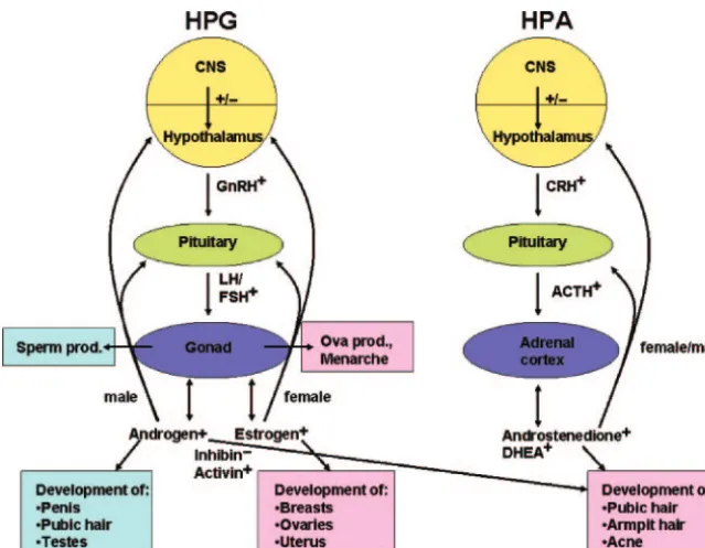
Puberty changes occur as a consequence of the acti-vation of the hypothalamic-pituitary-gonadal (HPG) axis and hypothalamic-pituitary-adrenal (HPA) axis. The HPG axis is under the control of both inhibitory and stimulatory mechanisms, as illustrated in Fig 1. In the human, the HPG is active in the midfetal, neonatal, and early infancy periods but becomes relatively quiescent
during childhood. Puberty is marked by the reactivation of the HPG axis manifested by an increase in frequency and amplitude of gonadotropin releasing hormone (GnRH) pulses in the hypothalamus, leading to a rise in pulsatile secretion of the gonadotropins LH and FSH from the anterior pituitary. Gonadotropin release stim-ulates the gonads. In girls, such maturation of reproduc-tive neuroendocrine function eventually leads to the onset of ovulatory menstrual cycles. Appropriate gonad-otropin regulation of ovarian function results in ovarian follicle secretion of ovarian androgens from theca cells and estradiol from granulosa cells before ovulation and secretion of progesterone from the corpus luteum and estradiol after ovulation. In boys, LH initiates the secre-tion of androgens (testosterone and androstenedione) from the testicular Leydig cells. Such onset of gonadal endocrine function is termed gonadarche. The initial molecular triggers of puberty onset remain unknown, although genetic and environmental factors are sus-pected.
Puberty is also associated with an independent phys-iologic event, adrenarche, or adrenal activation, that typically occurs between 6 and 8 years of age in both genders. Adrenarche leads to pubarche, the secondary sexual changes including pubic hair development, acne, and body odor. Adrenarche is marked by increased 17␣ -hydroxylase (17,20 lyase) activity of the P450c17 en-zyme and increased cytochrome b activity resulting in increased dehydroepiandrosterone, dehydroepiandros-terone sulfate, and androstenedione production. These initial hormonal increases rise over time, resulting in a cumulative dosage of androgens. Adrenarche is a strictly primate phenomenon.4 Although little information is available, there is some evidence that adrenarche occurs before puberty in chimpanzees and during the neonatal period in rhesus macaques and baboons.5
FIGURE 1
Recently, much has been learned about the central mechanisms underlying the initiation of mammalian pu-berty. Rodents, nonhuman primates, and humans have been and remain the main species used to study the basic components and regulatory hierarchies that control the process of puberty. Traditionally assessed pubertal onset end points in rodents include the age of male preputial separation (PPS), an androgen-mediated event, and, in females, the age of female vaginal opening (VO), an estrogen-mediated event. Age at first estrus is an end point used to assess completion of the pubertal process in females because it occurs the day after the first preovu-latory surge of gonadotropins.6 Some studies have as-sessed the age of breast cell differentiation events in the mouse and rat.7,8 Estrogen-mediated pubertal events that occur in nonhuman female primates include men-arche, sex skin swelling and reddening, and nipple growth.9,10Epiphyseal closure and cessation of long bone growth also have been assessed.11 In male monkeys, increases in testicular volume indicate onset of male puberty and reflect both gonadotropin- and testoster-one-mediated events.12Measurement of changes in tes-ticular size seems to be a reliable, noninvasive procedure to detect the initiation of puberty in both human and nonhuman primates.13
In addition to differences in the time frame, progres-sion, and phenotypic landmarks of puberty,14there is a fundamental difference in the neuroendocrine process that controls the initiation of puberty in rodents versus primates. In rodents and sheep,15 GnRH secretion is maintained at low levels during juvenile development by strong steroid inhibitory control, whereas a similar reduction in GnRH secretion is achieved in primates (including humans) by central mechanisms that operate independent of gonadal regulatory inputs.16,17Thus, data indicate that the peripubertal regulation of the GnRH-releasing system is a more centrally controlled process in primates than in rodents that exhibit “gonadal” control of GnRH secretion. Despite this operational difference, the pubertal activation of GnRH release in rodents and primates is initiated and regulated by similar excitatory and inhibitory pathways, providing input to GnRH neu-rons (reviewed by Ojeda and Terasawa18and Ojeda and Skinner6). These inputs include both trans-synaptic and glia-to-neuron communication pathways. As neuronal networks that use excitatory amino acids and the newly discovered kisspeptin peptide for neurotransmission or neuromodulation become activated, there is a concom-itant reduction in the activity of those neurons using the inhibitory neurotransmitters ␥ amino butyric acid and opioid peptides for transsynaptic communication. In ad-dition to this neuron-to-neuron flow of information, several trophic molecules have been identified as com-ponents of the cell– cell signaling process used by glial cells to regulate GnRH secretion. Prominent among these regulatory systems is a signaling complex that uses epidermal growth factor–like ligands and members of the epidermal growth factor receptor family for glia-to-neuron communication (reviewed by Ojeda et al19). Al-though some gene products that are involved in glia and neuronal communication to regulate puberty onset have
been identified, gene(s) upstream that provide the initial trigger of puberty timing remain largely unknown. The integrated use of genomics and proteomics using trans-genic rodent models, nonhuman primates, and humans with alterations in the onset of puberty is beginning to uncover some of the common evolutionary conserved molecular components that are used by the neuroendo-crine brain to control GnRH secretion and, in turn, the initiation of puberty in both rodents and higher pri-mates.20–23An example is the homeodomain geneTTF1, a transcription factor that was recently shown to be involved in the neuroendocrine control of both primate and rodent puberty.24 In addition, recent findings sug-gest that structural remodeling of GnRH neurons may play a key role in the onset of puberty.25
Although the initiating trigger(s) for pubertal onset is unknown for rodents, nonhuman primates and humans, earlier molecular markers have been identified, such as the newly described kisspeptin-GPR54 regulatory sys-tem. Humans who carry mutations of the GPR54 recep-tor22,23 and mice that lack this receptor23,26 each fail to reach puberty, indicative that activation of this signaling complex is essential for pubertal onset. Peptides that are produced by peripheral tissues, such as leptin and insu-lin-like growth factor 1, seem to be more important in maintaining pubertal progression rather than in its ini-tiation despite that insulin-like growth factor 1 has been reported to accelerate pubertal initiation in rodents and monkeys (reviewed by Ojeda and Terasawa18).
Understanding the mechanisms underlying the initi-ation, progression, and regulation of normal human pu-berty is essential for assessing the effect(s) of EDCs on human development. With limited empirical popula-tion-based data, it is important to consider other sources of data, such as those that arise from clinical populations. Although external validity may be limited from these data sources, useful information about potential cause is possible.
CLUES FROM CLINICAL RESEARCH THAT SUPPORT ENVIRONMENTAL INFLUENCES
A number of clinical conditions offer clues regarding the influence on environmental factors, such as nutrition and body size, on puberty. In fact, a recent clinical report attributed topical application of lavender and tea tree oils to gynecomastia in prepubescent boys on the basis of in vitro evidence supporting their weak estrogenic and anti-androgenic activities.27A brief overview of environ-mental influences follows in relation to precocious pu-berty, delayed pupu-berty, and polycystic ovary syndrome (PCOS).
Precocious Puberty
combined. Examples of genetic forms of peripheral pre-cocious puberty are McCune Albright syndrome in girls and testotoxicosis (or familial male precocious puberty) in boys.
Premature thelarche and premature adrenarche may be regarded as partial forms of precocious puberty, in which the full spectrum of pubertal changes is absent and only specific changes occur.28Premature thelarche is usually slowly progressing or even self-limiting but can occasionally transform into overt central precocious pu-berty. Although not precocious puberty, per se, prema-ture breast development and gynecomastia after expo-sure to estrogen-containing products have been reported in girls and boys, respectively, with reversal on cessation of the exposure.29–33
Pubertal Delay
Delayed puberty, defined as the absence of breast devel-opment by 13 years of age in girls or testicular volume
⬍4 mL by 14 years of age in boys, can occur as a result of primary hypothalamic-pituitary dysfunction or pri-mary gonadal failure. The most common cause of pu-bertal delay is constitutional, representing a transient delay in sexual development that is a variant of normal. Permanent hypogonadotropic hypogonadism may occur as an isolated hypothalamic-pituitary deficiency (eg, Kallman’s with or without cleft lip and/or palate) and may be associated with a variety of molecular genetic defects. All are congenital anomalies that are associated with multiple pituitary hormone deficiencies, not just those that affect puberty onset. Not all cases of delayed puberty have a known genetic cause, such as those that arise from head trauma or infection. Primary gonadal failure is associated with hypergonadotropism and is caused by numerous congenital or acquired disorders with a genetic (eg, gonadal dysgenesis, Klinefelter syn-drome, molecular genetic defects of the steroidogenic pathway, galactosemia) or environmental cause (eg, go-nadotoxic chemotherapy [alkylating chemotherapy im-pairs spermatogenesis but rarely affects Leydig cell func-tion], radiotherapy, surgery). Autoimmune disorders can cause delayed puberty through 2 different mecha-nisms: hypogonadotropic hypogonadism as a result of hypophysitis or primary gonadal failure as a result of autoimmune gonadal destruction.
Polycystic Ovary Syndrome
PCOS is a common heterogeneous disorder that affects 5% to 10% of reproductive-aged women. Insulin resis-tance/hyperinsulinemia occurs in 50% to 60% of PCOS-affected women compared with a prevalence of 10% to 25% in the general population. Criteria for diagnosing PCOS vary and were defined by the 1990 National In-stitutes of Health–National Institute of Child Health and Human Development conference as including chronic anovulation and hyperandrogenism (including acne, hirsutism, and male pattern baldness) after exclusion of other disorders, such as congenital adrenal hyperplasia (CAH), hypercortisolism, and hyperprolactinemia.34The more recent “Rotterdam criteria” for PCOS35 expanded
the National Institutes of Health definition to include 2 of 3 of the following: (1) clinical or biochemical evidence of hyperandrogenism; (2) intermittent or absent men-strual cycles; and (3) polycystic ovary morphology as visualized by ultrasound.
Puberty timing is altered in some girls with PCOS, dependent, in part, on androgen secretion/exposure and nutritional status. Premature pubarche has been re-ported among some but not all girls with PCOS and is believed to arise from increased adrenal androgen secre-tion secondary to premature adrenal pubertal matura-tion in the absence of other known causes (eg, CAH, androgen-secreting tumors, Cushing syndrome). Andro-gen concentrations are elevated for chronologic age but appropriate for the stage of pubic hair development. Skeletal maturation may be normal or slightly advanced. The presence of insulin resistance/hyperinsulinemia, dyslipidemia, exaggerated GnRH-stimulated ovarian thecal cell androgen secretion in the early stages of pu-berty, and subsequent development of hyperandrogenic chronic anovulation during the later stages of puberty suggest that premature pubarche is related to the devel-opment of PCOS.36In addition, the age at menarche is typically ⬃6 months earlier in girls with PCOS than in unaffected girls.37
The relation between PCOS and adult-onset diseases is now recognized. For example, girls and women with PCOS are at increased risk for developing impaired glu-cose tolerance (IGT) and type 2 diabetes in comparison with unaffected women; however, the clinical manifes-tations of hyperandrogenemia (anovulation, hirsutism, acne, and androgenic alopecia) differentiate PCOS from type 2 diabetes and obesity. Obese adolescent girls with PCOS have greater insulin resistance than obese girls without PCOS.38 In PCOS, IGT, and type 2 diabetes, impaired metabolic activity leads to suppression of insu-lin-mediated glucose uptake and lipolysis, leading to compensatory hyperinsulinemia.
Potential mechanisms underlying PCOS include (1) early origins or in utero programming stemming from exposures during critical windows of development,39 (2) LH hypersecretion, (3) primary ovarian abnormal-ity, (4) dysregulation of fat metabolism and adipogen-esis, and/or (5) dysregulation of the inflammatory axis. Data arising from girls who present clinically with CAH40 and nonhuman female primates that are exposed in utero to androgens39,41,42 are suggestive of prenatal programming of gonadotropin secretory dy-namics and PCOS. Women with CAH and PCOS have accelerated episodic release of LH and increased hy-pothalamic release of GnRH.40
metabo-lism, and pancreatic-cell insulin secretion, leading to the development of insulin resistance with compensa-tory hyperinsulinemia. Oxidative stress may also con-tribute to the development of insulin resistance. Activa-tion of the NF-B/IB-␣/IKK- pathway, a major pathway for inflammatory processes, by free fatty acid provides a potential link among coronary artery disease, type 2 diabetes, insulin resistance, and PCOS.45Familial clustering of IGT, type 2 diabetes, and PCOS suggests a role for genetic determinants of this metabolic pheno-type or in utero events such as decrements in fetal growth as measured by reduced birth size.46The hetero-geneity of PCOS may account for the lack of candidate genes identified to date.47
ENVIRONMENTAL INFLUENCES ON PUBERTY
Consistent with our definition, environmental factors are defined to include both EDCs and body shape and adiposity. Consistent with the focus of the workshop, this review focuses on single chemicals by mode of ac-tion. A more comprehensive review describing the tox-icology of puberty in rodents is found elsewhere.48,49 Last, this review urges caution in assessing an individual pubertal marker given the highly interrelated and time-dependent nature of pubertal onset and progression as orchestrated by gonadal hormones under the control of gonadotropins. In the presence of exogenous endocrine-active agents, pubertal markers can be independently induced or blocked so that they are not necessarily in synchrony with centrally regulated pubertal maturation. For example, peripubertal exposure to the gonadotropin agonist Lupron and the estrogenic agent diethylstilbestrol both delayed menarche in monkeys, but other markers of puberty were delayed or accelerated depending on the agent.9,50Therefore, the relative timing of all puberty-onset markers is the most informative, which argues for a com-plete pubertal assessment and related deviations in lieu of extreme categorization such as precocious puberty.
Animal Studies
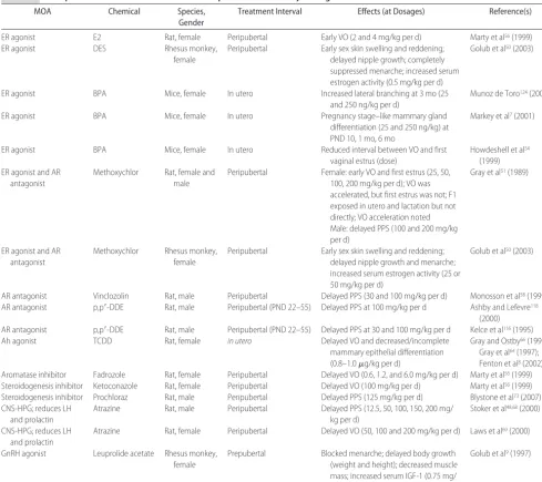
A summary of selected examples of single environmen-tal chemicals whose exposure at specific dosages may lead to puberty-timing alterations is presented in Table 1. This table is organized by mode of action (MOA). Estrogen receptor (ER) agonists (also called estrogen agonists or estrogens) and ER antagonists compete with the endogenous estrogen hormones estradiol and es-trone for binding to the classical ER. Such agonists and antagonists bind to the ER and activate or repress, re-spectively, transcription of ER-dependent genes and thereby enhance or diminish estrogenic activity in vivo (eg, induce uterine weight increase/decrease, accelerate/ delay puberty). After in utero exposure to a potent es-trogen agonist, gross reproductive malformations may result in female rats, presumably by altering the devel-opment of reproductive organs that are responsive to estrogen action. Examples of estrogen agonists that are present as environmental contaminants include 2,2-bis(p-hydroxyphenyl)-1,1,1-trichloroethane (HPTE), a metabolite of the pesticide chemical methoxychlor, and
some natural products such as isoflavonoids, flavonoids, stilbenes, lignans, equol, and phytoestrogens (eg, genistein, daidzein, coumestrol, 8-prenylnaringenin, resveratrol). Gestational and lactational maternal oral exposure to dosages as low as 25 mg/kg per day me-thoxychlor resulted in an earlier age of VO and first estrus in the female rat.51This was found to be pseudo-precocious rather than true pseudo-precocious puberty because VO was accelerated but the onset of estrous cyclicity was not affected. Similarly, prenatal subcutaneous injections of BPA,52,53 genistein,53 resveratrol,53 zearalenone,53 or DES53 accelerated VO in mice. Although prenatal oral exposure to BPA did not significantly accelerate the age at VO or first estrus in either the rat or mouse, prenatal oral treatment of mice with BPA significantly reduced the number of days between VO and first vaginal es-trus.54 When female rats were exposed after weaning throughout puberty to xenoestrogens like methoxy-chlor51 or 17 beta estradiol,56 early puberty onset was induced as measured by age at VO in the female rat.55In addition, prenatal BPA exposure resulted in altered mat-uration of the mammary gland at puberty and increased sensitivity to endogenous estrogens.7
Androgen receptor (AR) antagonists (also called an-drogen antagonists) compete with natural hormones testosterone and dihydrotestosterone for binding to the AR. Thus, a decrease in binding between the natural ligand and receptor occurs, which in turn inhibits AR-dependent gene expression in vivo. AR antagonist ex-posure in vivo can induce malformations in male repro-ductive tract and delay puberty in the male rat. AR antagonists include insecticides, herbicides, fungicides, toxic chemicals, and combustion byproducts. HPTE has also been shown to act as an AR antagonist.57 Peripuber-tal exposure to the AR antagonists methoxychlor or vinclozolin led to a delayed puberty in males as mea-sured by the age of PPS in the rat.51,58
Conversely, AR agonists (also called androgen ago-nists) compete with the endogenous hormones testos-terone and dihydrotestostestos-terone for AR, leading to an increase in AR-DNA binding in vitro and AR-dependent gene expression in vivo. Such environmental androgen contaminants induce malformations in the female repro-ductive tract59,60; accelerate male puberty in the rat61; and have been associated with the masculinization of female fish in the United States, Sweden, and New Zealand.62
respectively,64–67 and a delayed or incomplete differentia-tion of mammary epithelium.8
Some environmental chemicals affect puberty via MOAs that are involved in the central nervous system/ HPG axis, altering brain, hypothalamic, and/or pituitary function via direct interactions with the central nervous system neuroendocrine function. These include the pes-ticides thiram, molinate, metam sodium, chlordime-form, amitraz, triazoles, dichloroacetic acid, atrazine, propazine, simazine, methanol, and linuron. Prepubertal exposure to atrazine causes delayed puberty in the male and the female, as evidenced by an older age at acqui-sition of PPS68 and VO,69 respectively. The modes of action for atrazine’s reproductive effects include puberty timing and a reduction in circulating LH and prolactin.70 Perinatal exposure of mice to BPA results in a decrease of tyrosine hydroxylase neurons in the rostral
periven-tricular preoptic area at 22 to 24 days of age (prepuber-tal), an important brain region that regulates estrous cyclicity and estrogen-positive feedback.71
Chemicals that inhibit the synthesis of endogenous hormones (eg, testosterone, 17-estradiol, adrenal ste-roids) typically do so by competitively inhibiting the activity ofⱖ1 P450 steroidogenic enzymes. The predom-inant effects are mediated through the steroidogenic enzymes cholesterol side chain cleavage enzyme, steroid 17,20 lyase, and aromatase (which converts androgens to estrogens). Some affect estrogen synthesis preferen-tially over testosterone or adrenal hormones, and others affect adrenal and liver function. Examples of environ-mental and pharmaceutical aromatase inhibitors include conazole and imadazole fungicides (eg, prochloraz, fe-narimol, ketoconazole, fadrozole). Peripubertal expo-sure to the aromatase inhibitor fadrozole at dosages as TABLE 1 Examples of Published Studies of Chemical Exposures With Puberty-Timing Effects in Animals
MOA Chemical Species, Gender
Treatment Interval Effects (at Dosages) Reference(s)
ER agonist E2 Rat, female Peripubertal Early VO (2 and 4 mg/kg per d) Marty et al56(1999)
ER agonist DES Rhesus monkey,
female
Peripubertal Early sex skin swelling and reddening; delayed nipple growth; completely suppressed menarche; increased serum estrogen activity (0.5 mg/kg per d)
Golub et al50(2003)
ER agonist BPA Mice, female In utero Increased lateral branching at 3 mo (25 and 250 ng/kg per d)
Munoz de Toro124(2005)
ER agonist BPA Mice, female In utero Pregnancy stage–like mammary gland differentiation (25 and 250 ng/kg) at PND 10, 1 mo, 6 mo
Markey et al7(2001)
ER agonist BPA Mice, female In utero Reduced interval between VO and first vaginal estrus (dose)
Howdeshell et al54 (1999) ER agonist and AR
antagonist
Methoxychlor Rat, female and male
Peripubertal Female: early VO and first estrus (25, 50, 100, 200 mg/kg per d); VO was accelerated, but first estrus was not; F1 exposed in utero and lactation but not directly; VO acceleration noted Male: delayed PPS (100 and 200 mg/kg per d)
Gray et al51(1989)
ER agonist and AR antagonist
Methoxychlor Rhesus monkey, female
Peripubertal Early sex skin swelling and reddening; delayed nipple growth and menarche; increased serum estrogen activity (25 or 50 mg/kg per d)
Golub et al50(2003)
AR antagonist Vinclozolin Rat, male Peripubertal Delayed PPS (30 and 100 mg/kg per d) Monosson et al58(1999) AR antagonist p,p⬘-DDE Rat, male Peripubertal (PND 22–55) Delayed PPS at 100 mg/kg per d Ashby and Lefevre118
(2000) AR antagonist p,p⬘-DDE Rat, male Peripubertal (PND 22–55) Delayed PPS at 30 and 100 mg/kg per d Kelce et al116(1995) Ah agonist TCDD Rat, female in utero Delayed VO and decreased/incomplete
mammary epithelial differentiation (0.8–1.0g/kg per d)
Gray and Ostby66(1995); Gray et al64(1997); Fenton et al8(2002) Aromatase inhibitor Fadrozole Rat, female Peripubertal Delayed VO (0.6, 1.2, and 6.0 mg/kg per d) Marty et al56(1999) Steroidogenesis inhibitor Ketoconazole Rat, female Peripubertal Delayed VO (100 mg/kg per d) Marty et al56(1999) Steroidogenesis inhibitor Prochloraz Rat, male Peripubertal Delayed PPS (125 mg/kg per d) Blystone et al73(2007) CNS-HPG; reduces LH
and prolactin
Atrazine Rat, male Peripubertal Delayed PPS (12.5, 50, 100, 150, 200 mg/ kg per d)
Stoker et al48,68(2000)
CNS-HPG; reduces LH and prolactin
Atrazine Rat, female Peripubertal Delayed VO (50, 100 and 200 mg/kg per d) Laws et al69(2000)
GnRH agonist Leuprolide acetate Rhesus monkey, female
Prepubertal Blocked menarche; delayed body growth (weight and height); decreased muscle mass; increased serum IGF-1 (0.75 mg/ kg per month)
Golub et al9(1997)
low as 0.6 mg/kg per day led to a delayed VO in female rats.56Peripubertal exposure to 100 mg/kg per day of the steroidogenesis inhibitor ketoconazole also led to a de-layed VO.56 The MOA of prochloraz is the inhibition of aromatase72 and 17,20 lyase73,74apparently directly in-hibiting steroidogenesis of estrogens and androgens. Pro-chloraz also delays puberty in male rats.73The MOA of the fungicide chemical ketoconazole is inhibition of side-chain cleavage enzyme (SCC), progesterone, and adre-nal steroid synthesis. The MOA of many phthalate es-ters, including diethylhexyl phthalate, dibutyl phthalate, and benzyl butyl phthalate, is to inhibit fetal testis Leydig cell steroidogenesis and insl3 synthesis, leading to male developmental and reproductive system malformations (eg, retained nipples, reduced or absent reproductive or-gans, undescended testes, hypospadias, delayed PPS) with in utero exposure and delays in puberty with peripubertal exposure.75Administration of phthalates during puberty to marmosets induced ovarian and uterine alterations in fe-males and reduced peripubertal testosterone levels in males76; however, the effect on androgen levels was not significant because of the extreme variability in this mea-sure in this species.
Human Studies
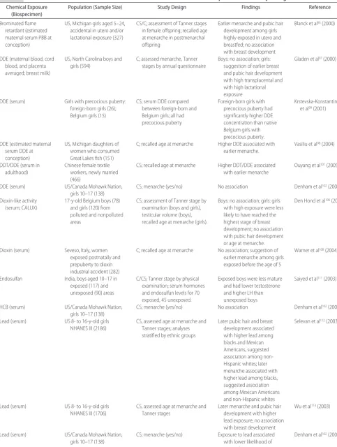
Many human studies have shown a positive relation between body fat, primarily measured by BMI or skin-fold thickness, and onset of puberty, as determined by the onset of the growth spurt, breast development, or menarche.77–84 Similar findings are observed in boys as measured by growth spurt or genital and pubic hair mat-uration.81,85–87Body size measures also have been associ-ated with early puberty,82,86,88as have dietary and physical activity exposures and pubertal timing.79,89–94 Environmen-tal exposure to persistent halogenated organic chemicals such as PCBs, dichlorodiphenyltrichloroethane/dichlorodi-phenyldichloroethylene (DDT/DDE), and brominated flame retardants have been associated with pubertal alter-ations, as have dioxin, hexachlorobenzene (HCB), en-dosulfan, and heavy metals, but to a lesser extent (Table 2). The first study to report that timing of puberty was associated with developmental exposure to persistent organic pollutants was conducted among a cohort of individuals who were exposed to brominated flame re-tardants.95An industrial accident resulted in the contam-ination of cattle feed with polybrominated biphenyls and widespread human exposure through consumption of meat and dairy products. Daughters of women who consumed contaminated farm products were inter-viewed or completed a self-administered mailed ques-tionnaire. Age at menarche and Tanner stages (for those who were younger than 18 years) were assessed. Ma-ternal serum levels of polybrominated biphenyl at the time of conception were estimated from a decay model using serum concentrations that were measured before and/or after the pregnancy.96 Girls who were exposed in utero to high concentrations (ⱖ7 ppb) and who were breastfed reported menarche 1 full year earlier than unex-posed girls (⬍1 ppb, the limit of detection) or girls who were exposed in utero but not breastfed. Breastfed girls with high in utero exposure also reported more advanced
pubic hair development compared with unexposed girls or girls who were not breastfed. There was no association found with Tanner stage of breast development.
Various studies have evaluated pubertal development among children who were exposed to DDT and its pri-mary metabolite DDE. Two studies specifically assessed developmental exposure (in utero and/or postnatal via lactation) within a cohort. Gladen et al97 conducted a prospective cohort study of boys and girls who resided in North Carolina in relation to DDE concentrations that were previously measured in the mothers’ serum, cord serum, and the placenta (averaged for in utero expo-sure). Concentrations in breast milk also were deter-mined (lactational exposure). Puberty-timing measures, Tanner stages (boys and girls), and menarche were as-sessed by annual questionnaires. For boys, no associa-tion was observed for either in utero or lactaassocia-tional DDE exposure. Among girls, there was no association with age at menarche; however, there was a suggestion of an association of higher in utero or lactational exposure and earlier breast and pubic hair development that was not statistically significant. Vasiliu et al98 also evaluated in utero exposure to DDT/DDE that was estimated using a decay model based on maternal measurements among daughters of a Michigan angler cohort. The authors ob-served a significantly earlier menarche among girls with an increased in utero exposure. Specifically, menarche was 1 year earlier for every 15-g/L increase in in utero exposure. No association was seen with breastfeeding and age at menarche.
TABLE 2 Human Studies That Assessed Associations Between Environmental Chemical Exposures and Puberty Timing
Chemical Exposure (Biospecimen)
Population (Sample Size) Study Design Findings Reference
Brominated flame retardant (estimated maternal serum PBB at conception)
US, Michigan girls aged 5–24, accidental in utero and/or lactational exposure (327)
CS/C; assessment of Tanner stages in female offspring; recalled age at menarche in postmenarchal offspring
Earlier menarche and pubic hair development among girls highly exposed in utero and breastfed; no association with breast development
Blanck et al95(2000)
DDE (maternal blood, cord blood, and placenta averaged; breast milk)
US, North Carolina boys and girls (594)
C; assessed menarche, Tanner stages by annual questionnaire
Boys: no association; girls: suggestion of earlier breast and pubic hair development with high transplacental and with high lactational exposure
Gladen et al97(2000)
DDE (serum) Girls with precocious puberty: foreign-born girls (26); Belgium girls (15)
CS; serum DDE compared between foreign-born and Belgium girls; all had precocious puberty
Foreign-born girls with precocious puberty had significantly higher DDE concentration than native Belgium girls with precocious puberty.
Krstevska-Konstantinova et al99(2001)
DDE (estimated maternal serum DDE at conception)
US, Michigan daughters of women who consumed Great Lakes fish (151)
C; recalled age at menarche Higher DDE associated with earlier menarche.
Vasiliu et al98(2004)
DDT/DDE (serum in adulthood)
Chinese female textile workers, newly married (466)
CS; recalled age at menarche Higher DDT/DDE associated with earlier menarche
Ouyang et al101(2005)
DDE (serum) US/Canada Mohawk Nation, girls 10–17 (138)
CS; menarche (yes/no) No association Denham et al102(2005)
Dioxin-like activity (serum; CALUX)
17-y-old Belgium boys (78) and girls (120) from polluted and nonpolluted areas
CS; assessment of Tanner stage by examination (boys and girls), testicular volume (boys), recalled age at menarche (girls).
Boys: no association; girls: girls with high exposure were less likely to have reached the highest stage of breast development; no association with pubic hair development or age at menarche.
Den Hond et al106(2002)
Dioxin (serum) Seveso, Italy, women exposed postnatally and prepuberty to dioxin industrial accident (282)
C; recalled age at menarche No association; suggestion of earlier menarche among girls exposed before the age of 5
Warner et al108(2004)
Endosulfan India, boys aged 10–17 in exposed (117) and unexposed (90) areas
C/CS; Tanner stage by physical examination; serum hormones and endosulfan levels for 70 exposed, 45 unexposed.
Exposed boys were less mature and had lower testosterone and higher LH than unexposed boys
Saiyed et al111(2003)
HCB (serum) US/Canada Mohawk Nation, girls 10–17 (138)
CS; menarche (yes/no) No association Denham et al102(2005)
Lead (serum) US 8- to 16-y-old girls NHANES III (2186)
CS, assessed age at menarche and Tanner stages; analyses stratified by ethnic groups
Later pubic hair and breast development associated with higher lead among blacks and Mexican Americans, suggested association among non-Hispanic whites; later menarche associated with higher lead among blacks, suggested association among Mexican Americans and non-Hispanic whites
Selevan et al112(2003)
Lead (serum) US 8- to 16-y-old girls NHANES III (1706)
CS, assessed age at menarche and Tanner stages
Later menarche and pubic hair development with higher lead exposure; no association with breast development
Wu et al113(2003)
Lead (serum) US/Canada Mohawk Nation, girls 10–17 (138)
CS; menarche (yes/no) Exposure to lead associated with lower likelihood of menarche
along the United States/Canadian border to assess serum concentrations and self-reported menarchal status (yes/ no). No association was observed between DDT
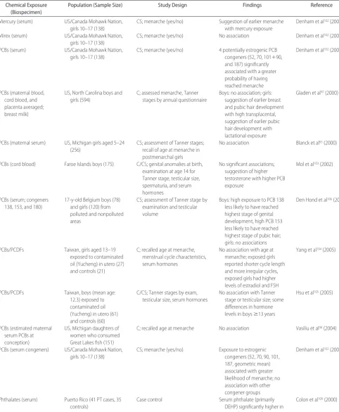
expo-sure and menarche despite the authors’ assumption that current serum concentrations were indicative of in utero and lactational exposures, given the long-standing con-TABLE 2 Continued
Chemical Exposure (Biospecimen)
Population (Sample Size) Study Design Findings Reference
Mercury (serum) US/Canada Mohawk Nation, girls 10–17 (138)
CS; menarche (yes/no) Suggestion of earlier menarche with mercury exposure
Denham et al102(2005)
Mirex (serum) US/Canada Mohawk Nation, girls 10–17 (138)
CS; menarche (yes/no) No association Denham et al102(2005)
PCBs (serum) US/Canada Mohawk Nation, girls 10–17 (138)
CS; menarche (yes/no) 4 potentially estrogenic PCB congeners (52, 70, 101⫹90, and 187) significantly associated with a greater probability of having reached menarche
Denham et al102(2005)
PCBs (maternal blood, cord blood, and placenta averaged; breast milk)
US, North Carolina boys and girls (594)
C; assessed menarche, Tanner stages by annual questionnaire
Boys: no association; girls: suggestion of earlier breast and pubic hair development with high transplacental, suggestion of earlier pubic hair development with lactational exposure
Gladen et al97(2000)
PCBs (maternal serum) US, Michigan girls aged 5–24 (256)
CS; assessment of Tanner stages; recall of age at menarche in postmenarchal girls
No association Blanck et al95(2000)
PCBs (cord blood) Faroe Islands boys (175) C/CS; genital anomalies at birth, examination at age 14 for Tanner stage, testicular size, spermaturia, and serum hormones
No significant associations; suggestion of higher testosterone with higher PCB exposure
Mol et al103(2002)
PCBs (serum; congeners 138, 153, and 180)
17-y-old Belgium boys (78) and girls (120) from polluted and nonpolluted areas
CS; assessment of Tanner stage by examination and testicular volume
Boys: high exposure to PCB 138 less likely to have reached highest stage of genital development, high PCB 153 less likely to have reached highest stage of pubic hair; girls: no associations
Den Hond et al106(2002)
PCBs/PCDFs Taiwan, girls aged 13–19 exposed to contaminated oil (Yucheng) in utero (27) and controls (21)
C; recalled age at menarche, menstrual cycle characteristics, serum hormones
No association with age at menarche; exposed girls reported shorter cycle length and more irregular cycles, exposed girls had higher levels of estradiol and FSH
Yang et al104(2005)
PCBs/PCDFs Taiwan, boys (mean age: 12.3) exposed to contaminated oil (Yucheng) in utero (61) and controls (60)
C/CS; Tanner stages by exam, testicular size, serum hormones
No association with Tanner stage or testicular size; some differences in hormone levels in boysⱖ13 years
Hsu et al105(2005)
PCBs (estimated maternal serum PCBs at conception)
US, Michigan daughters of women who consumed Great Lakes fish (151)
C; recalled age at menarche No association Vasiliu et al98(2004)
PCBs (serum congeners) US/Canada Mohawk Nation, girls 10–17 (138)
CS; menarche (yes/no) Exposure to estrogenic congeners (52, 70, 90, 101, 187, geometric mean) associated with greater likelihood of menarche; no association with other congener groups
Denham et al102(2005)
Phthalates (serum) Puerto Rico (41 PT cases, 35 controls)
Case control Serum phthalate (primarily DEHP) significantly higher in cases than controls
Colon et al109(2000)
cern regarding consumption of contaminated fish among this population.
Five studies evaluated developmental PCB exposures in relation to pubertal development. Gladen et al97 re-ported no relation between in utero or lactational PCB exposure and age at menarche among girls. Higher lac-tational exposure was associated with earlier Tanner staging in boys and girls, although the results did not achieve statistical significance. Mol et al103established a prospective cohort study of boys in the Faroe Islands, where PCB levels are known to be relatively high. PCB levels were measured in cord serum and tissue. Boys were examined at 14 years of age for Tanner staging, testicular size, spermaturia (sperm in urine), and serum hormones. No statistically significant associations were found between PCB concentrations and measures of pubertal development. A suggestion of a positive associ-ation between testosterone and PCB exposure was re-ported. Two cohort studies previously described95,98also evaluated in utero exposure to PCBs among daughters of women who were exposed. Neither study found an as-sociation between PCB exposure and age at menarche. Last, Yang et al104and Hsu et al105described no pubertal developmental differences among boys or girls with and without exposure to PCB- and polychlorinated dibenzo-furan– contaminated cooking oil.
Two cross-sectional studies of PCB exposure have been conducted. Den Hond et al106measured PCB con-geners in a sample of boys and girls who were aged 17 years and recruited from 1 of 2 exposed areas in Belgium: residence near a lead smelter or waste incinerator or a referent rural area. Puberty timing was measured using the Tanner staging as a part of a physical examination. Odds ratios were estimated for PCB concentration and odds for being in a puberty stage that indicated that puberty was not yet complete (ie, lower than stage 5). Exposed boys had completed pubertal development later, because odds ratios for higher PCB exposures (for both specific congeners and total PCBs) were associated with stage⬍5 for genital and pubic hair development. Because this study evaluated 17-year-olds, it is not clear whether puberty began later or the peripubertal duration was longer. No pubertal delays were seen in girls with PCB exposure. Among Akwesasne Mo-hawk Nation Girls, exposure to a group of 4 potentially estrogenic PCB congeners (52, 70, 101⫹90, and 187) was associated with a significantly greater likelihood of having reached menarche.102
Two studies assessed childhood exposure to dioxins and puberty. Den Hond et al106 measured dioxin-like activity with the chemically activated luciferase gene expression assay107 and reported an association with Tanner stage ⬍5 breast development; however, pubic hair stage in boys and girls and genital stage in boys was not significantly associated with dioxin-like activity. Warner et al108examined pubertal development among girls who were exposed postnatally or during childhood to dioxin in Seveso, Italy. TCDD was not associated with age of menarche (self-reported at interview). None of the women was exposed prenatally.
Colon et al109studied phthalate esters exposure and premature thelarche. Forty-one girls with thelarche
were compared with 35 control girls with regard to serum pesticides and phthalate esters. Significantly higher phthalate levels were found among the girls with thelarche than comparison girls. The authors concluded that the findings were suggestive of a possible associa-tion between exposures and premature breast develop-ment in girls. Interpretation of these findings may be limited by residual confounding and concerns about the analytical method of measuring the phthalate parent compound instead of the metabolites.110 Saiyed et al111 found that boys who were exposed to endosulfan in India were less mature and had lower testosterone and higher LH than unexposed boys.
Three cross-sectional published articles assessed the relation between lead exposure and pubertal measures using either the Third National Health and Nutrition Examination Survey112,113 or the Mohawk Nation Study.102 In the Third National Health and Nutrition Examination Survey analyses, blood lead levels were analyzed in relation to age at onset for Tanner stage 2 for breast and pubic hair and menarche among US girls aged 8 to 18 years.113Selevan et al112estimated odds ratios for lead levels in relation to progression of puberty (stages 2–5) among girls. Combined, these studies suggested an inverse relation between blood lead levels and the onset and progression of puberty in girls, especially for sub-groups within the population. An association also was found between higher blood lead level and delayed menarche, but this association was statistically signifi-cant only for black girls. The cross-sectional analysis among Mohawk nation girls also found later puberty among girls with higher lead exposures than girls with lower exposure.
Cross-species Concordance of Pubertal Timing Effects
pubertal development,112–115 suggesting a similar mecha-nism for rodents and humans. P,p⬘-DDE exposure in “for-eign-born” girls was associated with earlier puberty relative to native-born Belgians99and, possibly, increasing DDE in utero exposure with earlier puberty in girls97,98; however, no animal studies of DDE exposure and female puberty timing were identified, limiting additional interpretation of the human data. In male rats, peripubertal DDE exposure delays PPS,116–118although no relation to lower exposures was found in boys after in utero DDE exposure.97
The weight of animal and human data, although sug-gestive, remains inconclusive for establishing a causal relation between EDCs and human pubertal distur-bances. As reviewed, much of the available literature focuses on a single chemical or class, ignoring the
mix-ture of compounds to which humans are exposed. Much of the data are derived from cross-sectional studies, which are unable to establish the causal ordering of environment in relation to puberty. To this end, timing and dose at critical windows of human development have not been purposefully assessed. Other relevant co-variates such as lifestyle, genetic determinants, and other xenobiotics in the chemical mixture have not been well studied, if at all, in the context of EDCs.
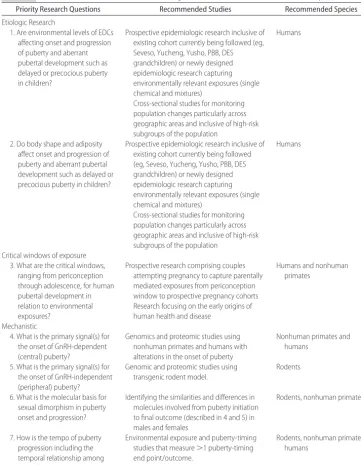
RESEARCH RECOMMENDATIONS
The panel was asked to make research recommendations that are responsive to critical data gaps regarding pur-ported environmental factors that may adversely affect puberty, to prioritize research initiatives including meth-TABLE 3 Research Recommendations for Focusing on Environmental Influences on Puberty
Priority Research Questions Recommended Studies Recommended Species
Etiologic Research
1. Are environmental levels of EDCs affecting onset and progression of puberty and aberrant pubertal development such as delayed or precocious puberty in children?
Prospective epidemiologic research inclusive of existing cohort currently being followed (eg, Seveso, Yucheng, Yusho, PBB, DES grandchildren) or newly designed epidemiologic research capturing environmentally relevant exposures (single chemical and mixtures)
Cross-sectional studies for monitoring population changes particularly across geographic areas and inclusive of high-risk subgroups of the population
Humans
2. Do body shape and adiposity affect onset and progression of puberty and aberrant pubertal development such as delayed or precocious puberty in children?
Prospective epidemiologic research inclusive of existing cohort currently being followed (eg, Seveso, Yucheng, Yusho, PBB, DES grandchildren) or newly designed epidemiologic research capturing environmentally relevant exposures (single chemical and mixtures)
Cross-sectional studies for monitoring population changes particularly across geographic areas and inclusive of high-risk subgroups of the population
Humans
Critical windows of exposure 3. What are the critical windows,
ranging from periconception through adolescence, for human pubertal development in relation to environmental exposures?
Prospective research comprising couples attempting pregnancy to capture parentally mediated exposures from periconception window to prospective pregnancy cohorts Research focusing on the early origins of human health and disease
Humans and nonhuman primates
Mechanistic
4. What is the primary signal(s) for the onset of GnRH-dependent (central) puberty?
Genomics and proteomic studies using nonhuman primates and humans with alterations in the onset of puberty
Nonhuman primates and humans
5. What is the primary signal(s) for the onset of GnRH-independent (peripheral) puberty?
Genomic and proteomic studies using transgenic rodent model.
Rodents
6. What is the molecular basis for sexual dimorphism in puberty onset and progression?
Identifying the similarities and differences in molecules involved from puberty initiation to final outcome (described in 4 and 5) in males and females
Rodents, nonhuman primates
7. How is the tempo of puberty progression including the temporal relationship among the different markers regulated?
Environmental exposure and puberty-timing studies that measure⬎1 puberty-timing end point/outcome.
ods development, and to identify avenues for research collaboration.
Environmental Factors Identified as Important for Human Puberty
After reviewing the weight of evidence described herein, the panel agreed that EDCs and body size or adiposity may be associated with pubertal disturbances in hu-mans. The panel further recognizes that the 2 broad exposures may be interrelated in that exposure to EDCs can affect fat metabolism (eg, organotin compounds and diethyl hexyl phthalate act as peroxisome proliferator– activated receptor agonists in adipose tissue). Although humans can do little to minimize exposure to ubiquitous EDCs other than change dietary practices, particularly with regard to fish consumption and preparation, body size is potentially amenable to behavioral intervention. Carefully designed epidemiologic research, particularly that with longitudinal capture of exposures and relevant covariates at critical windows and the sensitive and spe-cific measurement of markers of pubertal onset, progres-sion, or deviations from expected normative standards, is needed to answer remaining questions about the role of environmental influences on developmental mile-stones. Such studies are rightfully complex and require a priori consideration and capture of other environmental determinants of puberty such as those that are believed to be attributed to body size and composition and pos-sible genotypes and phenotypes that may emerge. Fur-ther complicating future research will be the challenges afforded by environmental mixtures, including those that arise from “natural” sources such as phytoestrogens.
Research Priorities and Future Directions
The panel identified 7 priority research questions reflect-ing 3 broad research domains: etiologic, exposure assess-ment, and mechanistic (Table 3). The panel strongly supported additional etiologic work aimed at answering lingering questions about the impact of EDCs and body size and adiposity on the onset and progression of pu-berty and aberrant clinical manifestations (ie, delayed or precocious puberty). The natural history of puberty re-quires delineation and ordering of a multitude of factors such as lipids, body shape, and diet in relation to puber-tal onset and progression. With regard to exposure as-sessment, continued work is needed to capture the tim-ing and dosage of maternal, paternal, or parental exposures across sensitive and critical windows of hu-man development to identify agents that may disrupt normal embryonic or fetal development resulting in fetal (re)programming with lifetime effects.119,120 Last, the panel continued to support basic research to identify the primary signal(s) for the onset of GnRH-dependent and GnRH-independent puberty in humans, nonhuman pri-mates, and other animals.
The increasing methodologic complexities that are as-sociated with capturing exposures, including environmen-tal mixtures, in the context of lifestyle and behavior across critical windows of human development argue for prospec-tive epidemiologic studies whose utility and feasibility have
been demonstrated.121,122Ideally, such studies would incor-porate longitudinal biospecimen collection at timed inter-vals for the quantification of chemical mixtures and life-style exposures that may affect growth and development inclusive of pubertal milestones. Efforts to develop a suite of sensitive pubertal markers, rather than a sole marker, that are consistent with the manner in which humans are exposed are needed. Future research needs are discussed elsewhere in this issue but underscore the need to develop behavioral, psychosocial, and neurologic end points along with the social and psychological consequences of preco-cious or delayed puberty.11
Last, regardless of the research question, newly de-signed animal and human research must use the best available methods. With regard to animal research, care-ful attention to choice of the most appropriate animal model, relevant critical or sensitive windows of expo-sure, route and duration of expoexpo-sure, timing and rele-vant dosages, assumptions regarding the dose-re-sponse curve, and an analytic statistical plan that is responsive to the study design, particularly assessment of mixtures and litter effects, is needed. Epidemiologic research also needs to address these same issues while recognizing the highly correlated nature of reproduc-tive outcomes and the need to model appropriately previous history of adverse reproductive outcomes, a strong predictor, in the context of more subtle envi-ronmental effects.123
CONCLUSIONS
A concerted body of multidisciplinary research is needed to answer lingering questions about the impact of EDCs, body size, and adiposity on human puberty. This re-search can be grounded within a broader rere-search base inclusive of human reproduction given the highly interre-lated and timed series of critical events that are character-istic of human growth and development, of which puberty is 1 of many key milestones. Basic research targeting the primary signals for onset is critical so that epidemiologic investigators can design research that is sensitive to the capture and measurement of key biomarkers of pubertal onset. Additional basic research is needed regarding com-parative species physiology of puberty, the primary signal of puberty onset for central versus peripheral signaling, and the basis for sexual dimorphism.
ACKNOWLEDGMENTS
Support for “The Role of Environmental Factors on the Onset and Progression of Puberty” workshop was pro-vided by US Environmental Protection Agency coop-erative agreement 830774, the National Institute of Environmental Health Sciences, and Serono Inc.
Grumbach, Marcia Herman-Giddens, John Himes, Anders Juul, Paul Kaplowitz, Carole Kimmel, Peter Lee, Robert Lustig, Tony Plant, Ed Reiter, Steve Schrader, Sherry Selevan, Richard Sharpe, Thorkild Sørensen, and Patricia Whitten. We especially thank Anders Juul for critical reading of the manuscript.
REFERENCES
1. Rockett JC, Johnson CD, Buck GM. Biomarkers for assessing reproductive development and health: part 1—pubertal de-velopment.Environ Health Perspect.2004;112(1):105–112 2. Marshall WA, Tanner JM. Variations in pattern of pubertal
changes in girls.Arch Dis Child.1969;44(235):291–303 3. Marshall WA, Tanner JM. Variations in the pattern of
puber-tal changes in boys.Arch Dis Child.1970;45(239):13–23 4. Arlt W, Martens JW, Song M, Wang JT, Auchus RJ, Miller
WL. Molecular evolution of adrenarche: structural and func-tional analysis of p450c17 from four primate species. Endocri-nology.2002;143(12):4665– 4672
5. Conley AJ, Pattison JC, Bird IM. Variations in adrenal andro-gen production among (nonhuman) primates. Semin Reprod Med.2004;22(4):311–326
6. Ojeda SR, Skinner MK. Puberty in the rat. In: Neill JD, ed.The Physiology of Reproduction. 3rd ed. San Diego, CA: Academic Press/Elsevier; 2006:2061–2126
7. Markey CM, Luque EH, Munoz De Toro M, Sonnenschein C, Soto AM. In utero exposure to bisphenol A alters the devel-opment and tissue organization of the mouse mammary gland.Biol Reprod.2001;65(4):1215–1223
8. Fenton SE, Hamm JT, Birnbaum LS, Youngblood GL. Persis-tent abnormalities in the rat mammary gland following ges-tational and lacges-tational exposure to 2,3,7,8-tetrachloro-dibenzo-p-dioxin (TCDD).Toxicol Sci.2002;67(1):63–74 9. Golub MS, Styne DM, Wheeler MD, et al. Growth retardation
in premenarchial female rhesus monkeys during chronic ad-ministration of a GnRH agonist (leuprolide acetate). J Med Primatol.1997;26(5):248 –256
10. Terasawa E, Fernandez DL. Neurobiological mechanisms of the onset of puberty in primates. Endocr Rev. 2001;22(1): 111–151
11. Golub MS, Germann SL, Hogrefe CE. Endocrine disruption and cognitive function in adolescent female rhesus monkeys. Neurotoxicol Teratol.2004;26(6):799 – 809
12. Van Wagenen G, Simpson ME. Testicular development in the rhesus monkey.Anat Rec.1954;118(2):231–251
13. Gray LE Jr, Wilson V, Noriega N, et al. Use of the laboratory rat as a model in endocrine disruptor screening and testing. ILAR J.2004;45(4):425– 437
14. Foster DL, Padmanabhan V, Wood RI, Robinson JE. Sexual differentiation of the neuroendocrine control of gonadotro-phin secretion: concepts derived from sheep models.Reprod Suppl.2002;59:83–99
15. Plant TM, Witchel SF. Puberty in nonhuman primates and humans. In: Neill JD, ed.The Physiology of Reproduction. 3rd ed. San Diego, CA: Academic Press/Elsevier; 2006:2177–2230 16. Richter TA, Terasawa E. Neural mechanisms underlying the
pubertal increase in LHRH release in the rhesus monkey. Trends Endocrinol Metab.2001;12(8):353–359
17. Plant TM. The male monkey as a model for the study of the neurobiology of puberty onset in man.Mol Cell Endocrinol. 2006;254 –255:97–102
18. Ojeda SR, Terasawa E. Neuroendocrine regulation of puberty. In: Pfaff D, Arnold A, Etgen A, Fahrbach S, Moss R, Rubin R, eds. Hormones, Brain and Behavior. NY: Elsevier; 2002: 589 – 659Vol 4. New York
19. Ojeda SR, Prevot V, Heger S, Lomniczi A, Dziedzic B,
Mun-genast A. Glia-to neuron signaling and the neuroendocrine control of female puberty.Ann Med.2003;35(4):244 –255 20. Ojeda SR, Lomniczi A, Mungenast A, et al. Towards
under-standing the neurobiology of mammalian puberty: genetic, genomic and proteomic approaches. In: Kordon C, Gaillard R, Christen Y, eds.Hormones and the Brain. Berlin, Germany: Springer; 2005:47– 60
21. Ojeda SR, Lomniczi A, Mastronardi C, Heger S, Roth C, Parent AS. Mini-review: the neuroendocrine regulation of puber-ty—is the time ripe for a systems biology approach? Endocri-nology.2006;147(3):1166 –1174
22. de Roux N, Genin E, Carel J-C, Matsuda F, Chaussain J-L, Milgrom E. Hypogonadotropic hypogonadism due to loss of function of the KiSS1-derived peptide receptor GPR54.Proc Natl Acad Sci USA.2003;100(19):10972–10976
23. Seminara SB, Messager S, Chatzidaki EE, et al. The GPR54 gene as a regulator of puberty.N Engl J Med.2003;349(17): 1614 –1627
24. Mastronardi C, Smiley GG, Raber J, Kusakabe T, Kawaquchi A, Matagne V. Deletion of the Ttf1 gene in differentiated neurons disrupts female reproduction without impairing basal ganglia function.J Neurosci.2006;26(51):13167–13179 25. Cottrell EC, Campbell RE, Han SK, Herbison AE. Postnatal
remodeling of dendritic structure and spine density in gonado-tropin-releasing hormone neurons. Endocrinology. 2006; 147(8):3652–3661
26. Funes S, Hedrick JA, Vassileva G, et al. The KiSS-1 receptor GPR54 is essential for the development of the murine repro-ductive system. Biochem Biophys Res Commun. 2003;312(4): 1357–1363
27. Henley DV, Lipson N, Korach KS, Bloch CA. Prepubertal gynecomastia linked to lavender and tea tree oils. N Engl J Med.2007;356(5):479 – 485
28. Witchel SF, Plant TM. Puberty: gonadarche and adrenarche. In: Strauss JF, Barbieri RL, eds.Yen and Jaffe’s Reproductive Endocrinology. 5th ed. Philadelphia, PA: Elsevier Saunders; 2004:493–535
29. Hertz R. Accidental ingestion of estrogens by children. Pediat-rics.1958;21(2):203–206
30. Beas F, Vargas L, Spada RP, Merchak N. Pseudoprecocious puberty in infants caused by a dermal ointment containing estrogens.J Pediatr.1969;75(1):127–130
31. Edidin DV, Levitsky LL. Prepubertal gynecomastia associated with estrogen-containing hair cream.Am J Dis Child.1982; 136(7):587–588
32. Felner EI, White PC. Prepubertal gynecomastia: indirect ex-posure to estrogen cream. Pediatrics.2000;105(4). Available at: www.pediatrics.org/cgi/content/full/105/4/e55
33. Tiwary CM. Premature sexual development in children fol-lowing the use of estrogen- or placenta-containing hair prod-ucts.Clin Pediatr (Phila).1998;37(12):733–739
34. Zawadski J, Dunaif A. Diagnostic criteria for polycystic ovary syndrome: towards a rational approach. In: Dunaif A, Givens JR, Haseltine F, eds. Polycystic Ovary Syndrome. Oxford, England: Blackwell Science Ltd; 1992:377–384
35. The Rotterdam ESHRE/ASRM-Sponsored PCOS consensus workshop group: Revised 2003 consensus on diagnostic cri-teria and long-term health risks related to polycystic ovary syndrome (PCOS).Hum Reprod.2004;19(1):41– 47
36. Witchel SF. Puberty and polycystic ovary syndrome.Mol Cell Endocrinol.2006;254 –255:146 –153
37. Iban˜ez L, Virdis R, Potau N, et al. Natural history of premature pubarche: an auxological study.J Clin Endocrinol Metab.1992; 74(2):254 –257
39. Abbott DH, Barnett DK, Bruns CM, Dumesic DA. Androgen excess fetal programming of female reproduction: a develop-mental aetiology for polycystic ovary syndrome?Hum Reprod Update.2005;11(4):357–374
40. Barnes RB, Rosenfield RL, Ehrmann DA, et al. Ovarian hy-perandrogynism as a result of congenital adrenal virilizing disorders: evidence for perinatal masculinization of neuroen-docrine function in women. J Clin Endocrinol Metab. 1994; 79(5):1328 –1333
41. Dumesic DA, Schramm RD, Abbott DH. Early origins of poly-cystic ovary syndrome.Reprod Fertil Dev.2005;17(3):349 –360 42. Zhou R, Bird IM, Dumesic DA, Abbott DH. Adrenal hyper-androgenism is induced by fetal androgen excess in a rhesus monkey model of polycystic ovary syndrome.J Clin Endocrinol Metab.2005;90(12):6630 – 6637
43. Wickenheisser JK, Nelson-DeGrave VL, McAllister JM. Hu-man ovarian theca cells in culture.Trends Endocrinol Metab. 2006;17(2):65–71
44. Abbott DH, Padmanabhan V, Dumesic DA. Contributions of androgen and estrogen to fetal programming of ovarian dys-function.Reprod Biol Endocrinol.2006;4:17
45. Diamanti-Kandarakis E, Alexandraki K, Piperi C, et al. In-flammatory and endothelial markers in women with polycys-tic ovary syndrome.Eur J Clin Invest.2006;36(10):691– 697 46. Eriksson JG, Osmond C, Kajantie E, Forsen TJ, Barker DJ.
Patterns of growth among children who later develop type 2 diabetes or its risk factors. Diabetologia. 2006;49(12): 2853–2858
47. Sanders EB, Aston CE, Ferrell RE, Witchel SF. Inter- and intrafamilial variability in premature pubarche and polycystic ovary syndrome.Fertil Steril.2002;278:473– 478
48. Stoker TE, Parks LG, Gray LE, Cooper RL. Endocrine-disrupting chemicals: prepubertal exposures and effects on sexual maturation and thyroid function in the male rat—a focus on the EDSTAC recommendations. Endocrine Disruptor Screening and Testing Advisory Committee.Crit Rev Toxicol. 2000;30(2):197–252
49. Goldman JM, Laws SC, Balchak SK, Cooper RL, Kavlock RJ. Endocrine-disrupting chemicals: prepubertal exposures and effects on sexual maturation and thyroid activity in the fe-male rat—a focus on the EDSTAC recommendations.Crit Rev Toxicol.2000;30(2):35–196
50. Golub MS, Hogrefe CE, Germann SL, Lasley BL, Natarajan K, Tarantal AF. Effects of exogenous estrogenic agents on puber-tal growth and reproductive system maturation in female rhesus monkeys.Toxicol Sci.2003;74(1):103–113
51. Gray LE Jr, Ostby J, Ferrell J, et al. A dose-response analysis of methoxychlor-induced alterations of reproductive devel-opment and function in the rat.Fundam Appl Toxicol.1989; 12(1):92–108
52. Honma S, Suzuki A, Buchanan DL, Katsu Y, Watanabe H, Iguchi T. Low dose effect of in utero exposure to bisphenol A and diethylstilbestrol on female mouse reproduction.Reprod Toxicol.2002;16(2):117–122
53. Nikaido Y, Yoshizawa K, Danbara N, et al. Effects of maternal xenoestrogen exposure on development of the reproductive tract and mammary gland in female CD-1 mouse offspring. Reprod Toxicol.2004;18(6):803– 811
54. Howdeshell KL, Hotchkiss AK. Thayer KA, Vandenbergh JG, vom Saal FS. Exposure to bisphenol A advances puberty. Nature.1999;401(6755):763–764
55. Rubin BS, Murray MK, Damassa DA, King JC, Soto AM. Perinatal exposure to low doses of bisphenol A affects body weight, patterns of estrous cyclicity, and plasma LH levels. Environ Health Perspect.2001;109(7):675– 680
56. Marty MS, Crissman JW, Carney EW. Evaluation of the EDSTAC female pubertal assay in CD rats using
17beta-estradiol, steroid biosynthesis inhibitors, and a thyroid inhib-itor.Toxicol Sci.1999;52(2):269 –277
57. Maness SC, McDonnell DP, Gaido KW. Inhibition of androgen receptor-dependent transcriptional activity by DDT isomers and methoxychlor in HepG2 human hepatoma cells.Toxicol Appl Pharmacol.1998;151(1):135–142
58. Monosson E, Kelce WR, Lambright C, Ostby J, Gray LE Jr. Peripubertal exposure to the antiandrogenic fungicide, vin-clozolin, delays puberty, inhibits the development of andro-gen-dependent tissues, and alters androgen receptor function in the male rat.Toxicol Ind Health.1999;15(1–2):65–79 59. Hotchkiss A, Lambright CS, Ostby JS, Parks-Saldutti L,
Van-denbergh JG, Gray LE Jr. Prenatal testosterone exposure permanently masculinizes anogenital distance, nipple devel-opment and reproductive tract morphology in female Sprague-Dawley rats: testosterone-induced malformations in rats.Toxicol Sci.2007;96(2):335–345
60. Wilson VS, Lambright C, Ostby J, Gray LE Jr. In vitro and in vivo effects of 17beta-trenbolone: a feedlot effluent contam-inant.Toxicol Sci.2002;70(2):202–211
61. Shin JH, Kim HS, Moon HJ, et al. Effects of flutamide on puberty in male rats: an evaluation of the protocol for the assessment of pubertal development and thyroid function.J Toxicol Environ Health A.2002;65(5– 6):433– 445
62. Parks LG, Lambright CS, Orlando EF, Guillette LJ Jr, Ankley GT, Gray LE Jr. Masculinization of female mosquitofish in Kraft mill effluent-contaminated Fenholloway River water is associated with androgen receptor agonist activity.Toxicol Sci. 2001;62(2):257–267
63. Roman BL, Peterson RE. In utero and lactational exposure of the male rat to 2,3,7,8-tetrachlorodibenzo-p-dioxin impairs prostate development: 1— effects on gene expression.Toxicol Appl Pharmacol.1998;150(2):240 –253
64. Gray LE, Wolf C, Mann P, Ostby JS. In utero exposure to low doses of 2,3,7,8-tetrachlorodibenzo-p-dioxin alters reproduc-tive development of female Long Evans hooded rat offspring. Toxicol Appl Pharmacol.1997;146(2):237–244
65. Gray LE, Ostby JS, Kelce WR. A dose-response analysis of the reproductive effects of a single gestational dose of 2,3,7,8-tetrachlorodibenzo-p-dioxin in male Long Evans Hooded rat offspring.Toxicol Appl Pharmacol.1997;146(1):11–20 66. Gray LE Jr, Ostby JS. In utero
2,3,7,8-tetrachlorodibenzo-p-dioxin (TCDD) alters reproductive morphology and function in female rat offspring.Toxicol Appl Pharmacol.1995;133(2): 285–294
67. Gray LE Jr, Kelce WR, Monosson E, Ostby JS, Birnbaum LS. Exposure to TCDD during development permanently alters reproductive function in male Long Evans rats and hamsters: reduced ejaculated and epididymal sperm numbers and sex accessory gland weights in offspring with normal androgenic status.Toxicol Appl Pharmacol.1995;131(1):108 –118 68. Stoker TE, Laws SC, Guidici DL, Cooper RL. The effect of
atrazine on puberty in male Wistar rats: an evaluation in the protocol for the assessment of pubertal development and thyroid function.Toxicol Sci.2000;58(1):50 –59
69. Laws SC, Ferrell JM, Stoker TE, Schmid J, Cooper RL. The effects of atrazine on female Wistar rats: an evaluation of the protocol for assessing pubertal development and thyroid func-tion.Toxicol Sci.2000;58(2):366 –376
70. Cooper RL, Stoker TE, Tyrey L, Goldman JM, McElroy WK. Atrazine disrupts the hypothalamic control of pituitary-ovarian function.Toxicol Sci.2000;53(2):297–307