CELLULAR INTERACTIONS W ITH IRON CHELATORS
b y
Katharine Pippin H oyes
A thesis subm itted for the Degree o f D octor o f Philosophy of the U niversity o f L ondon.
Decem ber 1991 D epartm ent o f Haem atology
ProQuest Number: 10609060
All rights reserved INFORMATION TO ALL USERS
The qu ality of this repro d u ctio n is d e p e n d e n t upon the q u ality of the copy subm itted. In the unlikely e v e n t that the a u th o r did not send a c o m p le te m anuscript and there are missing pages, these will be note d . Also, if m aterial had to be rem oved,
a n o te will in d ica te the deletion.
uest
ProQuest 10609060
Published by ProQuest LLC(2017). C op yrig ht of the Dissertation is held by the Author.
All rights reserved.
This work is protected against unauthorized copying under Title 17, United States C o d e M icroform Edition © ProQuest LLC.
ProQuest LLC.
789 East Eisenhower Parkway P.O. Box 1346
A B S T R A C T
The experiments contained within this thesis have characterized the ability of desferrioxamine (DFO) and 3-hydroxypyridin-4-one (3-HP-4-one) iron chelators to enter cells and mobilise iron from intracellular iron pools.
Radiolabelled l,2,diethyl-3-hydroxypyridin-4-one (CP94) and DFO have been used to study their cellular uptake and subcellular distribution. CP94 had rapid passage into and out o f K562 erythroleukaemia cells in both the iron-free and iron-complexed form by simple diffusion. In contrast, DFO was found to enter cells by a facilitated mechanism, possibly along an electrochemical gradient, and accumulated within cells at concentrations in excess of that in the extracellular medium. Iron chelated as FO passed out o f the cell relatively slowly and it is proposed that the charged nature of FO may delay its egress from the cell. Whilst the major intracellular site o f chelation by both compounds was the low molecular weight cytosolic iron pool, CP94 was more efficient at mobilising iron from intracellular organelles than DFO.
The effects of such iron depletion on cell function have been examined using in vivo and in vitro models of cellular proliferation. In vitro DFO and CP94 inhibited cell growth in a dose dependent manner. At chelator concentrations above 30|iM this was concomitant with inhibition of DNA synthesis and ribonucleotide reductase activity. However, lower chelator concentrations exerted an inhibitory effect on cell growth that was independent of DNA synthesis suggesting that iron chelators may affect other intracellular iron dependent pathways necessary for proliferation. Unlike DFO, CP94 and other 3-HP-4-ones were found to act as potent cell cycle synchronisation agents. Inhibition of bone marrow progenitor growth in vitro was also dose dependent for DFO and the 3-HP-4-ones. Furthermore in vivo chronic administration of the chelators led to the suppression of haemopoiesis.
For Guy
in appreciation o f his constant support and encouragement.
'A discovery is rarely.... a sudden achievement, nor is it the work o f one man; a long series o f observations, each in turn received in doubt and discussed in hostility are familiarised by time and lead at last to the gradual disclosure o f truth.1
Sir Berkeley Moynihan (1865-1936) In: Surgery, Gynaecology and Obstetrics (1920) 31: 549
ACKNOWLEDGEMENTS
I wish to express my gratitude to my supervisor, Dr John Porter, for all the advice, help and encouragement he has given me during the course o f this study. I should also like to thank Professor Emie Huehns for his help and in particular for his guidance in the preparation of this thesis.
I am particularly grateful to Professor Bob Hider and Paul Dobbin from the D epartm ent o f P harm acy, K ings C ollege London fo r sup p ly in g the hydroxypyridinone iron chelators. I would also like to thank Dr Surinder Singh, of the same department, for carrying out the HPLC analysis. I am also indebted to Mark Jones, Department o f Haematology, University College London for his help and advice with the bone marrow progenitor colony forming assays.
IN D E X
PAGE
T IT L E PA G E 1
A B ST R A C T 2
D E D IC A T IO N 3
A C K N O W L E D G E M E N T S 4
IN D E X 5
L IST O F FIG U R E S 9
LIST O F TA B LES 12
L IST O F A B B R EV IA T IO N S 14
C H A PTE R 1; G EN ER A L IN T R O D U C T IO N 16
1.1. INTRODUCTION 17
1.2. IRON PHYSIOLOGY 18
1.2.1. Iron Balance 18
1.2.2. Transferrin and the Transferrin Receptor 19
1.2.3. Cellular Iron Uptake 22
1.2.4. Cellular Iron Pools 25
1.2.5. Storage Iron 31
1.2.6. Cellular Iron Homeostasis 32
1.3. IRON OVERLOAD 33
1.4. IRON CHELATION 38
1.4.1. General Principles of Iron Chelation 38
1.4.2. Sites of Iron Chelation 41
1.4.3. Potential Oral Chelators 42
1.4.4. Use of Iron Chelators In Conditions 50
Unrelated to Iron Overload
CHAPTER 2: GENERAL MATERIALS AND METHODS 55
2.1. INTRODUCTION 56
2.2 IRON CHELATORS 56
2.3. CELLS AND CELL CULTURE 58
2.3.1. Cell Culture Media 58
2.3.2. Culture of Cell Lines 59
2.3.3. Isolation and Culture of Human Peripheral 59
Blood Lymphocytes
2.3.4. Isolation and Culture of Haemopoietic 60
Progenitor Cells from Murine Bone Marrow
2.3.5. Cell Counting and Viability 61
2.3.6. Freezing and Thawing Cells 61
2.3.7. Cytospin Preparation 62
2.4. GENERAL METHODS 62
2.4.1. Transferrin Labelling 62
2.4.2. Cell Cycle Analysis 64
2.4.3. Assays Conducted on Fractionated Cells 64
2.4.4. Ribonucleotide Reductase Assay 66
2.4.5. Protein Assay 69
CHAPTER 3: CELLULAR UPTAKE OF CP94 AND DFO 71
3.1. INTRODUCTION 72
3.2. UPTAKE EXPERIMENTS WITH [14C]DFO AND [14C]CP94 73 3.2.1. Uptake of [14C]DFO and [14C]CP94 by K562 cells at 37°C 73 3.2.2. Initial Rate of Uptake of [14C]CP94 and [14C]DFO 74
3.2.3. Discussion 77
3.3. HPLC ANALYSIS OF INTRACELLULAR DFO 77
3.5. IRON MOBILISATION FROM K562 CELLS BY CP94 AND DFO 85
3.6. GENERAL DISCUSSION 88
C H A P T E R 4: SU B C ELLU LA R D IST R IB U T IO N O F 90 C P94 AND D FO
4.1. INTRODUCTION 91
4.2. ANALYTICAL SUBCELLULAR FRACTIONATION OF K562 CELLS 92 4.3. SUBCELLULAR DISTRIBUTION OF [14C]CP94 AND [14C]DFO 96
4.4. SUBCELLULAR DISTRIBUTION STUDIES USING 59Fe 101
4.4.1. Effect o f lOOftM IBE CP94 and DFO on the Subcellular 101 Distribution of 59Fe delivered by Receptor Mediated
Endocytosis.
4.4.2. Mobilisation of Low Molecular Weight Iron by CP94 and DFO 107
4.4.3. Discussion 109
4.5. EFFECT OF PREINCUBATION WITH CP94 AND DFO PRIOR TO 111 LOADING CELLS WITH 59Fe TRANSFERRIN
4.6. GENERAL DISCUSSION 114
C H A P T E R 5: E F F E C T O F DFO AND T H E H Y D R O X PY R ID IN O N E 116 ON C E L L C Y C L E AND P R O L IFE R A T IO N
5.1 .INTRODUCTION 117
5.2. EFFECT OF CP94 AND DFO ON CELL CYCLE KINETICS 118
5.2.1. Dose Response Effect of DFO and CP94 on 118
K562 and Daudi Cell Cycle.
5.2.2. Effect of DFO and CP94 on the Cell Cycle of 125 Mitogen Stimulated Peripheral Blood Lymphocytes
5.2.3. Discussion 125
5.3. PHYSICOCHEMICAL PROPERTIES REQUIRED FOR 127
EFFECTIVE CELL CYCLE ARREST
5.4.CELL CYCLE SYNCHRONISATION BY 128 3-HYDROXYPYRJODIN-4-ONES
5.5. INHIBITION OF RIBONUCLEOTIDE REDUCTASE 137
BY CP94 AND DFO
5.6. EFFECT OF CP94 AND DFO ON CELL PROLIFERATION 138
5.6.1. Effect of CP94 and DFO on Cell Growth and Viability 140 5.6.2. Effect of CP94 and DFO on DNA,RNA and Protein Synthesis 144
5.6.3. Discussion 146
5.7. GENERAL DISCUSSION 149
C H A P T E R 6: IN VIVO AND IN V ITR O E F F E C T S O F DFO AND 152 3-HP-4-ONES ON M U R IN E H A E M O P O IE S IS
6.1. INTRODUCTION 153
6.2. IN VIVO STUDY 153
6.3. IN VITRO STUDY 159
6.3.1. Effects of DFO, CP94 and CP20 on Haemopoietic 159 Colony formation
6.3.2. Effects of Iron-Complexed Chelators on Haemopoietic 162 Colony Formation
6.3.3. Effect of Transferrin Saturation on Inhibition by 165 Chelator Complexes
6.3.4. Relationship Between Complex Lipophilicity and 168 Colony Inhibition
6.3.5. Discussion 170
6.4. GENERAL DISCUSSION 171
C H A P T E R 7: C O N C L U SIO N S 174
7.2. CELLULAR IRON MOBILISATION 176
7.2.1. Transit Across Cell Membranes 176
7.2.2. Access to Intracellular Iron Pools 178
7.2.3. Removal Of Iron From Cells 179
7.3. EFFECT OF IRON CHELATION ON CELL FUNCTION 181
7.3.1. The Antiproliferative Effects of Iron Chelators 181 7.3.2. The Use of Iron Chelators as Anti-neoplastic Agents 183
7.4. IRON CHELATOR TOXICITY 185
7.5. CONCLUSIONS AND FUTURE PERSPECTIVES. 188
R E F E R E N C E S 190
A P P E N D IC E S 209
APPENDIX 1. MANUFACTURERS OF REAGENTS 210
APPENDIX 2. BUFFERS 215
APPENDIX 3. MEASUREMENT OF SERUM IRON LEVELS 216
L IS T O F FIG U R ES
FIGURE 1. Schematic Representation of the Human Transferrin Receptor. 20 FIGURE 2. The Receptor Mediated Transferrin to Cell Cycle. 23
FIGURE 3. Intracellular Iron Pools. 26
FIGURE 4. Schematic Desription of the Interconversion o f 29 the 4Fe and 3Fe Cluster of Aconitase.
FIGURE 5. Schematic Representation of Ribonucleotide Reductase. 30 FIGURE 6. Co-ordinate Regulation of the Expression of the 34
Transferrin Receptor and Feritin.
FIGURE 7. The Chain Reaction of Lipid Peroxidation. 37
FIGURE 8. The Structure of Desferrioxamine B. 46
FIGURE 9. Bidentate Hydroxypyridinone Structures. 48
FIGURE 9. Bidentate Hydroxypyridinone Structures. 48
FIGURE 10. Labelling Transferrin with 59Fe. 63
FIGURE 11. Measurement of Ferritin by ELISA. 67
FIGURE 12. Typical Standard Curve for the Ferritin ELISA. 68 FIGURE 13. Typical Standard Curve for the Protein Assay. 70 FIGURE 14. Uptake of [14C]CP94 and [14C]DFO by K562 cells. 75 FIGURE 15. Initial Uptake of [14C]CP94 and [14C]DFO by K562 76
cells.
FIGURE 16. Typical HPLC Chromatograms for DFO and FO. 79 FIGURE 17. Calibration Curves for the Measurement of DFO 80
and FO by HPLC.
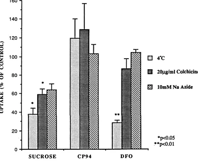
FIGURE 18. Effect of Temperature, Colchicine and Azide on 84 uptake of [14C]sucrose, [14C]CP94 and [14C]DFO
by K562 cells.
FIGURE 19. Mobilisation of 59Fe from K562 Cells by CP94 and DFO. 87
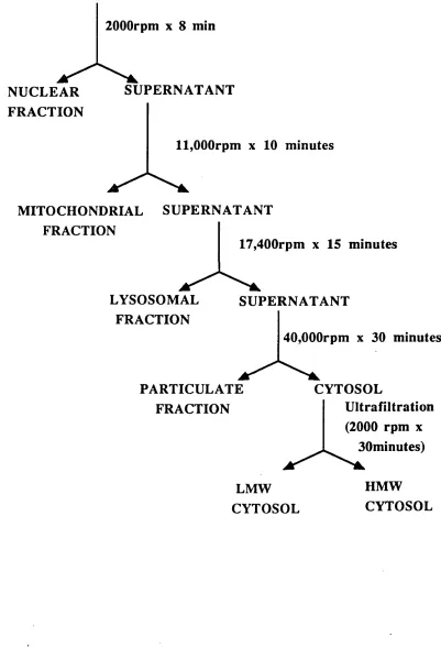
FIGURE 20. Fractionation Procedure. 95
FIGURE 21. Subcellular Distribution of [14C]CP94 and [14C]DFO 99 in K562 cells at 20 minutes.
FIGURE 22. Subcellular Distribution of [14C]CP94 and [14C]DFO 100 in K562 cells at 4 hours.
FIGURE 23. Pulse-chase Labelling of K562 cells with 59Fe. 102 FIGURE 24. Distribution of 59Fe in Control K562 Cells after 104
20 Minutes at 37°C.
FIGURE 25. Effect of lOOfiM IBE CP94 and DFO on Distribution 105 of 59Fe after 20 minutes.
FIGURE 26. Effect of 100|iM IBE CP94 and DFO on Distribution 106 of 59Fe after 4 hours.
Chelator Treated K562 cells.
FIGURE 28. LMW 59Fe in K562 Cells Treated with 100p.M IBE CP94 or DFO.
FIGURE 29. Distribution of 59Fe at 20 minutes following 4 hours Preincubation with CP94 and DFO.
FIGURE 30. Flow Cytometric DNA Histograms for Control K562 Cells. FIGURE 31. DNA Histograms for K562 Cells Treated with
IOOjiM IBE CP94 and DFO.
FIGURE 32. Relationship Between Partition Coefficient and Cell Cycle Arrest.
FIGURE 33. K562 Cell Cycle Arrest by DFO, CP130, CP94 and CP02.
FIGURE 34. Recovery o f K562 Cells From Cell Cycle Arrest by CP94 and DFO.
FIGURE 35. Recovery of Daudi Cells From Cell Cycle Arrest by CP94 and DFO.
FIGURE 36. Recovery of K562 cells from Cell Cycle Arrest After Addition of IOOjiM IBE Ferric Ammonium Citrate. FIGURE 37. Cell Cycle Synchronisation by 3-HP-4-ones. FIGURE 38. Inhibition of Ribonucleotide Reductase by CP94
and DFO.
FIGURE 39. K562 Cell Growth in the Presence of CP94 and DFO. FIGURE 40. Daudi Cell Growth in the Presence of CP94 and DFO. FIGURE 41. Concentration Dependent Inhibition of Cell Growth
by CP94 and DFO.
FIGURE 42. Concentration Dependent Inhibition of [3H]Thymidine Incorporation in K562 and Daudi Cells.
FIGURE 44.Concentration Dependent Inhibition of BFU-E Growth by Iron Chelators.
FIGURE 45. Effect of Addition of Iron on Inhibition of CFU-G+CFU-Mac by Iron Chelators.
FIGURE 46. Dose Response Effect o f FO, CP94-Fe and CP20-Fe on CFU-G+CFU-Mac Colony Growth. FIGURE 47. Relationship Between Complex Partition
Coefficient and Colony Inhibition.
L IS T O F TA B LES.
TABLE 1. Desirable Properties for an Iron Chelator. TABLE 2. Potential Oral Iron Chelators.
TABLE 3. Possible Applications for Iron Chelators in Conditions Unrelated to Iron Overload.
TABLE 4. Chelator Structure, Partition Coefficient and Stability Constant.
TABLE 5. HPLC Analysis of Intracellular DFO and FO.
TABLE 6. Distribution of Marker Enzynes Between Subcellular Fractions.
TABLE 7. Effects of CP94 and DFO on K562 Cell Cycle Kinetics. TABLE 8. Effects of CP94 and DFO on Daudi Cell Cycle Kinetics. TABLE 9. Effect of Low Concentrations of DFO and CP4 on the
Percentage of Polynuclear and Mitotic Forms.
TABLE 10. Effects of CP94 and DFO on Peripheral Blood Lymphocyte Cell Cycle Kinetics.
TABLE 11. Effect of DFO and CP94 on the Viability of Daudi and K562 Cells.
and DFO.
TABLE 13. Haematology for Mice Treated with 60 Doses of 156 200mg/kg Chelator.
TABLE 14. Bone Marrow Cellularity and Haemopoietic Colony 157 Formation for Mice Treated with 60 Doses of
200mg/kg Chelator.
TABLE 15. Haematology for Mice treated with 10 or 30 Doses of 158 200mg/kg Chelator.
TABLE 16. Transferrin Saturation and Inhibition of CFU-G+CFU-M 167 Colony Growth by Iron Free and Iron Complexed
CP94 and CP20.
L IS T O F A B BR EV IA TIO N S
ADP Adenosine diphosphate
AMP Adenosine monophosphate
ATP Adenosine triphosphate
BFU-E Burst forming unit - erythroid
BSA Bovine serum albumin
CDP Cytidine diphosphate
CFU-G Colony forming unit - granulocyte CFU-Mac Colony forming unit - macrophage
CPM Counts per minute
DFO Desferrioxamine
DNA Deoxyribonucleic acid
EDTA Ethylenediaminetetra-acetic acid
ELISA Enzyme linked immunoassay
FCS Foetal calf serum
FO Ferrioxamine
GI Gastrointestinal
GVHD Graft versus host disease
HMW High molecular weight
HPLC High performance liquid chromatography
HPO Hydroxypyridinone
3-HP-4-one 3-hydroxypyridin-4-one 3-HP-2-one 3-hydroxypyridin-2-one
IBE Iron binding equivalents
IC50 Inhibitory concentration 50%
IRE Iron regulatory element
IRE-BP Iron regulatory element binding protein
MCV Mean cell volume
NTBI Non-transferrin bound iron
PBS Phosphate buffered saline
PHA Phytohaemagglutinin
RA Rheumatoid arthritis
RNA Ribonucleic acid
SEM Standard error of the mean
UTR Untranslated region
1.1 INTRODUCTION
This thesis will investigate the actions o f iron chelators at a cellular level in order to further the understanding o f their mode of action, help in their future development and identify their toxic effects.
The ideal iron chelator has a demonstrable ability to prevent or modify iron mediated pathological processes without undue toxicity. The clinical development of novel chelating agents necessitates an evaluation of their efficacy and potential toxicides in a variety o f cellular and animal models. The use o f such models can also provide valuable information regarding the biological source of mobilised iron and any resultant alteration o f cell function.
Whilst the primary use of iron chelators has been alleviation of pathological iron overload, some attention has also been focused upon the ability o f such compounds to modify disease unrelated to iron loading. However, even the only clinically proven chelator desferrioxamine (DFO), which has been used therapeutically for nearly three decades, has significant toxicity (Porter and Huehns, 1989). This is particularly prevalent in patients with minimal iron loading, suggesting that such toxicity may result from the depletion of iron vital to cellular function rather than any effect o f DFO itself. This compound will be used in comparative studies with the hydroxypyridinones, a series o f iron chelators with potential for clinical use. Initial studies will focus upon the ability o f these chelators to enter cells and subcellular organelles and mobilise iron. Subsequent experiments seek to evaluate the effects o f such chelation on cell physiology, taking the key area of cell proliferation as a model.
The introduction that follows will provide an overview o f general iron physiology coupled with a discussion of the design and application of iron chelators in both iron overloaded and non-overloaded situations.
1.2 IRON PHYSIOLOGY
1.2.1 Iron balance
Eukaryotic cells have an obligate requirement for iron. This can be related to the critical role this metal plays in the function o f a wide variety o f biological redox systems, including the electron transport chains and ribonucleotide reductase. Consequently, the body's ability to maintain effective iron homeostasis is essential to health. Disturbances in iron balance are common but almost invariably result in a reduction in total body iron content, and iron deficiency affects 500-600 million people worldwide. In contrast, accumulation o f excessive iron within the body is a relatively rare condition.
In man the average iron content is normally 40-50mg Fe/kg body weight. Levels are generally higher in men than women. The major proportion o f body iron (approximately 30mg/kg) is bound in haemoglobin in circulating red cells. A further 4mg/kg is found in muscle as myoglobin and approximately 2mg/kg is in tissues as components of iron containing enzymes. Most o f the remaining iron is stored within the liver, spleen, bone marrow and muscle as ferritin and haemosiderin (Bothwell et al,
1979)
In women additional losses result from menstruation (0.6mg) and pregnancy (2.7mg) (Bothwell et al, 1979).
1.2.2 Transferrin and the Transferrin Receptor
In vertebrates serum transferrin provides the major physiological route for the extracellular transport of iron and its delivery to cells. The liver is the principal site of transferrin synthesis but other cell types including testicular Sertoli cells (Skinner and G riswold, 1982), cells in the central nervous system (Bloch et al, 1985) and lymphocytes (Soltys and Brody, 1970) also synthesize this protein.
Transferrin is a glycoprotein with a molecular weight of approximately 80,000. The molecule consists of a single polypeptide chain folded to form two globular domains termed N and C, relating to the N-terminal and C-terminal domains o f the protein. Binding of iron (III) to each of the N and C lobes requires concomitant binding of a bicarbonate anion (Schlabach and Bates, 1975). The amino acid residues involved in the specific binding o f iron by transferrin have been identified by X-ray crystallography. A t both the N and C-terminals iron is directly co-ordinated to two tyrosines, one histidine and one aspartic acid and indirectly co-ordinated to an arginine via the bicarbonate anion which acts as a bridging ligand (Bailey et al, 1988). Under physiological conditions the apparent stability constants for the N and C binding sites are approximately 1022M_1 but this decreases rapidly with pH.
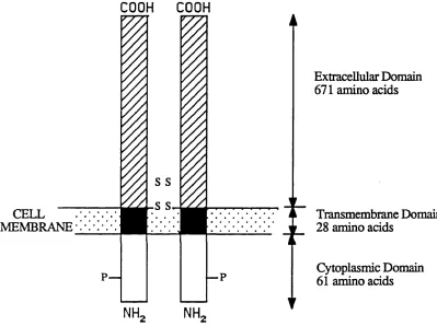
At the cell membrane the transferrin iron complex binds to specific receptors. The transferrin receptor is an integral membrane glycoprotein composed o f two identical 95,000 dalton subunits linked by disulphide bonds (Enns and Sussman, 1981a). Studies on transferrin receptors isolated from both normal and malignant tissues (Enns and Sussman, 1981b; Stein and Sussman, 1983) have demonstrated similar proteolytic digest maps indicating that the receptor is identical in all tissues. Each subunit is a transmembrane polypeptide o f 760 amino acids (McClelland et al, 1984; Schneider et al, 1984) consisting o f three dom ains (Figure 1). The
FIGURE 1: SCHEMATIC REPRESENTATION OF
THE HUMAN TRANSFERRIN RECEPTOR
CELL MEMBRANE
C O O H
C O O H
7
s s
p— —p
NH,
NH,
Extracellular Domain 671 amino acids
Transmembrane Domain 28 amino acids
Cytoplasmic Domain 61 amino acids
intracellular N-terminal domain comprises the first 61 residues. This is followed by a hydrophilic transmembrane domain of 28 amino acids. This sequence appears to act as a signal for insertion o f the receptor into the plasma membrane and its deletion confines the whole receptor to the cytoplasm (Zerial et al, 1986). The remaining 671 amino acids comprise the extracellular C terminal domain.
The cytoplasmic sequence contains a total o f 4 serines, all potential phosphorylation sites. However only serine 24 appears to be a target for protein kinase C mediated phosphorylation (Davis et al, 1986). There has been some controversy over whether phosphorylation is required for receptor internalisation (Zerial et al, 1987; May and Tyler, 1987) but studies using site directed mutagenesis to substitute Ser 24 by alanine show that the mutation has no effect on the rate o f transferrin receptor endocytosis (Davis and Meisner, 1987).
The transferrin receptor has very high affinity for diferric transferrin, with an estimated affinity constant of 2-7 xlO^M 'l at pH 7.4. Binding is pH dependent. At neutral pH the transferrin receptor has a higher affinity for diferric transferrin than for apotransferrin (Young, Bomford and Williams, 1984; Klausner et al, 1983; Dautry- Varsat et al, 1983). However at pH less than 7.0 the receptors affinity for apotransferrin increases to that o f diferric transferrin (Dautry-Varsat et al, 1982; Klausner et al, 1983).
Transferrin receptor expression appears to be linked to cellular iron requirements. Proliferating cells express transferrin receptors at a high density (Sutherland et al, 1981) and receptors are expressed at high levels in reticulocytes but not in mature red cells (Seligman, 1983). Furthermore, in placenta there is a direct correlation between levels of placental transferrin receptor and foetal iron requirements (Mcardle and Morgan, 1982).
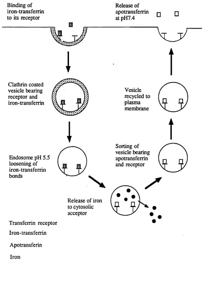
1.2.3 Cellular Iron Uptake
Many cells acquire iron by the receptor mediated endocytosis o f transferrin (Figure 2). This process is initiated by the binding o f transferrin to the transferrin receptor at the cell surface. The receptor-transferrin complexes localise in clathrin coated pits which bud off from the cell surface to form clathrin coated vesicles. The vesicles lose their coats, probably in an ATP dependent process, and fuse to form an endosome. Through the action o f a proton pumping ATPase o f the endosomal membrane, the vesicles interior is rapidly acidified (pH 5.5). The low pH facilitates the release o f iron from transferrin and the iron is transported across the endosomal membrane into the cytosol leaving apotransferrin tightly bound by its receptor at the acidic pH. The apotransferrin-receptor complex is recycled back to the plasma membrane, possibly through the Golgi complex (Stoorvogel et al, 1988),where exposure to neutral pH effects release of the apotransferrin from the receptor (Dautry- Varsat et al, 1983; Klausner et al, 1983).
From studies in preparations of endocytotic vesicles from rabbit reticulocytes (Nunez et al, 1990) a four step model has been proposed for the dissociation of iron from transferrin and its subsequent transfer across the endosomal
membrane:-(i) An acidification system which promotes iron (HI) dissociation from transferrin and requires vesicle acidification promoted by a variety o f membrane ATPases.
(ii) A reduction system in which the dissociated iron (III) bound to an iron binding moiety on the cis side of the membrane is reduced to iron (II).
(iii) A translocation system which moves iron (II) to the trans side o f the vesicular membrane along a specific transmembrane pathway.
(iv) A mobilization system in which iron (II) on the trans side o f the membrane is mobilised by cytosolic low molecular weight carriers.
FIGURE 2: THE RECEPTOR MEDIATED TRANSFERRIN-TO-CELL CYCLE
T
□
Binding of iron-transferrin to its receptor
I
Clathrin coatedvesicle bearing receptor and iron-transferrin
Release of apotransferrin at pH7.4
Vesicle recycled to plasma membrane
□
l
Sorting of vesicle bearing apotransferrin and receptor Endosome pH 5.5
loosening of iron-transferrin bonds
Release of iron to cytosolic acceptor
Iron-transferrin Apotransferin Iron
process than mere acidification. Furthermore, a recent study has indicated that the transferrin receptor itself may facilitate release o f iron from transferrin within the endosome (Bali et al, 1991).
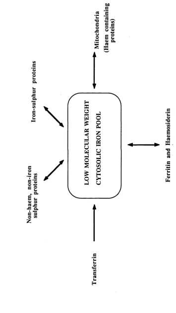
1.2.4 Intracellular Iron Pools
When iron initially enters cells it transiently enters a low molecular weight (LMW) cytosolic pool before incorporation into iron-containing enzymes and ferritin (Jacobs et al, 1977) (Figure 3). The existence of this pool was originally inferred from indirect studies with the iron chelator DFO. Administration of repeated doses o f DFO to normal rats provoked excretion of ferrioxamine (FO) in proportion to body iron stores. H owever, when the anim als were hypertransfused, the resulting inhibition of erythropoiesis led to reduced FO excretion. As there was no reduction in the animals' iron stores, this suggests that ferritin and haemosiderin were not the direct donors of iron to DFO and compounds on the pathway between ferritin and transferrin were probably the immediate iron source (Libschitz et al, 1971). Furthermore, release of haemoglobin iron by the reticuloendothelial system in phenylhydrazine induced haemolysis increased FO excretion (Cummings et al, 1967), and similarly FO excretion increased 4-8 hours after transfusion of nonviable erythrocytes but returned to normal after 20 hours despite the increase in storage iron (Samson et al 1977).
Subsequent studies with 5^Fe have supported the presence of a LMW iron pool in a number of cell types (White, 1976a, 1976b; Pippard et al, 1982; Bakkeren et al, 1985; Mulligan et al, 1986). The transient nature of this pool makes characterisation of the iron complex difficult, but it has been shown that iron in this pool is bound primarily to ATP (Weaver and Pollack, 1989).
In addition to the LM W cytosolic pool there is evidence o f a non-haem non-iron-sulphur mitochondrial iron pool which serves as a short term reserve for haem synthesis (Tangeras et al, 1980). Whilst maturing erythroid cells have the greatest capacity for haem synthesis, this takes place in virtually all cells due to the ubiquitous nature o f haemoproteins. The mitochondrial iron pool represents approximately 1 nmol/mg mitochondrial protein, roughly one third o f total mitochondrial iron (Tangeras et al, 1980). Its major function appears to be donation o f iron to ferrochelatase for
F IG U R E 3: IN T R A C E L L U L A R IR O N P O O L S V) 3 *3
o
u a u 3 J= Q. 3 CAi \
§
/
I - CA X
Z.S /
co
U XA
c c ©u
~ a.
E u
a> 3
w jz -C Q.
3 *3 © ^ .2 *u
■a
s
o
A w o c ‘2 • 2 ^© 3 o .5 U V■+ *
insertion into protoporphyrin IX, the final step in the haem biosynthetic pathway (Tangeras, 1985). In vivo this pool must be constantly resupplied with iron from a cytosolic source (Funk et al, 1986). Recent studies suggest that the major pathway by which iron is delivered to mitochondria may be through hydrolysis of cytosolic iron- ATP to iron-AMP which binds to specific mitochondrial receptors (W eaver and Pollack, 1990).
In addition to these endogenous pools, iron is an essential component of many cellular proteins. These may be classified into the haemoproteins, iron-sulphur proteins and non-haem non-iron-sulphur proteins.
The haem oproteins include the oxygen transporters haem oglobin and myoglobin. The role o f iron in the biology and chem istry o f haemoglobin and myoglobin has been extensively studied and reviewed (Dickerson and Geis, 1983; Crichton, 1991) and since these proteins are somewhat peripheral to the theme o f this thesis, they will not be discussed in detail here. Other haem containing proteins include the peroxidases, which constitute part of the microbicidal system in phagocytic cells (Klebanoff, 1975), catalase, which catalyses the dismutation o f hydrogen peroxide to oxygen and water, cytochrome oxidase and various electron transport enzymes and cytochromes of the electron transport chain.
The iron-sulphur proteins are molecules which contain iron atoms bound to sulphide forming a cluster linked to a polypeptide chain by the thiol groups of cysteine residues. Several types of iron-sulphur cluster are known. In the simplest form a single iron atom is tetrahedrally co-ordinated to the sulphydryl groups o f four cysteine residues o f the protein. A second kind denoted by [2Fe-2S] contains two iron atoms and two inorganic sulphides in addition to four cysteine residues. A third type is designated [3Fe-4S] and contains three iron molecules and four inorganic sulphides with the cysteine residues and the fourth type [4Fe-4S] contains four iron atoms four sulphides and four cysteine residues. The roles of iron-sulphur proteins are numerous and range from electron transport (such as components of the mitochondrial electron
transport chain) to enzymes with both redox and non-redox functions such as succinate dehydrogenase, and aconitase respectively.
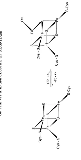
Aconitase is a mitochondrial enzyme of the citric acid cycle that catalyses the stereospecific dehydration and rehydration reactions that convert citrate to isocitrate via the intermediate cis-aconitate. Purified aconitase exists in an inactive [3Fe-4S]+ state which can be activated in vitro to the [4Fe-4S]2+ form by introduction o f a fourth Fe2+ under reducing conditions (Beinert and Kennedy, 1989) (Figure 4). This is distinct from other iron-sulphur proteins in which the Fe-S centre is relatively stable. Thus aconitase provides an example o f the possible way in which the activity o f a protein is responsive to changes in iron availability.
The third class of iron containing proteins consists o f a heterogeneous group of enzymes and proteins which contain non-haem,non-iron-sulphur iron. One notable member of this group is ribonucleotide reductase. The reduction of ribonucleotides to deoxyribonucleotides occurs through a series o f redox reactions catalysed by thioredoxin reductase and ribonucleotide reductase (Thelander and Reichard, 1979) with the overall stoichiometry
of:-NADPH + H+ + ribonucleoside diphosphate --- >
NADP+ + deoxyribonucleoside diphosphate + H2O
F
IG
U
R
E
4:
S
C
H
E
M
A
T
IC
D
E
S
R
IP
T
IO
N
OF
T
H
E
IN
T
E
R
C
O
N
V
E
R
S
IO
N
w co<
H Z c u<
to O w H w P to u a> to fn Q Z<
a» to Ti to S3 H to O Xo.
, 0 0 --- LLCD\ / l /
CD I COW a CO CO I <0
o
<D + to + CO Ia)
o
<o
iD
toa)
CO --- LL
i„/|
CD I4
- u
i s
/
</)
O
CO COc/>
>NO
COI</>
O
o ON ON■o
a>
Cca»
to■a
s
C3 l-a>
ja *3ffl
B©
u
T3a>
-4_»
Q .
C3
"O
<
FIGURE 5: SCHEMATIC REPRESENTATION OF
RIBONUCLEOTIDE REDUCTASE
Substrate Specificity site
C ontrol Sites
Activity Site
SH
SH
SH SH
C atalytic Site
3+
3+
B1 Subunit
B2 Subunit
1.2.5. Storage Iron
Any iron which is surplus to immediate functional needs is stored in cells as ferritin or haemosiderin. Ferritin is the major iron storage protein and is found in all living organisms with a consistent structure of an almost spherical protein shell surrounding an iron core o f variable size (Harrison et al, 1980). The iron core is in the form of a polymeric iron hydroxide to which some phosphate groups may be attached. The amount o f iron in the core is variable and ranges from zero (apoferritin) to a maximum of 4500 atoms. For iron to enter the ferritin molecule it must initially be in the ferrous form, as the protein subunits are arranged to leave a number o f channels through which ferrous iron may pass. However once inside the molecule the iron is oxidised to the ferric form and polymerises. Release o f iron in vitro requires the presence of reducing agents and is increased at low pH (Reviewed by Harrison, 1986; Crichton, 1991).
The protein shell is composed of 24 subunits o f which three types have been identified: H (heavy or heart subunit) consisting of 182 amino acids with a molecular weight of approximately 21,000; L (light or liver) consisting of 174 residues with a molecular weight of approximately 19,000 and the G (glycosylated) subunit with a molecular weight o f approximately 23,000. In human tissues H-rich isoferritins are more acidic and are found in cells which have high iron requirements such as heart muscle and nucleated red cells, and also in cells which are not involved in iron storage such as lymphocytes and monocytes. The more basic L-rich isoferritins appear to be required for iron storage and are found in liver, spleen and placenta (Wagstaff et al, 1978). The G subunit is found predominantly in serum. Serum ferritin is composed almost exclusively of L and G subunits (Cragg et al, 1981) containing little iron. The biological significance o f this ferritin is obscure. However serum ferritin levels can be used as a clinical indicator of iron stores (Worwood, 1986).
In pathological iron overload the bulk o f the excess iron is stored as haemosiderin (Selden et al, 1980). Haemosiderin is a heterogeneous compound
consisting o f insoluble ferric hydroxide with variable amounts of phosphate and protein. It is localized in membranous structures termed siderosomes, which represent enlarged lysosomes and contain lysosomal enzymes (Richter, 1978). Similarities in the composition o f the iron core of ferritin and haemosiderin led to the suggestion that haemosiderin is derived from lysosomal proteolysis o f ferritin followed by aggregation o f the naked cores (Fisbach et al, 1971). Subsequent electron microscopic studies of liver samples from iron overloaded patients and carbonyl-iron fed rats show that cytosolic ferritin appears to attain a maximum concentration, after which it aggregates and fuses with the lysosomal compartment, so form ing a siderosome (Iancu and Neustein, 1977; Iancu, 1989).
Further evidence for the derivation o f haemosiderin from ferritin comes from the observation that haemosiderin consistently contains peptides that may be ferritin degradation products (Weir et al, 1984a). H ow ever recent studies suggest that in certain pathological conditions haemosiderin may also be formed independently of ferritin (Mann et al, 1988; Dickson et al, 1988).
1.2.5 Cellular Iron Homeostasis
IRE-BP is recruited to the high affinity state. High affinity binding of this protein to a 3' IRE results in lower mRNA degradation and increased translation and protein synthesis. By contrast, binding to a 5' IRE results in repression o f translation and inhibition o f protein synthesis. Therefore iron deprivation leads to increased translation o f transferrin receptor mRNA and reduced translation of ferritin mRNA (Figure 6). The iron status o f the cell determines the affinity state o f the IRE-BP through an iron m ediated redox mechanism termed the sulphydryl switch (Klausner and Harford, 1989). Recently the cDNA for human IRE-BP has been cloned (Rouault et al, 1990). There is extensive amino acid sequence homology between IRE-BP and mitochondrial aconitase (Rouault et al, 1991; Hentze and Argos, 1991). This suggests that IRE-BP itself may be an iron-sulphur protein which may respond to changes in cellular iron levels by a redox sensitive conversion between the [3Fe-4S] and the [4Fe-4S] forms in a similar manner to aconitase (Section 1.2.3).
Transferrin receptor expression is also linked to cell proliferation. Pelosi-Testa et al (1986) proposed that intracellular iron regulation and cell proliferation regulate expression by different mechanisms. Rao et al (1986) showed that desfenioxam ine treatment o f K562 cells causes an early increase in transferrin receptor mRNA, whilst haemin had the opposite effect. Transcriptional assays with isolated nuclei indicated that this regulation was at a transcriptional level. Furthermore, deletion o f the 3' non coding region o f the cDNA abolishes the iron-dependent but not the proliferation dependent regulation of transferrin receptor expression (Owen and Kuhn, 1987).
1.3 IRO N OVERLOAD
Normal body iron stores are regulated between 0 and 2000mg depending on the balance between available dietary iron and the iron requirements of the individual (Bothwell et al, 1979). Iron overload can result when additional iron gains access to the body by increased oral absorption or parenteral iron administration.
F IG U R E 6 : CO ORD INA TE R E GU L AT IO N O F T H E E X P R E S S IO N O F T H E TRANSFERRIN RECEP TOR A N D F E R R IT IN Q W 00
<
U r t u w Q Zo
rt HH < rt D r t rt w u.s *= 'E
< < <
<L>
(50
C3
03
Wh O *-» C/3 e o
03
M o03
*oc «>
2 ^ S 2
W Q*
CL3 V u a <u o
C/3 C/3 C/3
3 s
m
<
< <<
W a ^T3 V
oo
CP
c z r t B < Zrt
£
rt
o
H
&
r tCJ
r tr t
J5*
z
2CO
r t to r t
s
r trt
s
O
C/5
Z < r tH
T3 <Ui3 £ *2
c H I
03 HH a
il rt £3
a r t M I E .a
03
03
0 3 *n<
<
<
<
00
§
s 1 03
03
03
O Q,
a- ^ 8 § 8 .§
l /
8 8 O o £ E
m a\o\
Z O r t r t
Increased iron absorption may occur as a result o f inappropriately large amounts o f iron absorbed from a diet containing normal amounts o f iron, as occurs in idiopathic haemochromatosis and the iron-loading anaemias (Cox, 1990). Alternatively oral iron overload may result from the consumption of large amounts of dietary or medicinal iron as in the South African Bantu population who drink large amounts o f acidic beer brewed in iron pots (Bothwell et al, 1964).
Parenteral iron overload is produced by repeated blood transfusions in individuals suffering from dyserythropoietic anaemias such as 6 thalassaemia major. Haemoglobin has an iron content of 0.33% by weight so each 500ml o f transfused whole blood represents 215mg of iron that cannot be excreted. Such patients require up to 50 units of blood annually depending on their age and size, therefore the burden of transfused iron is between 2.5 and l l g per year (Cox, 1990). Consequently patients who receive more than 100 units of blood usually develop iron overload. Furthermore, conditions such as B thalassaemia are characterised by a large degree o f ineffective erythropoiesis resulting in enhanced absorption o f dietary iron so contributing to the net positive balance o f iron (Pippard et al, 1977).
This progressive accumulation o f iron leads to cardiac, liver and endocrine damage, often w ith diabetes and failure to enter puberty. If this iron overload is untreated death often results from cardiac abnormalities before the end o f the second decade o f life (Moddell, 1979).
Biopsy samples from iron overloaded spleens from thalassaemic patients (Heys and Dormandy, 1981) and from livers o f iron overloaded animals (Hanstein et al, 1975) have suggested that one o f the primary mechanisms o f cell and tissue damage in iron overload is iron induced lipid peroxidation via the following mechanism:
-(1) Fe3+ + 0 2- ---* Fe2+ + 02
(2) Fe2+ + H202 ---* Fe3+ + OH + OH-(3) O2-+H2O2 --- * 02 + OH +
In this reaction Fe3+ is reduced to Fe2+ and m olecular oxygen by superoxide (1). Catalytic Fe2+ is then available for the Fenton reaction (2) generating hydroxyl radicals and restoring Fe3+ for another catalytic cycle. The sum of these reactions (3), generally known as the Haber Weiss reaction, produces molecular oxygen, the hydroxyl radical (OH) and the hydroxyl anion.
FIGURE 7: THE CHAIN REACTION OF LIPID PEROXIDATION
Polyunsaturated fatty acid
Molecular rearrangement
Oxygen uptake
O
0
I
H
(From Gutteridge and Haliwell, 1990)
Hydrogen abstraction
Conjugated diene
Peroxyl radical: propagates peroxidation by abstraction of H from another fatty acid.
Lipid hydroperoxide
1.4 TRON CHELATION
The only effective treatment for transfiisional iron overload is chelation therapy. Currently DFO is the only iron chelator o f proven clinical benefit. This drug is not effective when administered orally, and whilst the use o f overnight subcutaneous infusions o f DFO have markedly improved the life expectancy o f transfusion dependent patients, poor patient compliance is a major problem. This combined with the high cost of DFO highlights the need for an inexpensive orally active iron chelator.
1.4.1 General Principles of Iron Chelation
TABLE 1: DESIRABLE PRO PERTIES FOR AN IRON CHELATOR
a) Properties to m axim ize activity High rate of intestinal absorption High affinity for Fe^+
Penetration o f tissues, cells and subcellular sites o f abnormal iron accumulation
Long half life of free chelate
b) Properties to m inim ize toxicity High selectivity for Fe^+
Limited lipophilicity
Rapid excretion of iron-chelator complex No redistribution or reabsorption o f iron No facilitation o f micro-organism growth.
Such compounds should ideally display high affinity and selectivity for iron (III) and a low affinity for other transition metals such as copper and zinc. The structure of an iron chelator strongly influences its ability to bind iron. Iron (III) has a high charge density and tends to bind tightly to compounds with a similar charge density, in particular oxygen species such as hydroxamates, hydroxypyridinones and catecholates (Porter et al, 1989). The stability o f the chelator-iron complex is also dependent on the number of covalently linked arms on the chelator. Iron (III) is most stable when linked with 6 oxygen atoms, these may be supplied by three bidentate chelators, donating two oxygen atoms each or one hexadentate chelator donating all six. Consequently complexes formed from hexadentate ligands are more thermodynamically stable than their bidentate counterparts (Martell, 1989) and so are less likely to dissociate at low concentrations. Bidentate com plexes, however, can partially dissociate at low concentrations to form 2:1 and 1:1 complexes.
i.e Fe.L3 < ~ [Fe.L2]+ f T T~ [Fe.L]2+ * ~ Fe3 + + L
3:1 Complex 2:1 Complex 1:1 Complex
These complexes may generate hydroxyl radicals that can participate in free radical reactions. Therefore from a chemical viewpoint hexadentate chelators are more effective than bidentates.
However a key property of an oral iron chelator is the ability to cross biological membranes enabling absorption from the GI tract and penetration into intracellular iron pools. Absorption from the GI tract and passage across cell membranes will be impaired if the molecular weight exceeds 600, therefore low molecular weight bidentate chelators may have more access across the intestinal mucosa and into cells than relatively high molecular weight hexadentate compounds.
water at pH7.4 has been demonstrated to approximate to this partitioning (Leo et al, 1971). Kpart values close to 1 signify approximately equal solubility in each phase. It has been established that the optimal balance between toxicity and efficacy of an iron chelator is attained when the Kpart is between 0.2 and 1.0 (Gyparaki et al, 1987; Porter et al, 1988).
1.4.2 Sites o f Iron Chelation
Studies using a hypertransfused rat model have demonstrated the existence of two alternative pathways for in vivo iron chelation (Hershko and Weatherall, 1988). Iron may be chelated extracellularly in the plasm a or from the breakdown of reticuloendothelial cells, or intracellular iron may be chelated by direct penetration of the chelators into cells. In the normal situation plasma iron is bound to transferrin and is unavailable for chelation (Hallberg and Hedenberg, 1965; Hershko et al, 1973). However, as iron overload develops the transferrin iron binding capacity becomes saturated and non-transferrin bound iron (NTBI) appears (Hershko et al, 1978). The NTBI pool is quantitatively small but plays an important role in the generation o f free radical mediated lipid peroxidation of cell membranes (Gutteridge et al, 1985; Halliwell and Gutteridge, 1986). As NTBI is not bound to high affinity ligands, chelators in plasm a may act on this pool and produce beneficial effects by decreasing lipid peroxidation without actually decreasing the body's iron stores. Another source of extracellular iron is from the reticuloendothelial system. The release o f iron from macrophages in the liver, bone marrow and spleen following catabolism o f senescent red cells is approximately 30mg/24h in normal subjects (Finch et al, 1970). This forms a considerable pool of chelatable iron and water soluble chelators act mainly at this site. Iron chelators may also compete with transferrin at the site o f delivery o f iron to cells such as hepatocytes which may accumulate iron by a redox process at the cell surface (Section 1.2.3) (Thorstensen and Romslo, 1988). Iron chelated from these extracellular pools is then excreted in the urine.
In contrast, lipid soluble compounds may enter cells such as hepatocytes and chelate iron in situ which is then excreted in the bile. A high proportion of intracellular iron is in haemoglobin, myoglobin or haem-containing proteins and, therefore, is not available for chelation. Ferritin is the major form of intracellular iron storage and in a normal adult represents approximately lg of body iron and in iron overloaded subjects ferritin levels can reach 40-80g. However, direct chelation from ferritin occurs very slowly (Crichton et al, 1980). Evidence suggests that the m ajor source o f rapidly chelatable intracellular iron is the transient low molecular weight iron pool (White et al, 1976b) through which all intracellular iron traffic passes (Section 1.2.4). However other intracellular sites such as intralysosomal chelation may also be quantitatively important (Laub et al, 1985).
1.4.3 Potential Oral Chelators
Over the last decade many potential oral iron chelators have been screened using in vivo and in vitro models. The most prominent of these compounds are summarised in Table 2. The experiments in this thesis will investigate the actions of 2 major chelator classes - the hydroxamates, in particular DFO, and the hydroxypyridinones.
D esferrioxam ine (DFO )
T A B L E 2: P O T E N T IA L O R A L IR O N C H E L A T O R S H H U
<
< C* O C/3 ■e9^ ’fiR1 3 ^ o ^ 2 rg ;=S «* o
c u •s c3 a <u c o »2
<D
<U
Oh > •QO
<D '•OC/i O O
fe ^ 8 * 1
Qh ai •5
|fe
c >. o 1 Qh Qhr l *
Cx3 a>
a &
2 s
< o
s o
U
a> W a>
Z u
+ VQ <N (N 4^ irT + W -1 Qh £ c X! W x o ’S ^ ft ^
fia
*0O
<
0 ■§c £ Sffi^ Q >*o
.-S Q1 co (N
<
£
s a«3 &
*fi s
<u» o >.
•s s
w eQ &
T
A
B
L
E
2
(CONT'D)
F
U
N
C
T
IO
N
A
L
E
X
A
M
P
L
E
N
ET
C
H
A
R
G
E
O
R
A
L
AC
TIV
ITY
GR
OUP
Fr
ee
C
o
m
p
le
x
________________________
r-(30 On T3C
ed c/i 73. §
-S
ONO rT - a c3
o 03 ej a
>» w
JL* ■*-»
1 'C
c
S3 *—»S w
® 5/5 T3 O
S -*3
e<S OJO
C/5 b
7 3 .2
£3 03
.2 W)
S |
•g a
• S o o O*ed On
73 73
o s
<3
t:
o
0*
o
On OnC/5 i - H
"si ^
cj ^
.s ^
S g
s °
•a
.1 §
t M
?N M
I f i
s
f e<L>
c
o
N
ci
^3
K
I 8
T 3 -r-J
£ 1
j
tJ- oOn CM
( J O
CM
3 A c« —
°
2
os c
^ Q>
JC
Ocu
#e
jou
>»
a
>»
x
o
u
-a
►
»
03 ■m
N CVI
Z DC
O
■o G«
a>o>
J3i2
© X
s o
« JO
OS u cu a ( j■o o
Q
J
c X• mm s E
(A
da
pump has subsequently allowed overnight subcutaneous infusions o f this drug to become a standard therapy for transfusional iron overload. The major disadvantage of DFO is that it possessess a net charge o f +1 at neutral pH which limits intestinal absorption and is also susceptible to acid cleavage in the gastrointestinal tract and so orally inactive (Keberle, 1964). Furthermore the plasm a half life o f DFO is only 5-10 minutes (Summers et al, 1979) because it is rapidly m etabolized and also excreted in the free form by the kidneys. In order to achieve negative iron balance DFO has to be administered by slow subcutaneous infusion over 8-12 hours, five days a week. As a result non-compliance has become a major problem with many patients.
D FO is a hydroxam ate siderophore produced by Streptom yces p ilo su s (Keberle, 1964) (Figure 8) with high affinity and selectivity for iron (III) (binding constant =1031). DFO forms a neutral complex with iron to form feirioxamine (FO). FO has low lipid solubility and so will not easily penetrate cells, resulting in minimal redistribution of chelated iron to other sites in the body. W hilst DFO has proved relatively non-toxic, prolonged use has revealed a num ber o f adverse effects. The major concern surrounds ocular and auditory toxicities. These include retinal and visual field disturbances and high frequency hearing loss (Reviewed by Porter and Huehns, 1989). A further side effect of DFO treatment is the facilitation o f the growth of the pathogen Yersinia enterocolitica thus increasing the possibility o f enterocolitis in DFO treated patients (Robins-Browne, 1983).
W hilst DFO has several disadvantages, it remains the only clinically proven iron chelator and the standard against which any novel iron chelator should be compared. In view of this, DFO was selected for the studies that form the body o f this thesis.
T he H yd roxypyrid inon es
The hydroxypyridinones were designed by H ider and colleagues (Hider, K ontoghiorghes and Silver, 1982; Hider, K ontoghiorghes, Silver and Stockham, 1984a; and 1984b). These compounds are orally active due to their neutral charge in both the iron bound and iron free form and high chemical stability which protects against attack by digestive enzymes in the GI tract
The hydroxypyridinones are hybrid com pounds resem bling both natural catechol and hydroxamate chelators. Both bidentate and hexadentate compounds have been investigated. There are three classes o f bidentate hydroxypyridinones (Figure 9):- the 3-hydroxypyridin-2-ones (Hider, K ontoghiorghes and Silver, 1982), the 1- hydroxypyridin-2-ones (Hider, Kontoghiorghes, Silver and Stockham, 1984a) and the 3-hydroxypyridin-4-ones (Hider, Kontoghiorghes, Silver and Stockham, 1984b). O f these the 3-hydroxypyridin-4-ones (3-HP-4-ones) have the highest affinity for iron (ID), with a binding constant (Log 63) of 1036 (Taylor, Morrison and Hider, 1988) and so would be predicted to be the most active. This has been confirmed by studies on isolated ferritin (Brady et al, 1988) and in hepatocyte culture systems (Porter et al, 1988). The l-hydroxypyridin-2-ones have the low est affinity for iron (III), log 63 =1027 and have the additional disadvantage o f bearing a net charge o f -1 at pH 7.4 resulting in m inim al oral activity. The presence o f this charge also reduces the selectivity o f these compounds. The 3-hydroxypyridin-2-ones (3-HP-2-ones) have a stability constant o f 1032 and in iron m obilisation studies these com pounds were marked less active than the 3-hydroxypyridin-4-ones (Porter et al, 1988). Hence the bidentate l-hydroxypyridin-2-ones and 3-hydroxypyridin-2-ones are unlikely to be clinically useful in the treatment of iron overload.
The 3-hydroxypyridin-4-one compounds form neutral complexes with iron (ID) over the pH range 5.0-9.0. M odification o f the R i and R2 side chains allows the formation o f a num ber of compounds with a wide range o f partition coefficients. Studies with prim ary hepatocyte monolayer cultures have shown a strong correlation between iron mobilisation and the Kpart value o f the free ligand (r=0.94) (Porter et al,
FIGURE 9: BIDENTATE HYDROXYPYRIDINONE STRUCTURES
P H
R
3-HYDROXYPYRIDIN-4-ONE
P H
R
3-HYDROXYPYRIDIN-2-ONE
OH
1988). Compounds with intermediate lipid solubility (Kpart between 0.2 and 1.0) appear to have the optimal properties for iron mobilisation over a wide range of concentrations. Lipophilic chelators with K part o f greater than 1.0 were found to induce cell toxicity, whilst very hydrophilic com pounds were ineffective at iron mobilisation.
The oral activity o f 3-hydroxypyridin-4-ones has been demonstrated in an iron- overloaded 59Fe mouse model (Gyparaki et al, 1987; Porter et al, 1990), in rats pulsed w ith 59Fe ferritin (K ontoghiorghes et al, 1987a) and in overloaded rabbits (Kontoghiorghes and Hoffbrand, 1986). As in the in vitro isolated hepatocyte model, compounds with partition coefficients close to 1.0 displayed optimal oral activity whilst compounds with partition coefficients greater than 1.0 were toxic at relatively low doses (Porter et al, 1990).
1,2,dim ethyl 3-hydroxypyridin-4-one, CP20 (L I), has been selected for clinical studies in man (Kontoghiorghes et al, 1987b; 1987c ). This compound has been shown to promote urinary iron excretion but no significant increase in faecal iron excretion has been observed (Kontoghiorghes et al, 1990; O livieri et al, 1990). Furthermore, no reduction in serum ferritin has been reported, despite administration of this compound for up to 15 months. Transient agranulocytosis has been reported in one patient receiving long term L I treatment (Hoffbrand et al, 1989) and other possible toxicities include transient arthralgia (Bartlett et al, 1990) and drug induced systemic lupus erythematosus (Mehta et al, 1991). Due to its low partition coefficient (Table 4), CP20 (L I) may not be as active as other 3-hydroxypyridin-4-one chelators since more lipophilic compounds may be better absorbed and have more access into cells such as hepatocytes, resulting in the increased faecal iron excretion necessary for significant negative iron balance.
Several 3-hydroxypyridin-4-ones have been shown to be more active than CP20 (L I) in primary hepatocyte cultures (Porter et al, 1988) and in short and long term animal studies (Porter et al, 1990; 1991; Hershko et al, 1990; 1991a). In particular the di-ethyl derivative CP94 has been identified as possessing the best therapeutic
safety m argin (Porter et al, 1990). For this reason CP94 was selected as the lead compound for comparison to DFO in this thesis.
1.4.4 Use o f Iron Chelators In Conditions Unrelated To Iron Overload
Iron mediated free radical production has been implicated as a causative factor in the tissue damage associated with m any disease states. DFO has been shown to interfere with free radical production both by iron chelation and by direct scavenging of the superoxide and hydroxyl radical (Hoe et al, 1982). Therefore iron chelators may have a role in the treatment of free radical mediated tissue injury.
Rheumatoid arthritis (RA) is often associated with abnormalities in body iron m etabolism (Blake et al, 1981). The onset o f inflammation is accompanied by a rapid fall in serum iron levels follow ed by a drop in haemoglobin concentration and deposition o f ferritin in the synovial membrane (Muirden, 1966). Both low molecular w eight iron complexes and ferritin are also found in synovial fluid (Blake and Bacon, 1981) and these may catalyse free radical production and lipid peroxidation. Iron may also act as a focus for the migration o f lymphocytes and macrophages typically found in the synovium of affected joints (Blake et al, 1981).
Thus a viable rationale exists for the use o f iron chelators to inhibit the inflam m atory response in RA and prevent further tissue damage. In experim ental models o f RA daily administration o f DFO markedly reduced chronic inflammation in guinea pigs (Blake et al, 1983) and reduced the joint inflammation associated with adjuvent disease in rats (Andrews et al, 1987). H owever the results o f clinical studies with DFO in RA patients have been disappointing. N o significant anti-inflamm atory effect has been dem onstrated for DFO and the advent o f adverse effects in several patients has led to reduction of DFO dose to levels that are unlikely to have significant effects or the abandonment of chelation therapy (Blake et al, 1985; Poison e t al, 1986).
(ischaemia/reperfusion). It has been postulated that oxygen radicals generated during the reperfusion period have a major role in such tissue injury (McCord, 1985). Several studies have shown that DFO treatm ent may result in significant attenuation o f reperfusion injury. In isolated perfused hearts administration o f DFO at the tim e of postischaemic reflow resulted in greater recovery o f myocardial function and energy metabolism (Ambrosio et al, 1987). Similarly studies in a cardiac arrest m odel in experim ental dogs suggest that DFO plus a calcium antagonist can protect against reperfusion injury (Aust and White, 1985).
Free radical production on reperfusion may also im pair successful organ transplantation. Rabbit kidneys subjected to warm ischaemia by clamping the renal artery followed by reperfusion show increased lipid peroxidation and subsequent functional derangement (Green et al, 1986a). However administration o f intravenous D FO to the animal just before reperfusion effectively inhibited lipid peroxidation (Green et al, 1986b). Similarly single passage arterial flushing with saline containing 60mM DFO markedly reduced lipid peroxidation in kidneys stored at 0°C for 24 hours (Green et al, 1986c).
Tissue damage associated with the activation of complement and phagocytes is also closely related to hydroxyl radical production and may be inhibited by DFO. Infusion o f cobra venom into rats causes systemic complement activation leading to aggregation and activation o f neutrophils in pulmonary capillaries, ultimately resulting in injury to the endothelial cells o f the lung microvasculature (Ward et al, 1983). Such lung injury can be prevented by pretreatm ent with DFO but not FO. Conversley infusion o f iron greatly potentiates the lung damage. This model may approximate to the human adult respiratory distress syndrome (W ard et al, 1989) in which toxic oxygen metabolites from activated neutrophils may be responsible for the alveolar injury associated with this condition (Baldwin et al, 1986).
An entirely different therapeutic application for iron chelators is the reversible arrest o f cell proliferation by inhibition of ribonucleotide reductase (Section 1.2.4), the rate limiting enzyme in DNA synthesis. In vitro studies with a variety o f iron chelators
have shown reversible inhibition of proliferation in a range o f cell types (Reviewed by Cazzola et al, 1990; see also Chapter 5). In vitro D FO inhibits the proliferation o f mitogen and lectin stimulated T-cells and the induction o f cytotoxic T cell activity (Lederman et al 1984, B ierer and Nathan 1990). These studies suggest that iron chelators may have a role as immunomodulating agents. In an in vivo model o f murine pancreatic allografts DFO administration appeared to reduce chronic islet cell rejection (Bradley et al, 1986) and to reduce the severity and duration o f experimental allergic encephalitis in rats (Bowem et al, 1984). However it is unclear w hether the beneficial effects o f DFO in these in vivo studies were due to an immunosuppressive effect, or to the inhibition o f free radical production from the respiratory bust o f infiltrating phagocytes.
DFO has also been used to treat graft versus host disease (GVHD) in human bone marrow transplant allograft recipients (W einberg et al, 1988). DFO at a dose o f 50mg/kg/d resulted in prompt resolution o f GVHD in association with inhibition o f interleukin-2 receptor expression in lymphocytes. H ow ever when DFO was used for the prevention o f GVHD after allogeneic transplantation in patients with thalassaemia major, it was found to delay significantly engraftment so its use was discontinued after a few cases (Luccarelli et al, 1989).
The inhibition o f ribonucleotide reductase is probably responsible for the in vivo and in vitro antim alarial activity o f iron chelators. In vitro m icrom olar concentrations o f DFO and 3-HP-4-ones inhibit the growth o f Plasmodium falciparum (Hershko et al, 1991b). DFO and the 3-HP-4-ones also inhibit Plasmodium berghei in rats (Hershko et al, 1991b) and DFO has been found to inhibit P. vinckei and P. falciparum in mice and monkeys respectively (Fritch et al, 1985; Pollack et al, 1987).
Furthermore the phenolic aminocarboxylate chelator N ,N '-bis(2-hydroxylbenzoyl)- ethylenediam ine-N ,N '-diacetic acid (HBED) has also been dem onstrated to have potent antimalarial activity (Yinnon et al, 1989).
should be emphasised that all these observations are preliminary and much research is required before iron chelators can play a significant role in the clinical management of such conditions.

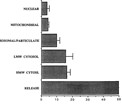
![FIGURE 22:SUBCELLULAR D ISTR IB U TIO N OF ['*C ]C P94](https://thumb-us.123doks.com/thumbv2/123dok_us/9163011.1454599/101.612.55.517.107.553/figure-subcellular-istr-ib-tio-of-p.webp)