RVC OPEN ACCESS REPOSITORY – COPYRIGHT NOTICE
This is the peer-reviewed, manuscript version of an article published in In Practice. The final version is available online via http://dx.doi.org/10.1136/inp.k4485.
The full details of the published version of the article are as follows:
TITLE: Laminitis in horses
AUTHORS: Nicola Menzies-Gow
JOURNAL TITLE: In Practice
PUBLISHER: BMJ Publishing Group
PUBLICATION DATE: 1 November 2018 (online)
1
Laminitis in Horses
Introduction
Laminitis is a common and painful condition of the adult equine, often resulting in permanent
lameness or euthanasia. Reported estimates of laminitis frequency to range from 1.5-34%,1
depending on the population studied (general practice or referral institutions), proportion of
ponies versus horses, presence of intercurrent diseases (particularly gastrointestinal disease)
and the geographic location (generally USA, UK or Australia). It may occur as a single episode
or, more commonly, as repeated bouts over a prolonged period (recurrent laminitis). The risk
of all-cause mortality was increased nearly 6-fold by the presence of laminitis in a population
of horses treated in first opinion practices in the UK (hazard ratio 5.94 vs. no chronic disease)
indicating the importance of this disease.1
Forms of Laminitis
Laminitis is now considered to be a clinical syndrome associated with systemic disease (sepsis
or systemic inflammatory response syndrome [SIRS] or endocrine disease) or altered weight
bearing rather than being a discrete disease entity.2 Thus, laminitis can be divided into three
forms:
1. Sepsis-associated laminitis
Laminitis that occurs secondary to the SIRS and in particular sepsis is termed sepsis-associated
laminitis. It occurs in association with severe gastrointestinal disease and endotoxemia. Two
models have been used to investigate the pathogenesis of this form of laminitis.3,4 In the
2 by intestinal bacteria resulting in a severe drop in intestinal pH, death of Gram-negative
bacteria and enterocolitis in which mucosal injury results in the systemic absorption of
numerous substances. However, the exact identity of the laminitis trigger(s) remains elusive.
The other experimental model that appears to mirror sepsis-related laminitis is the black
walnut extract model. These models have produced evidence of evidence of systemic
inflammation, endothelial activation, leucocyte adhesion and emigration, altered cytokine
expression and oxidative injury. These result in failure of critical laminar basal epithelial cell
functions and consequent failure of the epithelial adhesion molecules (hemidesmosomes),
which attach the epidermal cells to the basement membrane. Laminar separation follows.
2. Endocrinopathic laminitis
Endocrinopathic laminitis is the commonest form of laminitis, accounting for 90% of cases of
laminitis in two studies.5,6 It encompasses laminitis linked with insulin dysregulation (ID), as
occurs in association with equine metabolic syndrome (EMS), pituitary pars intermedia
dysfunction (PPID) and glucocorticoid administration. The key feature of EMS is ID, which may
manifest as hyperinsulinaemia, an excessive insulin response to oral carbohydrate
consumption or tissue insulin resistance.7 PPID is a progressive neurodegenerative disorder
associated with loss of the inhibitory dopaminergic input to the pituitary pars intermedia (PI).8
This results in increased production of the normal hormone products of the PI, some of which
may antagonise the actions of insulin resulting in ID in a subset of animals. Exogenous
corticosteroid administration is associated with an increased risk of laminitis in animals
possessing other laminitis risk factors,1 possibly due to antagonism of insulin by the
3 Prolonged experimental hyperinsulinaemia induced laminitis in healthy ponies9 and horses.10
In contrast to sepsis-associated laminitis, lamellar inflammation was not a major histological
feature11-13 and evidence of systemic or gastrointestinal inflammation was not apparent. The
lamellar histological changes were more consistent with stretching rather than separation of
the basement membrane, accompanied by increased mitotic activity and cellular
proliferation.11-13 Various theories have been postulated to explain the potential relationship
between hyperinsulinaemia/ID and laminitis since alterations in glucose uptake/metabolism
have been discounted.14 One possibility is that whilst at physiological concentrations insulin
preferentially binds to the insulin receptor (InsR), at high concentrations insulin can bind and
activate the insulin-like growth factor-1 receptor (IGF-1R). There are significant numbers of
IGF-1R in equine lamellar tissue.15 Thus, hyperinsulinaemia may directly overstimulate
IGF-1R-mediated cell proliferation, which could potentially weaken the lamellar suspensory
apparatus triggering the onset of clinical signs. Alternatively, it may lead to receptor
down-regulation via negative feedback.16 A significant proportion of endocrinopathic laminitis cases
occur at pasture; thus, consumption of pasture carbohydrate may exacerbate
hyperinsulinaemia, resulting in laminitis.
3. Supporting limb laminitis
Supporting limb laminitis (SLL) is uncommon; a UK study revealed a practice prevalence of
just 0.02%.17 However, it is a major contributor to treatment failure in painful limb conditions
such as fractures and refractory cases of synovial sepsis. Although the severity and duration
of lameness are considered risk factors,18 the development of supporting limb laminitis (SLL)
4 studies utilising tissue microdialysis suggest that cyclic loading of the feet plays an essential
role in digital homeostasis at rest and that decreased frequency of unloading of a limb,
combined with increased mean load bearing on that limb, can result in lamellar ischaemia. 20
Thus, SLL may be a consequence of lamellar ischaemia.
Stages of Laminitis
Acute laminitis can be divided into three stages. Firstly, there is a developmental or prodromal
phase that begins with contact with the pathophysiological trigger and ends with the onset
of lameness up to 72 hours later. This is followed by the acute phase during which the clinical
signs are seen. Thus, the clinical signs only become apparent once the lamellar tissues have
already been subjected to significant metabolic and degenerative changes and treatment
should be initiated as soon as possible. The acute phase is followed by either resolution of the
disease or entry into the chronic phase.
Risk factors for laminitis
A systematic literature review identified those risk factors with the most reliable evidence.21
These included being a pony,22,23 the spring and summer months,24 being female,22,24
increasing age,22 regional or generalised obesity,22 and endotoxemia.25 However, that this
review did not separate the three forms of laminitis. In a case-control study that included only
endocrinopathic laminitis, factors which increased the laminitis risk included weight gain in
the previous three months, the summer and winter months, new access to grass in the
previous four weeks, box rest in the previous week, owner-reported history of laminitis,
5 EMS) disease and increasing time since the last anthelmintic treatment.26 Factors associated
with a decreased laminitis risk were increasing height, feeding of additional supplements in
the previous week and transportation in the previous week.26 Similarly, cases of
endocrinopathic laminitis were significantly older and more likely to be pony breeds compared
to the general hospital population.5
Diagnosis
The diagnosis of laminitis is usually based on the clinical signs (Table 1; Figure 1).27 The
lameness can vary in severity from that which is only perceptible at the trot, through to
spending prolonged periods recumbent.
Further diagnostic tests are performed in those cases where an underlying endocrinological
abnormality is suspected. A diagnosis of EMS is based on a history of recurrent laminitis in an
animal often described by the owners as a good doer or easy keeper. However, generalised
or regional obesity are not a prerequisite of EMS (Figure 2). It develops in animals <15 years
old and there may be a genetic link with certain breeds being over-represented. The gold
standard test to identify ID is yet to be determined; however, there are tests designed to
detect the specific manifestations (Table 2).
PPID is more common in older animals and in ponies compared to horses. Clinical signs can
be divided into early and advanced (Table 3; Figure 3). Currently recommended further
diagnostic tests include resting ACTH concentrations (using seasonally adjusted laboratory
6 insulin dysregulation should be performed to identify the subset of animals with PPID that
have ID.
Lateromedial foot radiographs are taken in those cases where movement of the pedal bone
is suspected or in those cases which fail to respond to the initial treatment (Figure 4).
Treatment of acute laminitis
Treatment should be initiated as soon as possible and should be aimed at providing analgesia
and foot support. Additionally, cryotherapy is indicated in certain circumstances.
Analgesia
Laminitis is an extremely painful condition and non-steroidal anti-inflammatory drugs
(NSAIDs) are the first choice for analgesia (Figure 5). However, there is no evidence to suggest
that any one specific NSAID is superior to the next.28
If NSAIDs do not provide sufficient pain relief, then opiates can be used in addition, including
butorphanol, pethidine and morphine (Figure 6). Transdermal fentanyl was reported to be
effective in horses with pain refractory to NSAID analgesia, especially in animals of lower body
weight in one small clinical report.29 However, uptake of fentanyl from a transdermal patch is
highly variable in adult horses.30 Tramadol has been advocated, however it has a low oral
bioavailability (∼9%), a short half-life (∼2 hours)31 and did not alter hoof withdrawal or
skin-twitch latency to a thermal stimulus.32 Thus, current evidence does not support its use alone.
If single drugs do not provide adequate analgesia, then multimodal therapy can be used in
7 combination with a constant-rate infusion of lidocaine, ketamine, butorphanol, 2 agonists or
combinations thereof.
A neuropathic component to the pain associated with laminitis has been demonstrated33
making ketamine and gabapentin potentially suitable drugs. In one study, oral tramadol alone
provided little pain relief, but the combination of tramadol and ketamine resulted in
decreased blood pressure, decreased forelimb offloading frequency and increased forelimb
loading in horses with naturally occurring laminitis.34 Gabapentin improved hindlimb pain that
was probably associated with femoral neuropathy in one horse35 and has a relatively low
bioavailability, but no apparent adverse effects following oral administration in horses.36
Further work is needed to assess the clinical effect of gabapentin more objectively in horses
with clinical pain. Newer therapies, such as soluble epoxide hydrolase inhibitors and vanilloid
receptor antagonists may prove useful in the future, but again further work is needed.37
Foot Support
Supporting the foot is an essential part of the management of acute laminitis. The horse
naturally adopts a stance that bears most of the weight over the caudal part of the foot rather
than the painful toe region. Additional support should be applied to this region of the foot in
order to provide pain relief and to minimise the mechanical forces on the laminae and hence
pedal bone movement. The simplest method is to increase the depth of the bedding, ensuring
that the bedding extends to the door where the horse will spend a significant proportion of
its day standing using shavings, sand, peat or hemp based products as they pack beneath the
feet best (Figure 7). Extra support can be applied directly to the caudal two thirds of the foot
8 and sole supports (Figure 8). Currently, there is no evidence to suggest that any one foot
support method is superior.28 The supports should be left in place whilst the horse remains
acutely painful and can be replaced by more permanent alternatives once the horse is
comfortable if required.
Vasodilator Therapy
Vasodilator therapy was historically used based on laminitis being a consequence of digital
hypoperfusion. However, this pathogenesis concept is now outdated for two forms of the
condition. Nevertheless, the sedative effect of acepromazine may have the additional
beneficial effect of reducing movement or even resulting in increased periods of time spent
recumbent with the weight taken off the feet. In a single study evaluating the outcome of
equine pasture-associated laminitis managed in first opinion practice in the UK, there was a
trend toward use of acepromazine being associated with survival.28
Cryotherapy
Prophylactic continuous cooling (cryotherapy) of the equine digit effectively limited the
biochemical, histological and clinical abnormalities associated with experimentally-induced
sepsis-associated; most probably through interruption of inflammatory signaling pathways as
well local vascular and metabolic mechanisms.40 In addition, prophylactic digital cryotherapy
was associated with a decreased incidence of laminitis in horses with colitis41 and prevented
or limited lamellar failure in experimentally-induced sepsis-associated laminitis when
initiated after the onset of clinical signs of laminitis.42 Thus, there is evidence to support the
9 been recommended that the hoof temperature should be maintained at <10°C for 72 hours,
achieved by immersion of the foot and pastern region in ice and water.43 There is currently
no published evidence relating to the use of cryotherapy for the prevention or treatment of
endocrinopathic or supporting limb laminitis.
Diet
Animals with acute endocrinopathic laminitis should be removed from pasture and box
rested. A diet based on grass hay (or hay substitute) with low (<10%) non-structural
carbohydrate (NSC) content should be fed and cereals avoided. Ideally, the forage should be
analysed before it is fed. Some recommend soaking hay in water for 30 to 60 minutes before
feeding to leach water soluble carbohydrates and so circumvent the need for analysis;
however, this does not reliably decrease the NSC content to <10% in all cases.44 Forage-only
diets do not provide adequate protein, minerals, or vitamins; thus a low-calorie commercial
ration balancer product that contains high-quality protein and a mixture of vitamins and
minerals is recommended.
Treatment of Underlying Endocrinopathies
Additional therapies are indicated if an underlying endocrinopathy is confirmed.
Pituitary pars intermedia dysfunction (PPID)
The first choice treatment for PPID is the dopamine agonist pergolide, which replaces the lost
dopaminergic inhibition to the PI and so reduces hormone production. It is licensed for the
treatment of PPID in the horse in the UK (Prascend, Boehringer Ingleheim; Figure 9). The initial
dose is 2g/kg p.o. SID for 4-6 weeks. The dose is increased in increments of 1g/kg/day with
10 or laboratory response; or decreased slowly at 4-6 week intervals to the lowest apparently
effective dose. Within the first month of treatment there should be an improvement in
attitude, lethargy and control of hyperglycaemia and a decrease in PU/PD; improvement in
the other clinical signs will occur within one to twelve months. Reported side effects include
diarrhoea, depression, anorexia and colic; however only anorexia and depression are
reported with any frequency. Monotherapy with the serotonin antagonist cyproheptadine
(Periactin, Merk Sharp & Dohme Ltd) is not advocated; however, it can be used in conjunction
with pergolide if pergolide alone is not effective.
Equine metabolic syndrome (EMS)
Treatment of EMS should focus on management changes aimed at weight reduction, if the
animal has regional or generalised adiposity, and exercise, which additionally improves ID.
Weight reduction is achieved through feeding a diet high in fibre and low in NSC. Grain and
other concentrated sources of calories should be removed from the diet. Hay or hay
substitute should initially be provided at 1.5% of current body weight per day, with
subsequent further reductions in feed amount depending on the extent of weight loss. This
should be decreased to <1.0% of target body weight, as this may increase the risk for hindgut
dysfunction, stereotypical behaviours, ingestion of bedding, or coprophagy. The ration should
be divided into three to four feeds per day and strategies to prolong feed intake time should
be considered, such as use of multiple hay nets with small holes. The optimal amount of
exercise required has yet to be determined, but daily light exercise is probably best once the
11 If management changes are unsuccessful alone, then pharmacologic interventions can be
additionally used in the short term (3-6 months). Metformin was initially advocated to
improve insulin sensitivity; however, the bioavailability is very low (7%)45 and it does not have
insulin sensitising effects46 in the horse. Instead metformin reduces the glycaemic and
insulinaemic responses to oral carbohydrate ingestion;47 thus it may be more useful in
preventing post prandial hyperinsulinaemia associated with turn out to pasture or feed
consumption. Levothyroxine is advocated in animals with generalised or regional adiposity.
Weight loss is promoted through an increase in the metabolic rate; however, the diet has to
be strictly controlled because polyphagia may be a consequence of medication.
Prevention
Sepsis-associated laminitis
Prevention of sepsis-associated involves early and effective treatment of the cause of the
sepsis or SIRS, the use of appropriate anti-endotoxic therapy and the prophylactic use of
digital cryotherapy.
Supporting limb laminitis (SLL)
More research is necessary before specific recommendations for prevention of SLL can be
made. Some authors suggest that as limb cycling is an essential component of the circulation,
it would be prudent to institute, whenever practicable, measures to improve foot circulation
in horses at risk of SLL via either controlled exercise (walking) or physical therapy.19 However,
12
Endocrinopathic laminitis
Prevention of endocrinopathic laminitis centres on appropriately treating the underlying
endocrinopathy, maintaining an optimum body condition and limiting intake of pasture NSC
that may exacerbate ID.
A diet based on grass hay (or hay substitute) with low (<10%) NSC content should be fed and
cereals avoided. The NSC content of pasture fluctuates widely; thus, zero grazing should be
considered. However, if an animal is to be turned out, steps should be taken to minimise NSC
intake (Table 4). Forage-only diets do not provide adequate protein, minerals, or vitamins and
so a low-calorie commercial ration balancer product that contains high-quality protein and a
mixture of vitamins and minerals is recommended.
If weight gain is required or the animal is undertaking a large amount of exercise, then caloric
intake can be increased by adding unmolassed soaked sugar beet pulp to the diet
(0.2-0.7kg/day) or by feeding vegetable oil (100-225ml SID or BID up to a maximum of 100 ml/100
kg of body weight).
Several supplements containing magnesium, chromium or cinnamon and a variety of herbs
are marketed with claims for improved insulin sensitivity but scientific evidence of their
efficacy is lacking. A recent study demonstrated that a supplement containing chromium,
magnesium and other nutraceuticals had no effect on insulin sensitivity in laminitic obese
horses.48
Exercise is also essential in the prevention of laminitis as it has been shown to improve insulin
13 but that this probably needs to be maintained on a regular and possibly even daily basis for
the improvement to persist.
Conclusion
In conclusion, laminitis is a common and painful condition of the horse that is now considered
to be a clinical syndrome associated with systemic disease (sepsis-associated or
endocrinopathic laminitis) or altered weight bearing (supporting limb laminitis) rather than
being a discrete disease entity. Various risk factors have been identified for endocrinopathic
laminitis. Diagnosis is based on the history and clinical signs. Further diagnostic tests are
undertaken in cases where an underlying endocrinopathy is suspected and radiographs are
taken if pedal bone movement is suspected or the animal is not responding to appropriate
therapy. Analgesia and foot support are the mainstay of therapy. Digital cryotherapy is useful
in the treatment of sepsis-associated laminitis. Prevention involves prompt treatment of any
underlying disease (all forms of the disease), use of digital cryotherapy (sepsis-associated
laminitis), and maintaining an optimum body condition and limiting carbohydrate intake to
14
References
1. Welsh CE, Duz M, Parkin TDH, et al. Disease and pharmacologic risk factors for first and subsequent episodes of equine laminitis: A cohort study of free-text electronic medical records.
Prev Vet Med 2017;136:11-18.
2. Patterson-Kane JC, Karikoski NP, McGowan CM. Paradigm shifts in understanding equine laminitis. Vet J 2018;231:33-40.
3. van Eps AW, Pollitt CC. Equine laminitis induced with oligofructose. Equine Vet J 2006;38:203-208.
4. Belknap JK. Black walnut extract: an inflammatory model. Vet Clin North Am Equine
Pract 2010;26:95-101.
5. Karikoski NP, Horn I, McGowan TW, et al. The prevalence of endocrinopathic laminitis among horses presented for laminitis at a first-opinion/referral equine hospital. Domest Anim
Endocrinol 2011;41:111-117.
6. Donaldson MT, Jorgensen AJ, Beech J. Evaluation of suspected pituitary pars intermedia dysfunction in horses with laminitis. J Am Vet Med Assoc 2004;224:1123-1127.
7. Tadros EM, Frank N. Endocrine disorders and laminitis. Equine Veterinary Education 2013;25:152-162.
8. McFarlane D. Equine pituitary pars intermedia dysfunction. Vet Clin North Am Equine
Pract 2011;27:93-113.
9. Asplin KE, Sillence MN, Pollitt CC, et al. Induction of laminitis by prolonged hyperinsulinaemia in clinically normal ponies. Vet J 2007;174:530-535.
10. de Laat MA, McGowan CM, Sillence MN, et al. Equine laminitis: induced by 48 h hyperinsulinaemia in Standardbred horses. Equine Vet J 2010;42:129-135.
15 12. Karikoski NP, Patterson-Kane JC, Singer ER, et al. Lamellar pathology in horses with pituitary pars intermedia dysfunction. Equine Vet J 2016;48:472-478.
13. Karikoski NP, Patterson-Kane JC, Asplin KE, et al. Morphological and cellular changes in secondary epidermal laminae of horses with insulin-induced laminitis. Am J Vet Res 2014;75:161-168.
14. Asplin KE, Curlewis JD, McGowan CM, et al. Glucose transport in the equine hoof.
Equine Vet J 2011;43:196-201.
15. Kullmann A, Weber PS, Bishop JB, et al. Equine insulin receptor and insulin-like growth factor-1 receptor expression in digital lamellar tissue and insulin target tissues. Equine Vet J 2016;48:626-632.
16. de Laat MA, Pollitt CC, Kyaw-Tanner MT, et al. A potential role for lamellar insulin-like growth factor-1 receptor in the pathogenesis of hyperinsulinaemic laminitis. Vet J 2013;197:302-306. 17. Wylie CE, Newton JR, Bathe AP, et al. Prevalence of supporting limb laminitis in a UK equine practice and referral hospital setting between 2005 and 2013: implications for future epidemiological studies. Vet Rec 2015;176:72.
18. Peloso JG, Cohen ND, Walker MA, et al. Case-control study of risk factors for the development of laminitis in the contralateral limb in Equidae with unilateral lameness. J Am Vet Med
Assoc 1996;209:1746-1749.
19. van Eps A, Collins SN, Pollitt CC. Supporting limb laminitis. Vet Clin North Am Equine
Pract 2010;26:287-302.
20. Medina-Torres CE, Underwood C, Pollitt CC, et al. The effect of weightbearing and limb load cycling on equine lamellar perfusion and energy metabolism measured using tissue microdialysis. Equine Vet J 2014.
16 22. Alford P, Geller S, Richardson B, et al. A multicenter, matched case-control study of risk factors for equine laminitis. Prev Vet Med 2001;49:209-222.
23. Dorn CR, Garner HE, Coffman JR, et al. Castration and other factors affecting the risk of equine laminitis. Cornell Vet 1975;65:57-64.
24. Menzies-Gow NJ, Katz LM, Barker KJ, et al. An epidemiological study of pasture-associated laminitis and concurrent risk factors in the South of England. Vet Rec 2010.
25. Parsons CS, Orsini JA, Krafty R, et al. Risk factors for development of acute laminitis in horses during hospitalization: 73 cases (1997-2004). J Am Vet Med Assoc 2007;230:885-889.
26. Wylie CE, Collins SN, Verheyen KL, et al. Risk factors for equine laminitis: a case-control study conducted in veterinary-registered horses and ponies in Great Britain between 2009 and 2011.
Vet J 2013;198:57-69.
27. Dyson SJ. Diagnosis of laminitis In: M.W. Ross SJD, ed. Diagnosis and management of
lameness in the horse. 2nd ed. St Louis, MO, USA: Elsevier, 2011;371-372.
28. Menzies-Gow NJ, Stevens K, Barr A, et al. Severity and outcome of equine pasture-associated laminitis managed in first opinion practice in the UK. Vet Rec 2010;167:364-369.
29. Thomasy SM, Slovis N, Maxwell LK, et al. Transdermal fentanyl combined with nonsteroidal anti-inflammatory drugs for analgesia in horses. J Vet Intern Med 2004;18:550-554.
30. Orsini JA, Moate PJ, Kuersten K, et al. Pharmacokinetics of fentanyl delivered transdermally in healthy adult horses--variability among horses and its clinical implications. J Vet
Pharmacol Ther 2006;29:539-546.
31. Stewart AJ, Boothe DM, Cruz-Espindola C, et al. Pharmacokinetics of tramadol and metabolites O-desmethyltramadol and N-desmethyltramadol in adult horses. Am J Vet Res 2011;72:967-974.
17 33. Jones E, Vinuela-Fernandez I, Eager RA, et al. Neuropathic changes in equine laminitis pain. Pain 2007;132:321-331.
34. Guedes AG, Matthews NS, Hood DM. Effect of ketamine hydrochloride on the analgesic effects of tramadol hydrochloride in horses with signs of chronic laminitis-associated pain.
Am J Vet Res 2012;73:610-619.
35. Davis JL, Posner LP, Elce Y. Gabapentin for the treatment of neuropathic pain in a pregnant horse. J Am Vet Med Assoc 2007;231:755-758.
36. Terry RL, McDonnell SM, Van Eps AW, et al. Pharmacokinetic profile and behavioral effects of gabapentin in the horse. J Vet Pharmacol Ther 2010;33:485-494.
37. Guedes AG, Morisseau C, Sole A, et al. Use of a soluble epoxide hydrolase inhibitor as an adjunctive analgesic in a horse with laminitis. Vet Anaesth Analg 2013;40:440-448.
38. Hunt RJ, Brandon CI, McCann ME. Effects of acetylpromazine, xylazine, and vertical load on digital arterial blood flow in horses. Am J Vet Res 1994;55:375-378.
39. Gilhooly MH, Eades SC, Stokes AM, et al. Effects of topical nitroglycerine patches and ointment on digital venous plasma nitric oxide concentrations and digital blood flow in healthy conscious horses. Vet Surg 2005;34:604-609.
40. Van Eps AW, Leise BS, Watts M, et al. Digital hypothermia inhibits early lamellar inflammatory signalling in the oligofructose laminitis model. Equine Vet J 2012;44:230-237.
41. Kullmann A, Holcombe SJ, Hurcombe SD, et al. Prophylactic digital cryotherapy is associated with decreased incidence of laminitis in horses diagnosed with colitis. Equine Vet J 2014;46:554-559.
42. van Eps AW, Pollitt CC, Underwood C, et al. Continuous digital hypothermia initiated after the onset of lameness prevents lamellar failure in the oligofructose laminitis model. Equine Vet
J 2014;46:625-630.
18 44. Longland AC, Harker I, Harris PA. The loss of water-soluble carbohydrate and soluble protein from nine different hays submerged in water for up to 16 hours. . Proceedings of the Equine Science Society 2009.
45. Hustace JL, Firshman AM, Mata JE. Pharmacokinetics and bioavailability of metformin in horses. Am J Vet Res 2009;70:665-668.
46. Tinworth KD, Boston RC, Harris PA, et al. The effect of oral metformin on insulin sensitivity in insulin-resistant ponies. Vet J 2011.
47. Rendle DI, Rutledge F, Hughes KJ, et al. Effects of metformin hydrochloride on blood glucose and insulin responses to oral dextrose in horses. Equine Vet J 2013;45:751-754.
19
Tables
Table 1: Clinical signs of laminitis
Clinical signs associated with laminitis Lameness affecting two or more limbs
Characteristic stance of leaning back on the heels and taking weight off toes
Bounding digital pulses
Increased hoof wall temperature
Pain on hoof tester pressure at the region of the point of the frog
Palpable depression at the coronary band
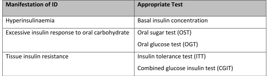
Table 2: Further diagnostic tests use to identify insulin dysregulation in clinical practice
Manifestation of ID Appropriate Test
Hyperinsulinaemia Basal insulin concentration
Excessive insulin response to oral carbohydrate Oral sugar test (OST) Oral glucose test (OGT) Tissue insulin resistance Insulin tolerance test (ITT)
Combined glucose insulin test (CGIT)
Table 3: Clinical signs associated with pituitary pars intermedia dysfunction
Early clinical signs Advanced clinical signs
Delayed haircoat shedding Regional hypertrichosis Lethargy
Regional adiposity
Change in body conformation Laminitis
Generalised hypertrichosis
Loss of seasonal haircoat shedding Skeletal muscle atrophy
Hyperhidrosis
Absent reproductive cycling/infertility Laminitis
Polyuria/polydipsia (PU/PD) Secondary infections
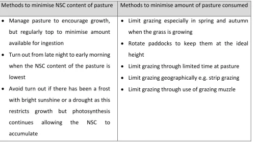
20 Table 4: Methods to minimise NSC ingestion from pasture
Methods to minimise NSC content of pasture Methods to minimise amount of pasture consumed
Manage pasture to encourage growth,
but regularly top to minimise amount
available for ingestion
Turn out from late night to early morning
when the NSC content of the pasture is
lowest
Avoid turn out if there has been a frost
with bright sunshine or a drought as this
restricts growth but photosynthesis
continues allowing the NSC to
accumulate
Limit grazing especially in spring and autumn
when the grass is growing
Rotate paddocks to keep them at the ideal
height
Limit grazing through limited time at pasture Limit grazing geographically e.g. strip grazing Limit grazing through use of grazing muzzle
Figure Headings
Figure 1: A New Forest gelding with the characteristic stance of leaning back on the heels
Figure 2: A Welsh mare with generalised obesity
Figure 3: A New Forest pony gelding with hypertrichosis and recurrent laminitis consistent with a diagnosis of PPID
Figure 4: A) Lateromedial radiograph of the foot showing pedal bone rotation and B) Gross appearance of the same foot post mortem
Figure 5: Various non-steroidal anti-inflammatory drugs are available to provide analgesia
Figure 6: Opiates and Ketamine can be used to provide additional analgesia
21 Figure 8: Various foot supports are commercially available