TwoTIR-like domain containing proteins in a newly
emerging zoonotic
Staphylococcus aureus
strain sequence
type 398 are potential virulence factors by impacting on the
host innate immune response
Nicholas J. Patterson1, Juliane Günther2, Amanda J. Gibson1, Victoria Offord1, Tracey J. Coffey3, Gary Splitter4, Ian Monk5, Hans-Martin Seyfert2 and Dirk Werling1*
1Molecular Immunology Group, Department of Pathology and Pathogen Biology, Royal Veterinary College, Hatfield, UK 2Leibniz Institute for Farm Animal Biology, Dummerstorf, Germany
3School of Veterinary Medicine and Sciences, Faculty of Medicine and Health Sciences, University of Nottingham, Sutton Bonington, UK 4Department of Pathobiological Sciences, University of Wisconsin–Madison, Madison, WI, USA
5Department of Microbiology and Immunology, Faculty of Medicine, Dentistry and Health Sciences, University of Melbourne, Melbourne, VIC, Australia
Edited by:
Hao Shen, University of Pennsylvania, USA
Reviewed by:
Lauren A. Zenewicz, The University of Oklahoma, USA
Hans-Joachim Schuberth, University of Veterinary Medicine Hanover, Germany
*Correspondence:
Dirk Werling, Molecular Immunology Group, Department of Pathology and Pathogen Biology, Royal Veterinary College, Hawkshead Lane, Hatfield, AL9 7TA, UK
e-mail: [email protected]
Staphylococcus aureus, sequence type (ST) 398, is an emerging pathogen and the leading cause of livestock-associated methicillin-resistantS. aureusinfections in Europe and North America. This strain is characterized by high promiscuity in terms of host-species and also lacks several traditional S. aureus virulence factors. This does not, however, explain the apparent ease with which it crosses species-barriers. Recently, TIR-domain containing proteins (Tcps) which inhibit the innate immune response were identified in some Gram-negative bacteria. Here we report the presence of two proteins, S. aureus TIR-like Protein 1 (SaTlp1) and S. aureusTIR-like Protein 2 (SaTlp2), expressed by ST398 which contain domain of unknown function 1863 (DUF1863), similar to the Toll/IL-1 receptor (TIR) domain. In contrast to the Tcps in Gram-negative bacteria, our data suggest that SaTlp1 and SaTlp2 increase activation of the transcription factor NF-κB as well as downstream
pro-inflammatory cytokines and immune effectors. To assess the role of both proteins as potential virulence factors knock-out mutants were created. These showed a slightly enhanced survival rate in a murine infectious model compared to the wild-type strain at one dose. Our data suggest that both proteins may act as factors contributing to the enhanced ability of ST398 to cross species-barriers.
Keywords:TLR signaling,Staphylococcus aureus, bacterial proteins,TCP, innate immune response, mouse model
INTRODUCTION
Staphylococcus aureus is an important commensal and pathogen of animal species causing a variety of diseases ranging from skin and soft tissue infections to more severe, life threatening infec-tions such as toxic shock syndrome in both human and animal species (Lowy, 1998). In addition,S. aureusis a major pathogen causing bovine mastitis, a disease of high economic importance (Holmes and Zadoks, 2011). Here, infections with S. aureus
mainly causes subclinical infections of the udder parenchyma, which is accompanied by the absent of the expression of early innate immune defense genes when compared to infection with
Escherichia coli (Petzl et al., 2008). Indeed, comparative kinetic analysis between E. coli and S. aureus infected primary bovine mammary epithelial cells (pbMECs) showed thatS. aureus infec-tion failed to induce tumor necrosis factor (TNF), interleukin (IL)-1 and IL-8 (CXCL8), which did not depend on the lack of recognition by bovine Toll-like receptor (TLR) 2 or 4 (Yang et al., 2008; Gunther et al., 2011). Treatment of S. aureus infections is complicated by its propensity to acquire antibiotic resistance determinants, most notably the emergence of methicillin-resistant
S. aureus(MRSA). Human MRSA infections can be classified as
mastitis cases due to ST398 has been reported in recent years, adding to the zoonotic risk (Holmes and Zadoks, 2011). In this regard, it is also worth mentioning that infection of the udder by
S. aureusdoes not seem to induce an appropriate innate immune response (Yang et al., 2008), potentially indicating some immune evasion strategies. Given these observations, the relative ease with which ST398 crosses the species-barrier and the lack of certain important S. aureus virulence factors such as panton-valentine leukocidin (PVL), toxic shock syndrome toxin-1 (TSST-1), and leukotoxin M (LukM;Vanderhaeghen et al., 2010) it is possible that ST398 may possesses additional and non-traditional virulence factors. One such group of virulence factors could be represented by Toll/IL-1 receptor (TIR)-domain containing proteins (Tcps), which have been identified in various bacteria (Spear et al., 2009). Whereas Tcps may have a normal function in protein–protein interaction (Spear et al., 2009), the presence of some Tcps in Gram-negative bacteria has been shown to shut-down TLR sig-naling (Spear et al., 2009). TLRs are vital components of the innate immune system, recognizing microbe-associated molec-ular patterns (MAMPs), leading to activation of transcription factors important for initiating the immune response (Akira et al., 2006). The intracellular TIR domain of TLRs is responsible for recruitment and activation of TIR-domain containing adaptor molecules such as myeloid differentiation factor 88 (MyD88), TIR-domain containing adaptor inducing interferon-β (TRIF), TIR-domain containing adaptor protein (TIRAP), and sterile-α
and HEAT/Armadillo motifs-containing protein (SARM; McGet-trick and O’Neill, 2004). Tcps share low sequence identity to their mammalian counterparts, and both are characterized by the presence of three boxes of conserved residues set in a core sequence ranging from 135 to 160 amino acids (Rana et al., 2011). In addition to Tcps, several additional protein classes have recently been identified that may interact and interfere with TLR signal-ing. A conserved region has been identified in transmembrane receptors including similar expression to fibroblast-growth factor (SEF) and IL17Rs in eukaryotes and bacteria. This region shows structural homology to TIR domains, the key functional subdo-main used by the Toll/IL-1 family receptors. A similar dosubdo-main is also found in Act1/CIKS, an essential adaptor downstream of IL-17R family members that is required for activation of NF-κB and other signals. Thus, the SEF/IL17R/CIKS/Act1 homology (SEFIR) domain is observed in both transmembrane and cytoplas-mic proteins (Onishi et al., 2010). Sequence and structure-based fold prediction server results support the distant similarity of the SEFIR domain to the TIR structure, except for the most C-terminal region. SEFIR and TIR domains are related, belong-ing to a new STIR domain superfamily consistbelong-ing of SEFIR, TIR and the recently identified TIR-like domain of unknown function (DUF) 1863 (Novatchkova et al., 2003). In Toll/IL-1R-like path-ways, the cytoplasmically localized TIR domain of a receptor and the TIR domain of a soluble adaptor interact physically and acti-vate signaling. The similarity between the SEFIR and TIR domains involves the above mentioned conserved boxes 1 and 2 of the TIR domain that are implicated in homotypic dimerization, but there is no sequence similarity between SEFIR domains and the TIR sequence box 3. By analogy, it was suggested that SEFIR-domain proteins function as signaling components of Toll/IL-1R-similar
pathways and that their SEFIR domain mediates physical protein– protein interactions between pathway components (Wu et al., 2012).
Several studies have revealed that a diverse range of both pathogenic and non-pathogenic microorganisms express pro-teins containing STIR domains (Newman et al., 2006; Cirl et al., 2008;Spear et al., 2012). Despite low sequence identity, the crys-tal structure of Paracoccus denitrificans (PdTIR) is structurally similar to the TIR domain of human TLR1. PdTIR interacts with both MyD88 and the TLR4 TIR domains, however, no inhibitory function has been demonstrated (Low et al., 2007;
Chan et al., 2009). In contrast, only Tcps identified in Gram-negative bacteria have been shown to inhibit MyD88-dependent TLR signaling. TlpA from Salmonella enterica inhibits activa-tion of the transcripactiva-tion factor NF-κB (Newman et al., 2006). Similarly TcpB, present in a range ofBrucella species including
B. melitensis, B. ovisandB. abortus,and TcpC fromE. coliinhibits NF-κB activation by direct interaction with MyD88, subsequently aiding bacterial macrophage entry and enhancing degradation of MAL (Radhakrishnan et al., 2009, 2011; Radhakrishnan and Splitter, 2010). In contrast, theYersinia pestisTIR-domain pro-tein (YpTdp) has been shown to interact with MyD88 and down-regulate activation of IL-1β; although the presence of this protein did not affect the phenotype of deletion mutants in a mouse infection model (Rana et al., 2011; Spear et al., 2012). There is less information available regarding SEFIR- (Wu et al., 2012) and DUF1863-domain containing proteins within the STIR superfamily.
The hypothesis of the current work was to assess whether proteins belonging to these families are in general present in Gram-positive bacteria, and specifically in ST398, which may explain the ease with that this specific S. aureuscrosses into other species. Here we present data identifying two DUF1863-domain contain-ing proteins, namedS. aureusTIR-Like Proteins 1 and 2 (SaTlp1 and SaTlp2) in ST398S. aureus, which affect TLR-dependent
NF-κB activation in HEK293 and pbMECs. Initial experiments showed that a knock-out (KO) mutation of ST398 lacking both proteins seemed to enhanced survival of mice in anin vivoinfection model using a defined colony forming unit (CFU), although the data did not reach a level of significance. These proteins may be important virulence factors and contribute to the ability of ST398 to cross species-barriers.
MATERIALS AND METHODS
CELLS CULTURE CONDITIONS
expression, their culture in RPMI 1640 (Gibco) on collagen-coated plates, and general challenge conditions were previously described (Gunther et al., 2011).
BACTERIAL STRAINS, GROWTH CONDITIONS, AND MLST
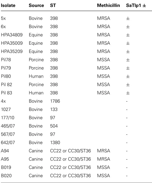
S. aureusstrains isolated from various species and disease presen-tations (Table 1), were grown on 5% Sheep blood agar (Oxoid) and cultured in brain heart infusion medium (Oxoid) at 37◦C. Bacterial genomic DNA was isolated using a Wizard® Genomic DNA Purification Kit (Promega) following manufacturer’s guide-lines. Polymerase chain reaction (PCR) was performed with Easy-A High-Fidelity PCR Mastermix (Agilent) using the primers stated on http://saureus.mlst.net/ (Aanensen and Spratt, 2005) for the genes arcC, aroE, glpF,gmk, pta, tpi, and tpi. Each primer pair amplified an internal fragment of the housekeeping gene and allowed accurate sequencing of ∼450-bp fragments of each gene on both strands. For each locus, the sequences obtained from all isolates were compared and the different sequences were assigned allele numbers. For each isolate, the alleles at each of the seven loci defined the allelic profile that corresponded to a ST.
IDENTIFICATION AND CLONING OF BACTERIAL Tcps
Identification of Tcp-like proteins within S. aureus sequences were performed by searching for homologs of mammalian and bacterial STIR domains in the non-redundant protein sequences database1; using the PSI-BLAST algorithm (Altschul
et al., 1997). Identified proteins were screened initially for struc-tural similarities to known Tcp using SMART (Schultz et al., 19982) and the similarity of their tertiary structure compared to other proteins using the BioInfoBank Meta Server3. Subse-quently, primers (Table 2) were used to amplify the sequences
of SaTlp1 and SaTlp2 (GenBank Acc. No. CAQ50581.1 and
CAQ50581.2, respectively) which also introduced a mutation to alter the start codons to ATG, allowing transcription in eukaryotic cells. PCR products were excised from 1% agarose gels using a GenElute Gel Extraction Kit (Sigma), cloned into pGEM-T Easy (Promega) and inserts sequenced. Subsequently,
SaTlp1, SaTlp2, and TcpB were cloned into the pcDNA3.3
TOPO plasmid (Invitrogen). All plasmids were propagated in TOP10E. coli(Invitrogen) bacteria, grown in Luria Broth supple-mented with 100µg ml−1ampicillin (Invivogen), purified using a PureYield Plasmid Miniprep kit (Promega) and subsequently used for transfection into HEK-boTLR2, 293-hTLR5, or pbMEC cells.
COMPUTATIONAL MODELING OF SaTlp1
The secondary structure prediction and 3D template suggestions for structural SaTlp1, TcpB, and TLR2 were performed by HHpred (Soding et al., 2005) and used to generate a manually adjusted structural alignment. The PdTIR structure is comprised of four chains with differing structures which were manually aligned with anArabidopsis thalianaTIR-like protein and human TIR domains
1http://blast.ncbi.nlm.nih.gov
2http://smart.embl-heidelberg.de/
3http://bioinfo.pl/
Table 1 | Names, species isolated from, MLST group, methicillin-sensitivity (when relevant) and presence of SaTlp1 of the Staphylococcus aureusstrains.
Isolate Source ST Methicillin SaTlp1±
5x Bovine 398 MRSA ±
6x Bovine 398 MRSA ±
HPA34809 Equine 398 MRSA ±
HPA35009 Equine 398 MRSA ±
HPA35209 Equine 398 MRSA ±
Pil78 Porcine 398 MSSA ±
Pil79 Porcine 398 MSSA ±
Pil80 Human 398 MSSA ±
Pil 82 Porcine 398 MSSA ±
Pil 83 Human 398 MSSA ±
4x Bovine 1786
-1027 Bovine 133
-177/10 Bovine 97
-465/07 Bovine 504
-567/07 Bovine 97
-642/07 Bovine 1380
-A94 Canine CC22 or CC30/ST36 MRSA
-A95 Canine CC22 or CC30/ST36 MRSA
-B019 Canine CC22 or CC30/ST36 MSSA
-B020 Canine CC22 or CC30/ST36 MSSA
-from TLR2 (1FYW), TLR10 (1FYX), and MyD88 (2JS7) to the target sequences. The PDB structure 3LD8 was used to span the BB loop insertion which was not present in any of the TIR pro-tein template sequences. MODELER version 9.10 (Fiser and Sali, 2003a) was used to generate 100 models and the poorly defined loop region (residues 43–49) of the highest ranking model refined by MODLOOP (Fiser and Sali, 2003b). Models were validated using PROCHECK (Morris et al., 1992), Verify3D (Eisenberg et al., 1997), and ProQ (Wallner et al., 2003). Sequence and structural similarity were analyzed by PDBeFold (Krissinel and Henrick, 2007).
NF-κB LUCIFERASE ASSAY
The generation of HEK cells expressing TLR2, the main recep-tor for recognition of Gram-positive bacteria as well as the NF-κB reporter gene assay have been described recently ( Will-cocks et al., 2013). HEK cells expressing either boTLR2 or TLR5 (293-hTLR5; Invivogen) were seeded at a density of 2.5×10⁵ and were transfected after 24 h with 250 ng NF-kB-Luc (Firefly luciferase gene under the control of NF-κB promoter; Promega) and 1250 ng pSaTlp1-3.3, pSaTlp2-3.3, or pTcpB-3.3, using TurboFect™in vitroTransfection Reagent (Fermentas) or Lipo-fectamine 2000 (Invitrogen) following manufacturers’ guidelines and returned to the incubator for 24 h. A plasmid whereTcpB
had been ligated into pcDNA3.3 TOPO in the reverse orienta-tion (pTcpB3.3INV) was used as a control. Cells were then split
Table 2 | Primers used for cloning and subsequent screening forSaTlp1 andSaTlp2, and subsequent cloning into pcDNA3.3 and pIMAY.
Primer Sequence
pcDNA3.3 cloning
SaTlp1-5′+Kozak GCG ATG GAA AGA CAA CAA AC
SaTlp1-3′ CTA ATC TGA AAA AGC CTC
SaTlp2-5′+Kozak GAA ATG GCG CGT AAA ACA TTT
SaTlp2-3′ TTA TTT TCT TCT ACA GAT
TcpB-5′+Kozak GCG ATG TCT AAA GAG AAA CAA GCC
TcpB-3′ TCA GAT AAG GGA ATG CAG T
pIMAY cloning
A atatGGTACCGCTTGGAAATGATTTCTGAGTGTGGAATGG
B CACAATTATATTCCACTGGTATGGAAATAATCGC
C ATTTCCATACCAGTGGAATATAATTGTGTAAAAAACT
ATAACTAAAATTGTAAGTCAATTTTAAC
D atatGAGCTCTGTTGTCGTACAGTTTCTGCAGAATACC
OUT-F TATTGCCATTTTCTCAAGATTCAAGTGG
OUT-R TTACTCTCTCACCCTCTTGAAACTTTTCC
Underlined: restriction sites.
and three wells of cells stimulated with either 100 ng ml−1 of
the TLR2/6 ligand FSL-1 (Invivogen), 500 ng ml−1of the TLR5
ligand flagellin (Invivogen) or 30 µg ml−1 heat-killed E. coli. (FBI Dummerstorf). After 24 h gene activation was analyzed using the Luciferase® Reporter Assay System (Promega) follow-ing manufacturer’s guidelines. The cell lysates were made usfollow-ing passive lysis buffer, centrifuged at 16,000×g for 5 min and the OD 280 nm measured using a ND-1000 spectrophotometer (Nan-odrop) to allow for normalization, as previously described (Liu et al., 2011).
ASSESSMENT CYTOKINE PRODUCTION BY QUANTITATIVE PCR
To measure the effect of intracellular SaTlp1 or TcpB proteins on the production of inflammatory response genes in pbMEC, their mRNA concentration was determined for those cells transfected with the respective expression vector or the negative control plas-mid. The pbMECs were grown to 80% confluence into 9 cm plates and were subsequently transfected with 1000 ng of the respective expression vector using Lipofectamine as described above. After an overnight recovery the transfected cells of each 9 cm plate were split into two wells of a six well plate (in order to obtain dupli-cates for the following inflammatory challenge). Subsequent to an additional recovery of 1 day the pbMEC were stimulated with 30µg ml−1heat-killedE. colifor three hours. After the
stimula-tion total RNA was extracted using Trizol (Invitrogen) and RNA quality was determined as previously described (Gunther et al., 2011). For cDNA synthesis, 1.5µg RNA was primed with a mix-ture of gene specific reverse oligonucleotide primers (Table 3) as described (Goldammer et al., 2004). After cDNA purification with HiPure columns (Roche) relative mRNA concentrations were determined with quantitative real-time PCR (RT-qPCR) using the LightCycler instrument and the SYBR green Plus kit (both
from Roche), essentially as described previously (Goldammer et al., 2004;Gunther et al., 2011). Sequences of the oligonucleotide primers for the Chloride intracellular channel protein 1 (CLIC1; house-keeping gene), the proinflammatory cytokinesTNF-α, IL-1β,IL-6,andCXCL8, inducible Nitric oxide synthase 2 (iNOS2), the bactericidal β-defensin lingual antimicrobial peptide (LAP) and the acute phase protein Serum amyloid A (SAA3)are given in
Table 3.
ASSESSMENT OF CYTOKINE PRODUCTION BY ELISA
To measure the effect of intracellular SaTlp1 or TcpB pro-teins on the production of inflammatory response genes in pbMEC, the corresponding supernatants from pbMEC used to generate mRNA were analyzed for the presence of CXCL8 and TNF using ELISA systems as described recently (Met-calfe et al., 2014). IL-1β was analyzed using a bovine IL-1β
ELISA kit (Pierce Protein Biology Products) as per manufacture’s recommendation.
GENERATION OF!saTlp1/!saTlp2MUTANT
In order to deleteSaTlp1andSaTlp2from the ST398 genome the pIMAY plasmid and protocols were used as previously described
Table 3 | Sequences of primer-pairs and probe used for qPCR reactions.
Gene Tested Primer Sequence
IL-1β cDNA 5′-TGCCAGTCCTTGGGGTTATT
Forward 5′-AACCGAGAAGTGGTGTTCTGC
Reverse 5′-TTGGGGTAGACTTTGGGGTCT
IL-6 cDNA 5′-GGGAGCCCCAGCTACTTCAT
Forward 5′-GGAGGAAAAGGACGGATGCT
Reverse 5′-GGTCAGTGTTTGTGGCTGGA
CXCL8 cDNA 5′-GGCCCACTCTCAATAACTCTC
Forward 5′-CCTCTTGTTCAATATGACTTCCA
Reverse 5′-CATGGAACAATGTACATGCGAC
iNOS2 cDNA 5′-CCGGGGTCCTATGGTCAAA
Forward 5′-ACAGGATGACCCCAAACGTC
Reverse 5′-TCTGGTGAAGCGTGTCTTGG
LAP cDNA 5′-TTTTTTTTTTTTTTTTTTTTN
Forward 5′-AGGCTCCATCACCTGCTCCTT
Reverse 5′-CCTGCAGCATTTTACTTGGGCT
SAA3 cDNA 5′-GCCAGCAGGTCTGAAGTGG
Forward 5′-CTTTCCACGGGCATCATTTT
Reverse 5′-CTTCGGGCAGCGTCATAGTT
TNFα cDNA 5′-CTGTGAGTAGATGAGGTAAAGC
Forward 5′-CTTCTGCCTGCTGCACTTCG
Reverse 5′-GAGTTGATGTCGGCTACAACG
CLIC1 cDNA 5′-GATCCCCTCATCCTCAGCAC
Forward 5′-AGAACAACCGCAGGTCGAAT
(Monk et al., 2012). Deletion constructs were produced by ampli-fying regions 500-bp upstream and downstream of the genes using primer pairs A/B and C/D respectively. These PCR prod-ucts were used as templates for the spliced overlap extension (SOE) PCR using A/D primers. The PCR product was purified and cleaved at endonuclease sites added by the A and D primers and cloned into pIMAY using standard protocols. Allelic exchange was achieved by transforming electrocompetent ST398 with the pIMAY construct and plating on brain heart infusion agar (BHIA) with chloramphenicol (Cm) at 28◦C (Monk et al., 2012). Poten-tial mutants were identified after antisense secY induction by colony PCR with OUT-F/OUT-R primers. The double mutant was confirmed by sequencing the deleted region from isolated genomic DNA.
MURINE INFECTION MODEL
The murine peritoneal S. aureus infection is generally consid-ered by be a good model to assess invasiveness and subsequently infectivity ofS. aureus. The study was performed at the AAALAC-accredited Michigan State UniversityIn vivoFacility (East Lansing, MI, USA, study number 14IVF-IF-1003, IACUC approval No. 01/13-013-004). The study was conducted in accordance with the current guidelines for animal welfare (Guide for the Care and Use of Laboratory Animals, 8th Edn, 2011). The procedures used in this study were reviewed and approved by the Institu-tional Animal Care and Use Committee. Six- to eight-week-old female CD-1 mice (Charles River, Portage, MI, USA) with an aver-age weight of 23.8 gm were randomly assigned into 13 groups (n = 6 per group). Animals were housed three per cage and provided food and water ad libitum. Animals were acclimated 8 days prior to use. One day prior to infection a parent culture
of S. aureusST398, and the KO strain ∆SaTlp1/∆SaTlp2 were
removed from frozen storage and streaked out onto trypticase soy agar supplemented with 5% lysed sheep blood cells (TSA-5% SB). Cultures were grown overnight at 37◦C, at ambient atmo-sphere. On the day of infection, the cultures were removed from the incubator and used to prepare an inoculum of each organ-ism in trypticase soy broth (TSB). Bacterial concentrations were measured in the broth by forward light scatter measurement at 600 nm (O.D.600) and adjusted to provide a target value of 0.600. A 1:100 dilution was prepared, using TSB supplemented with 10% gastric hog mucin (GHM) as the diluent, to gener-ate a high dose concentration for challenge of 7.0 log10 bacteria in a dose volume of 0.5 mL. Serial 1:10 dilutions of the high concentration were made into TSB-10% GHM to prepare 6.0 log10, 5.0 log10, 4.0 log10, 3.0 log10, and 2.0 log10 challenge doses of each strain, and the inoculum concentration of each dosing preparation was confirmed by plating serial dilutions for CFU enumeration. Animals received mucin control, wild-type
S. aureus, or KOS. aureusas a single intraperitoneal injections.
Animals were monitored at least twice daily for signs of mor-bidity. During periods of expected high mortality, mice were monitored a minimum of four times per day. Animals exhibit-ing clinical signs listed as criteria for implementation of early
4http://www.aaalac.org/accreditedorgsdirectorysearch/aaalacprgms.cfm
FIGURE 1 | (A)Alignment of the DUF1863 domains of SaTlp1, SaTlp2, and TcpB with TIR domains of known structure. The regions corresponding to the TIR motifs boxes 1, 2, and 3 are indicated by the rectangles above the alignment, highlighted in gray. Secondary structure elements corresponding to the known proteins are annotated above with conservation from high (red) to low (blue) shown below.(B)Predicted tertiary structure of (a) SaTlp1, (b) TcpB and (d) boTLR2 and crystal structure of (c) PdTIR.
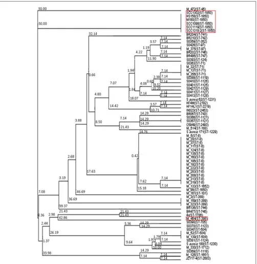
FIGURE 2 | Neighbor joining tree ofS. aureus isolates. Strains falling under “Bovine Mastitis” disease classification on MLST.net and ST398 and ST1850. Red boxes indicate STs in which SaTlp1 and SaTlp2 have been identified. The tree was computed by using concatenated
nucleotide sequences of seven housekeeping genes and was constructed using the tool provided by the MLST database (http:// saureus.mlst.net/sql/uniquetree.asp?). Bootstrap values are indicated above the branches.
STATISTICAL ANALYSIS
All in vitro experiments were performed at least three times,
with samples run in at least duplicates. Data are presented as averages of all experiments, and results were assessed for statis-tical significance by a two-way ANOVA followed by a Bonferroni t-test between treatment groups using Excel and GraphPad Prism
software packages (version 5; GraphPad Software, Inc., La Jolla, CA, USA).
FIGURE 3 | Impact of SaTlp1 and SaTlp2 on NF-κB activation in two
different cell types.Cells were transfected as described, and relative luciferase units were analyzed. Graphs showing the RLU perµg protein in lysates from(A)HEK293-boTLR2 cells stimulated with 100 ng ml−1FSL-1 and(B)primary bovine mammary epithelial cells (pbMECs) stimulated with 30µg ml−1heat-killedE. coli.Cells were transfected with plasmids indicated. Error bars represent the standard error of the mean for each condition (±SEM) of three independent repeats performed in triplicates. Control: medium alone; NFkB alone: cells transfected with NFkB coding plasmid only; invTcpB: cells transfected with a plasmid in which the TcpB sequence was inserted in reverse direct; SaTlp1: cels transfected with plasmid coding for SaTlp1; SaTlp2: cells transfected with plasmid coding for SaTlp2; SaTlp1+2: cells transfected with both plasmids. Significant fold changes are denoted by asterisks in the figure (*p<0.05; ***p<0.001).
atp<0.05. LD50 curves were generated using GraphPad Prism 5.03. A sigmoidal Emax dose response model derived from the Hill equation (four-parameter logistic equation with variable slope using GraphPad Prism v5.03) was used to determine the rela-tionship between the antibacterial drug dose and the efficacy, as determined by bacterial burden: Y=D+(A-D)/(1+10(log C–
X)*α). For the four-parameter logistic equation,Yis the observed
effect,Dis the bottom,Ais the top,Cis the LD50 or 50% of the observed maximum effect,Xis the log of the challenge concentra-tion andαis the Hill slope. The bottom value (D) was constrained
FIGURE 4 | Impact of SaTlp1 and SaTlp2 on NF-κB activation in HEK293-hTLR5 cells.Graphs showing the RLU perµg protein in lysates of cells stimulated with stimulated with 500 ng ml−1flagellin. Cells were transfected with plasmids indicated. Control: medium alone; NFkB alone: cells transfected with NFkB coding plasmid only; SaTlp1: cells transfected with plasmid coding for SaTlp1; SaTlp2: cells transfected with plasmid coding for SaTlp2; SaTlp1+2: cells transfected with both plasmids. Error bars represent the standard error of the mean for each condition (±SEM) of three independent repeats performed in triplicates. Significant fold changes are denoted by asterisks in the figure (***p<0.001).
to 0 and the top value (C) was left unconstrained. Data are shown as Kaplan–Meier Survival Curves.
RESULTS
SaTlp1 AND SaTlp2 SHARE STRUCTURAL HOMOLOGY TO KNOWN TIR DOMAINS
Initially we tried to identify Tcp inS. aureus using a bioinfor-matics approach. As Tcps in other bacteria have been shown to show similarities to mammalian TLR TIR domains, we com-pared different TLR TIR domains against all availableS. aureus
genomes (taxid:1280) using BLAST. This resulted in the identifi-cation of two immediately adjacent proteins (e=8×10⁻⁶ and e=3×10⁻⁵, respectively) containing a DUF1863 domain, located
within a recently identified putative transposon. SMART anno-tation confirmed that these proteins, namedS. aureus Tcp-like protein 1 and 2 (SaTlp1 and SaTlp2) contain DUF1863 domains.
SaTlp1 and SaTlp2 share less than 20% sequence identity with other members of the TIR superfamily. However, despite low similarity at the sequence level, secondary structure prediction suggests that at least four of the central beta strands common to TIR-like domains are present in these bacterial proteins. The clos-est structural homologs identified by HHpred are matches to the DUF1863 (PF08937) and TIR or TIR_2 (PF13676 and PF01582) domain-containing families and proteins sharing a Flavodoxin-like fold (SCOP 31128 and 31130). The closest secondary structure match to both SaTlp1 and TcpB was PdTIR (PDB: 3H16) while the
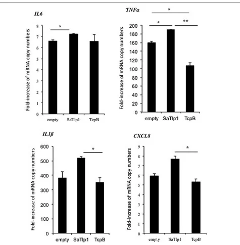
FIGURE 5 | Average fold increase of mRNA levels of the pro-inflammatory genesIL-1β, IL-6, TNF, CXCL8 in pbMEC
transfected with the empty plasmid, or plasmids coding for TcpB and SaTlp1 and stimulated with 30 µg ml–1 heat-killed E. coli for
three hours.Error bars represent the standard error of the mean for each condition (±SEM) of three independent repeats performed in triplicates. Significant fold changes are denoted by asterisks in the figure (*p <0.05; **p <0.01).
Within eukaryotic TIR domains, three conserved regions called boxes 1, 2, and 3 are proposed as putative adaptor binding sites, essential for signaling. The box 1 motif can be clearly identified in all of the TIR-like domains included in the alignment (Figure 1A). However, both SaTlp1 and SaTlp2 appear to have a helical inser-tion within the BB loop (box 2) and, unlike the other bacterial proteins shown, seem to entirely lack box 3. Computational mod-eling of SaTlp1 predicts the conserved TIR-like tertiary structure of four/five central beta strands surrounded by helices (Figure 1B).
While the modeled structure of TcpB is most similar to PdTIR (∼0.5 Å), SaTlp1 shows closer structural homology to TLR TIR domains (∼1.9 Å).
SaTlp1ANDSaTlp2ARE PRESENT IN ALL ST398 ISOLATES AND CC75
Staphyloccoccus aureus
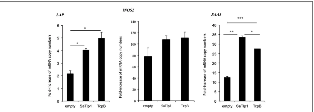
FIGURE 6 | Average fold increase of mRNA levels of the secondary response genesiNOS-2 LAPandSAA3in pbMEC transfected with the empty plasmid or plasmids coding for TcpB and SaTlp1 and
stimulated with 30µg ml–1heat-killedE. colifor three hours.Error
bars represent the standard error of the mean for each condition (±SEM) of three independent repeats performed in triplicates. Significant fold changes are denoted by asterisks in the figure (*p<0.05; **p<0.01; ***p<0.001).
created by MLST analysis, and the presence ofSaTlp1andSaTlp2 in these strains determined (Table 1). Interestingly,SaTlp1and
SaTlp2 were identified in all ST398 isolates tested, regardless of
the species of isolation, clinical symptoms of the host-species or methicillin-sensitivity status. Additionally, BLAST searches on 89 ST398 isolates were performed which revealed that all of these con-tained these genes. In addition to ST398, BLAST searches revealed these genes were present inS. aureusclonal complex (CC) 75 (also known as S. argenteus), however, they were not detected in any otherS. aureuslineage. ST398 and CC75 are only distantly related (Figure 2) and published work on CC75 states that it is a highly divergent lineage, showing an average seven fold greater nucleotide divergence in orthologous genes with typicalS. aureusstrains than other strains (Holt et al., 2011). Considering this divergent rela-tionship but the similarity of these genes it seems highly likely that the transposon containing them was transferred between the strains post-dating their divergence.
BOTH SaTlp1 AND SaTlp2 UP-REGULATE NF-κB ACTIVATION IN HEK-boTLR2 AND pbMEC
To assess whether the presence of the Tcp-like proteins in this
S. aureusstrain interferes with the activation of the innate immune response, we tested whether the identified proteins would exhibit similar effects as described for Tcps of other bacteria which seem to inhibit TLR signaling. We analyzed whether transfection of HEK cells expressing boTLR2 with SaTlp1 and SaTlp2 affected the downstream activity of the transcription factor NF-κB after stimulation with the TLR2 ligand FSL-1 (Figure 3A). The levels of NF-κB-induced luciferase were significantly up-regulated follow-ing transfection withSaTlp1andSaTlp2plasmids (bothp<0.001) when compared to the negative control. Simultaneous transfection of both plasmids also up-regulated NF-κB activation (p<0.001). In the HEK-boTLR2 system these plasmids up-regulated NF-κ B-dependent luciferase between 4.1 (SaTlp1) and 6.1 (SaTlp1/2) fold following stimulation compared to the negative control. In
contrast, transfection of HEK-boTLR2 cells withTcpBwhich had been shown before to block TLR-dependent NF-κB activation (Radhakrishnan et al., 2009) did not induce significant NF-κB stimulation, confirming these data. FSL-1 failed to activate NF-κB in HEK293 not expressing TLR2 (Data not shown).
As the HEK cell system is a somewhat artificial system, we next assessed the effects of SaTlp1 using pbMECs which express TLR2 and have been used before to assess the effect of mastitis caus-ing pathogens on the innate immune response (Yang et al., 2008;
Gunther et al., 2011). As seen in HEK293 cells, transfection of
SaTlp1(p=0.006) caused increased NF-κB activation in pbMEC in response to heat-killedE. coli. As before transfection of pbMECs with TcpB beforeE. colistimulation did not activate NF-κB sig-naling (p<0.001). However, this up-regulation was overall lower compared to HEK293 cells withSaTlp1up-regulating NF-κB by 1.4 fold compared to the negative control (Figure 3B).
THE EFFECT OF SaTlp1 AND SaTlp2 IS NOT LIMITED TO ONE SPECIES OR A SPECIFIC TLR
To assess whether the responses seen in both cell types would be unique to the interaction with boTLR2, we examined the effect of SaTlp1 and SaTlp2 in HEK cells transfected with hTLR5 cells stimulated with flagellin (Figure 4). Transfection ofSaTlp1 SaTlp2 (bothp<0.001) andSaTlp1/2(p<0.001) led to an up-regulation of NF-κB activity of between 2.0 (SaTlp2) and 2.5 (SaTlp1) fold. In contrast, transfection of HEK cells expressing hTLR5 withTcpB down-regulated its activation in response to flagellin (p<0.001). These results are therefore similar to those for boTLR2 expressing cells, suggesting the action of both proteins may not be limited to a specific TLR.
UP-REGULATION OF NF-κB ACTIVATION BY SaTlp1 INCREASES mRNA AND PROTEIN EXPRESSION OF PRIMARY AND SECONDARY RESPONSE GENES
FIGURE 7 | Values of the pro-inflammatory TNF (A), IL-1β(B), and
CXCL8 (C) analyzed in the supernatants of pbMEC transfected with the empty plasmid, or plasmids coding for TcpB and SaTlp1 and stimulated with 30µg ml–1heat-killedE. colifor three hours.Error
bars represent the standard error of the mean for each condition (±SEM) of three independent repeats performed in triplicates. Significant fold changes are denoted by asterisks in the figure (*p<0.05).
level for NF-κB-dependent cytokine production. We analyzed expression of the primary response genes IL-1β, IL-6, CXCL8, andTNFαas well as the secondary response genesiNOS-2, LAP, and SAA3 by qPCR, similar to that described before (Gunther et al., 2011). Stimulation of pbMEC transfected with either empty plasmids or plasmids containingSaTlp1followed byE. coli stim-ulation resulted in the up-regstim-ulation ofIL-1β,IL-6,CXCL8,and
TNF-αmRNA, levels to various extents (Figure 5). In contrast, transfection of pbMEC with a plasmid coding for TcpB in gen-eral reduced expression of these genes after E. coli stimulation (Figure 5). Transfection of pbMEC with plasmids coding for SaTlp1 and TcpB followed by subsequent stimulation withE. coli
increased mRNA expression for the secondary response genes
iNOS-2, LAP andSAA3 (Figure 6), withLAP expression being
effected more by TcpB.
Corresponding supernatants were analyzed for the presence of CXCL8, TNF-α, and IL-1β. The secretion pattern followed in general the results obtained for qPCR, with values obtained for all three cytokines being in generally low. Similar as to the data obtained by qPCR, supernatants of pbMEC transfected with SaTlp1 before stimulation withE. colicontained the largest amount of all three cytokines (Figures 7A–C), whereas no dif-ferences were seen between untransfected pbMEC and pbMEC transfected with TcpB.
A∆saTlp1/∆saTlp2 ST398 POTENTIALLY IMPACTS ON SURVIVAL RATE IN AStaphylococcus aureusMICE CHALLENGE MODEL
As the presence of SaTlp1 and SaTlp2 seemed to increase NF-κB activation and pro-inflammatory cytokine production in response to subsequent stimulation, we next assessed whether the absence of the genes has an impact on the virulence of S. aureusST398
in vivo. After creation of a∆saTlp1/∆saTlp2ST398 KO mutant,
the growth rate or colony appearance of both wild-type and mutantS. aureuswere compared and showed no difference (data not shown). In order to determine whether there are differences in the in vivopathogenicity of wild-type and∆saTlp1/∆saTlp2 ST398 a murine sepsis model was used in order to calculate the median lethal dose (MLD) of the bacteria. CD-1 mice were challenged via i.p. injection with 102–107 CFU of either
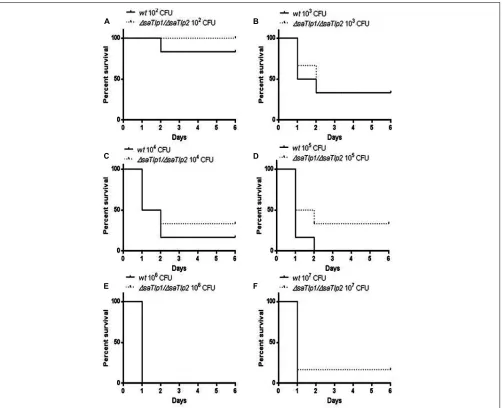
wild-type or mutantS. aureusafter which the time until their death was monitored. The data showed a trend for increased survival rates for mice infected with the∆saTlp1/∆saTlp2mutant, most noticeably in the case of infection with 105 CFU when 2 of 6
mice challenged with mutant bacteria survived until day 6 of the experiment whereas all mice infected with wild-type had died by day 2 (Figure 8). Despite the fact that none of the observed differences reach level of significance, the lowestp-value (p=0.13) was obtained for 105CFU. Considering the data from
all challenge concentrations, the MLD of the strains was calcu-lated using the Reed-Meunch equation. This showed the MLD of
∆saTlp1/∆saTlp2ST398 to be 3630 CFU compared to one of 575
for the wild-type ST398 indicating that in order for 50% of chal-lenged mice to be killed by the mutant, an infectious dose of over 6.3 times that of wild-type ST398 is required. This suggests that although differences between wild-type and ∆saTlp1/∆saTlp2 ST398 are not significant at individual concentrations, when is considered together there does appear to be genuine differences in their pathogenicity.
DISCUSSION
FIGURE 8 | Kaplan–Meier survival curves for groups of mice infected with different CFU of either wild-type or∆saTlp1/∆saTlp2ST398 S. aureus.Each figure represent six mice challenged by intra-peritoneal injection with 0.5 mL containing wild-type ST398 and six mice challenged
with∆saTlp1/∆saTlp2KO mutant which were then monitored for 6 days and deaths recorded. Figures show survival rates for mice challenged with (A)102CFU(B)103CFU(C)104CFU(D)105CFU(E)106CFU and(F) 107CFU.
activity of the transcription factor NF-κB in boTLR2, HEK-hTLR5, and pbMEC cells, as well as mRNA expression levels of pro-inflammatory primary and secondary response genes. However, in contrast to work published on the function of Tcps identified in Gram-negative bacteria, which were shown to down-regulate NF-κB activation as well as subverting the ensuing innate immune response (Radhakrishnan et al., 2009;Yadav et al., 2010;Rana et al., 2011), our data seems to suggest that these pro-teins activate NF-κB signaling and the production of inflammatory mediators. Although there is still a certain amount of debate as to whether TIR, SEFIR, and DUF1863 domains should be classified separately or as a single domain, our data suggests that they may have distinct functions within the same signaling pathways.
In addition to the differences seen in activation of NF-κB, this study is the first to investigate these presence of such proteins
from Gram-positive bacteria, as all published work on bacterial Tcps has been in Gram-negative bacteria such and B. meliten-sis (TcpB), E. coli (TcpC), and P. denitrificans (PdTIR). It is unclear whether NF-κB down-regulation is associated with Gram-negative and up-regulation with Gram-positive bacteria or if there are examples of up- and down-regulation for Tcp present in both. Further to this it must be considered that TIR domains are most likely to have more general protein/protein interac-tion funcinterac-tions, as reviewed recently (Spear et al., 2009), and modulation of TLR signaling by these proteins may not be the rule.
strain CC75, isolated in Northern Australia, also known aS. argen-teus(Holt et al., 2011). There is conflicting evidence as to whether the genetic element containing these genes is ancestral or acquired and further study would determine if there are any notable pathogenic similarities between these two strains, particularly in the context of ST398. Strains of this group are notable for their high promiscuity, with the ability to easily cross species-barriers, having been isolated from a range of livestock animals and also humans. Furthermore ST398S. aureusare often missing several traditional staphylococcal virulence factors such as PVL, suggest-ing that these bacteria may have acquired alternative factors that allow for their successful host colonization. It could be hypothe-sized that these Tlps may represent novel staphylococcal virulence factors, which may be unique to this specific MLST. Indeed, and in contrast to our data, the interference ofS. aureuswith MyD88-dependent TLR signaling was recently suggested (Gunther et al., 2011), with the authors demonstrating an inhibitory effect of
S. aureuson TNF-αand IL-1 mRNA production. The fact that our data show that the identified proteins are influencing the pro-inflammatory response system on both, mRNA and protein level indicates that both proteins are potentially interesting vir-ulence factors that warrant further investigation. This is of even more interest as not only SaTlp1, but also TcpB fromB. meli-tiensisseemed to affect mRNA expression levels of the secondary response genesLAPandSAA3. These requirede novosynthesis and have been shown to need chromatin-remodeling (Liu et al., 2011) or the transcription factor C/EBPδ(Litvak et al., 2009), and were differently expressed compared to the pro-inflammatory genes.
We are fully aware that thein vivomodel used may not be the most appropriate one to assess the function of the two identi-fiedS. aureusproteins. However, murine models forS. aureusare still considered the appropriate standard for invasiveness/survival (Kim et al., 2014;Zhang et al., 2014). Thein vivoinfection data from a murine sepsis model provided data which suggests a trend toward increased survival time for mice infected with the
∆saTlp1/∆saTlp2mutant compared to the wild-type ST398 strain although differences between these were not statistically signifi-cant at any individualS. aureusconcentration. The level where the greatest differences are apparent was when mice were infected with 105CFU. This is with ap-value of 0.1292 according to the Log-rank
test which, although not significant, would certainly merit further investigation considering the relatively small sample size at each CFU concentration (n=6). It is possible that at CFU concentra-tions lower than 105there may be less effect as the mice may be
more able to fight the infection regardless, whereas at higher con-centrations the effects of these proteins may be negated due to the sheer number of bacteria present. Around the 105mark may repre-sent something of a sweet spot where effect of these proteins can be seen. Additionally the small sample size is clearly susceptible to nat-ural variation between mice and challenge doses. This is especially important as while large differences in survival of infected mice were not observed there may be more subtlein vivoeffects of these proteins similar to those proposed for YpTlp (Spear et al., 2012). Subtlein vivoeffects would very likely require relatively large sam-ple sizes in order to be able to produce statistically significant results. The exact mechanisms used by these proteins are yet to be elucidated, as a direct interaction with host proteins has not been
identified. It should also be noted that there is still no clear picture as to how these bacterial proteins would reach any cellular target, as they do not contain domains associated with export from the bacterium. However, these findings are similar to TcpB ofBrucella. Despite this, TcpB exhibits lipid-binding properties ( Radhakr-ishnan and Splitter, 2010), and interacts with phosphoinositides through its N-terminal domain, leading to co-localization with the plasma membrane and components of the cytoskeleton ( Radhakr-ishnan et al., 2009). Furthermore thisBrucellaTIR protein has the property of cell permeability which would provide a mechanism for virulence and could mean that it influences nearby cells not infected with the bacteria, as well as contributing to trafficking within the cell to influence host cell machinery.
In conclusion, SaTlp1 and SaTlp2 are potential novel viru-lence factors in ST398S. aureus which appear to interact with the innate immune signaling machinery of host cells in novel ways. The presence of these proteins may account for some of the unusual characteristics of strains of S. aureuswithin ST398 and are therefore interesting targets for understanding these characteristics.
ACKNOWLEDGMENTS
We thank W. Petzl and H. Zerbe (Centre of Clinical Veterinary Medicine of the Ludwig-Maximilians-University Munich) and E. Statis (RVC) for providing the different Staphylococci strains, and G. Splitter for TcpB and control plasmids. The authors declare no conflict of interest. This project was supported by a BBSRC iCASE grant (BB/G018073/1) with Zoetis Animal Health to Dirk Werling. This manuscript represents publication No PID_00483 of the RVC.
REFERENCES
Aanensen, D. M., and Spratt, B. G. (2005). The multilocus sequence typing network: mlst.net. Nucleic Acids Res. 33, W728–W733. doi: 10.1093/nar/ gki415
Akira, S., Uematsu, S., and Takeuchi, O. (2006). Pathogen recognition and innate immunity.Cell124, 783–801. doi: 10.1016/j.cell.2006.02.015
Altschul, S. F., Madden, T. L., Schaffer, A. A., Zhang, J., Zhang, Z., Miller, W., et al. (1997). Gapped BLAST and PSI-BLAST: a new generation of protein database search programs.Nucleic Acids Res.25, 3389–3402. doi: 10.1093/nar/25.17.3389 Armand-Lefevre, L., Ruimy, R., and Andremont, A. (2005). Clonal
com-parison of Staphylococcus aureus isolates from healthy pig farmers, human controls, and pigs. Emerg. Infect. Dis. 11, 711–714. doi: 10.3201/eid1105. 040866
Chan, S. L., Low, L. Y., Hsu, S., Li, S., Liu, T., Santelli, E., et al. (2009). Molecular mimicry in innate immunity: crystal structure of a bacterial TIR domain.J. Biol. Chem.284, 21386–21392. doi: 10.1074/jbc.C109.007591
Cirl, C., Wieser, A., Yadav, M., Duerr, S., Schubert, S., Fischer, H., et al. (2008). Subversion of Toll-like receptor signaling by a unique family of bacterial Toll/interleukin-1 receptor domain-containing proteins.Nat. Med.14, 399–406. doi: 10.1038/nm1734
Eisenberg, D., Luthy, R., and Bowie, J. U. (1997). VERIFY3D: assessment of protein models with three-dimensional profiles.Methods Enzymol.277, 396–404. doi: 10.1016/S0076-6879(97)77022-8
Fiser, A., and Sali, A. (2003a). Modeller: generation and refinement of homology-based protein structure models.Methods Enzymol.374, 461–491. doi: 10.1016/S0076-6879(03)74020-8
Fiser, A., and Sali, A. (2003b). ModLoop: automated modeling of loops in protein structures.Bioinformatics19, 2500–2501. doi: 10.1093/bioinformatics/btg362 Fitzgerald, J. R. (2012). Livestock-associated Staphylococcus aureus: origin,
Foster, T. J. (2005). Immune evasion by staphylococci.Nat. Rev. Microbiol.3, 948– 958. doi: 10.1038/nrmicro1289
Goldammer, T., Zerbe, H., Molenaar, A., Schuberth, H. J., Brunner, R. M., Kata, S. R., et al. (2004). Mastitis increases mammary mRNA abundance of beta-defensin 5, toll-like-receptor 2 (TLR2), and TLR4 but not TLR9 in cattle.Clin. Diagn. Lab. Immunol.11, 174–185.
Graveland, H., Duim, B., Van Duijkeren, E., Heederik, D., and Wagenaar, J. A. (2011). Livestock-associated methicillin-resistantStaphylococcus aureusin ani-mals and humans.Int. J. Med. Microbiol.301, 630–634. doi: 10.1016/j.ijmm.2011. 09.004
Graveland, H., Wagenaar, J. A., Heesterbeek, H., Mevius, D., Van Duijkeren, E., and Heederik, D. (2010). Methicillin resistantStaphylococcus aureusST398 in veal calf farming: human MRSA carriage related with animal antimicro-bial usage and farm hygiene.PLoS ONE5:e10990. doi: 10.1371/journal.pone. 0010990
Gunther, J., Esch, K., Poschadel, N., Petzl, W., Zerbe, H., Mitterhuemer, S., et al. (2011). Comparative kinetics ofEscherichia coli- andStaphylococcus aureus-specific activation of key immune pathways in mammary epithelial cells demonstrates thatS. aureuselicits a delayed response dominated by interleukin-6 (IL-interleukin-6) but not by IL-1A or tumor necrosis factor alpha.Infect. Immun.79, 695–707. doi: 10.1128/IAI.01071-10
Holmes, M. A., and Zadoks, R. N. (2011). Methicillin resistant S. aureus in human and bovine mastitis.J. Mammary Gland Biol. Neoplasia16, 373–382. doi: 10.1007/s10911-011-9237-x
Holt, D. C., Holden, M. T., Tong, S. Y., Castillo-Ramirez, S., Clarke, L., Quail, M. A., et al. (2011). A very early-branchingStaphylococcus aureuslineage lacking the carotenoid pigment staphyloxanthin.Genome Biol. Evol.3, 881–895. doi: 10.1093/gbe/evr078
Jamrozy, D. M., Fielder, M. D., Butaye, P., and Coldham, N. G. (2012). Comparative genotypic and phenotypic characterisation of methicillin-resistant Staphylococ-cus aureusST398 isolated from animals and humans.PLoS ONE7:e40458. doi: 10.1371/journal.pone.0040458
Kim, H. K., Missiakas, D., and Schneewind, O. (2014). Mouse models for infectious diseases caused byStaphylococcus aureus.J. Immunol. Methods410, 88–99. doi: 10.1016/j.jim.2014.04.007
Krissinel, E., and Henrick, K. (2007). Inference of macromolecular assemblies from crystalline state.J. Mol. Biol.372, 774–797. doi: 10.1016/j.jmb.2007.05.022 Lindsay, J. A. (2010). Genomic variation and evolution ofStaphylococcus aureus.Int.
J. Med. Microbiol.300, 98–103. doi: 10.1016/j.ijmm.2009.08.013
Litvak, V., Ramsey, S. A., Rust, A. G., Zak, D. E., Kennedy, K. A., Lampano, A. E., et al. (2009). Function of C/EBPdelta in a regulatory circuit that discriminates between transient and persistent TLR4-induced signals.Nat. Immunol.10, 437–443. doi: 10.1038/ni.1721
Liu, S., Shi, X., Bauer, I., Gunther, J., and Seyfert, H. M. (2011). Lingual antimicrobial peptide and IL-8 expression are oppositely regulated by the antagonistic effects of NF-kappaB p65 and C/EBPbeta in mammary epithelial cells.Mol. Immunol.
48, 895–908. doi: 10.1016/j.molimm.2010.12.018
Low, L. Y., Mukasa, T., Reed, J. C., and Pascual, J. (2007). Characterization of a TIR-like protein fromParacoccus denitrificans.Biochem. Biophys. Res. Commun.
356, 481–486. doi: 10.1016/j.bbrc.2007.03.003
Lowy, F. D. (1998).Staphylococcus aureusinfections.N. Engl. J. Med.339, 520–532. doi: 10.1056/NEJM199808203390806
McGettrick, A. F., and O’Neill, L. A. (2004). The expanding family of MyD88-like adaptors in Toll-like receptor signal transduction.Mol. Immunol.41, 577–582. doi: 10.1016/j.molimm.2004.04.006
Metcalfe, H. J., La Ragione, R. M., Smith, D. G., and Werling, D. (2014). Func-tional characterisation of bovine TLR5 indicates species-specific recognition of flagellin.Vet. Immunol. Immunopathol.157, 197–205. doi: 10.1016/j.vetimm. 2013.12.006
Monk, I. R., Shah, I. M., Xu, M., Tan, M.-W., and Foster, T. J. (2012). Transforming the untransformable: application of direct transformation to manipulate genet-icallyStaphylococcus aureusandStaphylococcus epidermidis.MBio3:e00277–11. doi: 10.1128/mBio.00277-11
Morris, A. L., Macarthur, M. W., Hutchinson, E. G., and Thornton, J. M. (1992). Stereochemical quality of protein structure coordinates.Proteins12, 345–364. doi: 10.1002/prot.340120407
Newman, R. M., Salunkhe, P., Godzik, A., and Reed, J. C. (2006). Identification and characterization of a novel bacterial virulence factor that shares homology
with mammalian Toll/interleukin-1 receptor family proteins.Infect. Immunol.74, 594–601. doi: 10.1128/IAI.74.1.594-601.2006
Novatchkova, M., Leibbrandt, A., Werzowa, J., Neubuser, A., and Eisenhaber, F. (2003). The STIR-domain superfamily in signal transduction, develop-ment and immunity.Trends Biochem. Sci. 28, 226–229. doi: 10.1016/S0968-0004(03)00067-7
Onishi, R. M., Park, S. J., Hanel, W., Ho, A. W., Maitra, A., and Gaffen, S. L. (2010). SEF/IL-17R (SEFIR) is not enough: an extended SEFIR domain is required for il-17RA-mediated signal transduction. J. Biol. Chem.285, 32751–32759. doi: 10.1074/jbc.M110.121418
Petzl, W., Zerbe, H., Gunther, J., Yang, W., Seyfert, H. M., Nurnberg, G., et al. (2008).Escherichia coli, but notStaphylococcus aureustriggers an early increased expression of factors contributing to the innate immune defense in the udder of the cow.Vet. Res.39, 18. doi: 10.1051/vetres:2007057
Radhakrishnan, G. K., Harms, J. S., and Splitter, G. A. (2011). Modulation of microtubule dynamics by a TIR domain protein from the intracellu-lar pathogenBrucella melitensis. Biochem. J.439, 79–83. doi: 10.1042/BJ20 110577
Radhakrishnan, G. K., and Splitter, G. A. (2010). Biochemical and functional analysis of TIR domain containing protein fromBrucella melitensis.Biochem. Biophys. Res. Commun.397, 59–63. doi: 10.1016/j.bbrc.2010.05.056
Radhakrishnan, G. K., Yu, Q., Harms, J. S., and Splitter, G. A. (2009). Bru-cellaTIR domain-containing protein mimics properties of the Toll-like receptor adaptor protein TIRAP. J. Biol. Chem. 284, 9892–9898. doi: 10.1074/jbc. M805458200
Rana, R. R., Simpson, P., Zhang, M., Jennions, M., Ukegbu, C., Spear, A. M., et al. (2011).Yersinia pestisTIR-domain protein forms dimers that inter-act with the human adaptor protein MyD88. Microb. Pathog.51, 89–95. doi: 10.1016/j.micpath.2011.05.004
Schultz, J., Milpetz, F., Bork, P., and Ponting, C. P. (1998). SMART, a simple modular architecture research tool: identification of signaling domains.Proc. Natl. Acad. Sci. U.S.A.95, 5857–5864. doi: 10.1073/pnas.95.11.5857
Smith, T. C., Male, M. J., Harper, A. L., Kroeger, J. S., Tinkler, G. P., Moritz, E. D., et al. (2009). Methicillin-resistantStaphylococcus aureus(MRSA) strain ST398 is present in midwestern U.S. swine and swine workers.PLoS ONE4:e4258. doi: 10.1371/journal.pone.0004258
Soding, J., Biegert, A., and Lupas, A. N. (2005). The HHpred interactive server for protein homology detection and structure prediction.Nucleic Acids Res.33, W244–W248. doi: 10.1093/nar/gki408
Spear, A. M., Loman, N. J., Atkins, H. S., and Pallen, M. J. (2009). Microbial TIR domains: not necessarily agents of subversion?Trends Microbiol. 17, 393–398. doi: 10.1016/j.tim.2009.06.005
Spear, A. M., Rana, R. R., Jenner, D. C., Flick-Smith, H. C., Oyston, P. C., Simpson, P., et al. (2012). A Toll/interleukin (IL)-1 receptor domain protein fromYersinia pestisinteracts with mammalian IL-1/Toll-like receptor pathways but does not play a central role in the virulence ofY. pestisin a mouse model of bubonic plague.Microbiology158, 1593–1606. doi: 10.1099/mic.0.055012-0
Vanderhaeghen, W., Hermans, K., Haesebrouck, F., and Butaye, P. (2010). Methicillin-resistantStaphylococcus aureus(MRSA) in food production animals.
Epidemiol. Infect.138, 606–625. doi: 10.1017/S0950268809991567
Wallner, B., Fang, H., and Elofsson, A. (2003). Automatic consensus-based fold recognition using Pcons, ProQ, and Pmodeller.Proteins53(Suppl. 6), 534–541. doi: 10.1002/prot.10536
Wassenberg, M. W., Bootsma, M. C., Troelstra, A., Kluytmans, J. A., and Bonten, M. J. (2011). Transmissibility of livestock-associated methicillin-resistant Staphy-lococcus aureus(ST398) in Dutch hospitals.Clin. Microbiol. Infect.17, 316–319. doi: 10.1111/j.1469-0691.2010.03260.x
Willcocks, S., Offord, V., Seyfert, H. M., Coffey, T. J., and Werling, D. (2013). Species-specific PAMP recognition by TLR2 and evidence for species-restricted interaction with Dectin-1.J. Leukoc. Biol.94, 449–458. doi: 10.1189/jlb.0812390 Witte, W., Strommenger, B., Stanek, C., and Cuny, C. (2007). Methicillin-resistant
Staphylococcus aureusST398 in humans and animals, Central Europe.Emerg. Infect. Dis.13, 255–258. doi: 10.3201/eid1302.060924
Wu, B., Gong, J., Liu, L., Li, T., Wei, T., and Bai, Z. (2012). Evolution of prokaryotic homologues of the eukaryotic SEFIR protein domain.Gene492, 160–166. doi: 10.1016/j.gene.2011.10.033
independent effects onEscherichia colivirulence.PLoS Pathog.6:e1001120. doi: 10.1371/journal.ppat.1001120
Yang, W., Zerbe, H., Petzl, W., Brunner, R. M., Gunther, J., Draing, C., et al. (2008). Bovine TLR2 and TLR4 properly transduce signals fromStaphylococcus aureus
andE. coli, butS. aureusfails to both activate NF-kappaB in mammary epithe-lial cells and to quickly induce TNFalpha and interleukin-8 (CXCL8) expression in the udder. Mol. Immunol. 45, 1385–1397. doi: 10.1016/j.molimm.2007. 09.004
Zhang, B., Hua, Y., Yu, B., Lau, C. C., Cai, J., Zheng, S., et al. (2014). Recombinant ESAT-6-like proteins provoke protective immune responses against invasiveStaphylococcus aureusdisease in a murine model.Infect. Immun.doi: 10.1128/IAI.02498-14 [Epub ahead of print].
Conflict of Interest Statement:The authors declare that the research was conducted in the absence of any commercial or financial relationships that could be construed as a potential conflict of interest.
Received: 23 June 2014; accepted: 14 November 2014; published online: 09 December 2014.
Citation: Patterson NJ, Günther J, Gibson AJ, Offord V, Coffey TJ, Splitter G, Monk I, Seyfert H-M and Werling D (2014) Two TIR-like domain containing proteins in a newly emerging zoonotic Staphylococcus aureus strain sequence type 398 are potential virulence factors by impacting on the host innate immune response. Front. Microbiol.
5:662. doi: 10.3389/fmicb.2014.00662
This article was submitted to Microbial Immunology, a section of the journal Frontiers in Microbiology.