Correspondence
Simon Cook, Queen Mother Hospital for Animals, Royal Veterinary College, Hawkshead Lane, AL9 7TA London, UK. Tel: +44 (0)1707 666366;
Fax: +44 (0)1707 649384; E-mail: [email protected]
Funding Information
No sources of funding were declared for this study.
Received: 20 April 2017; Revised: 15 August 2017; Accepted: 12 September 2017
Clinical Case Reports2017; 5(12): 1995–1999
doi: 10.1002/ccr3.1243
Key Clinical Message
Although resection of multilobular tumors of bone can be associated with a good prognosis and long disease-free intervals in dogs, osteosarcomatous trans-formation should be considered a cause for rapid recurrence of clinical signs.
Keywords
Craniotomy, neurology, osteosarcom, veterinary
Case Report
A 2-year and 11-month-old male neutered Jack Russell Terrier presented to the Queen Mother Hospital for Ani-mals, the Royal Veterinary College, University of London for further evaluation of focal seizures and cervical hyper-esthesia of 7-day duration. General physical examination was within normal limits. Neurological examination revealed obtundation, bilaterally absent menace responses, and cervical hyperesthesia. No other abnormalities were noted during the neurological examination. A multifocal neuroanatomical localization with forebrain and cervical spinal cord involvement was considered most likely. Results of a complete blood count and a biochemistry profile were within normal limits. The dog was premedi-cated with methadone 0.1 mg/kg IV. Diazepam (0.3 mg/ kg IV) was administered prior to induction of anesthesia with propofol (4-6 mg/kg IV dosed to effect). Anesthesia was maintained with sevoflurane vaporized in oxygen. Magnetic resonance imaging (MRI; 1.5T Intera, Philips Medical Systems, Eindhoven, the Netherlands) of the head included sagittal and transverse plane T2-weighted
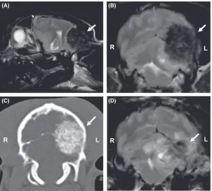
MX8000 IDT, Philips Medical Systems) study of the tho-rax and abdomen was within normal limits, while CT of the head demonstrated the mass to have a similar attenu-ation to bone (Fig. 1C). Although the largest portion of the mass was within the cranial vault, an extracranial por-tion was evident, with distorpor-tion of the lateral aspect of the occipital and parietal bones. Multilobular tumor of bone, osteosarcoma, and chondroma was considered the most likely differential diagnoses at this time. The dog was anesthetized the next day with the aforementioned protocol and positioned in sternal recumbency. A com-bined left-sided rostrotentorial and caudal fossa cran-iotomy were performed, which allowed removal of the well-defined bony mass en bloc (Fig. 2). The wound was closed routinely without performing a cranioplasty. The dog recovered uneventfully from anesthesia. Intraopera-tive analgesia consisted of an IV constant rate infusion of remifentanil (0.15–0.4lg/kg/min), while early postopera-tive analgesia consisted of a combination of paracetamol (10 mg/kg IV q12 h), methadone (0.1–0.2 mg/kg IV q4 h), and dexamethasone (0.2 mg/kg IV q24 h). The dog demonstrated ambulatory cerebellar ataxia and
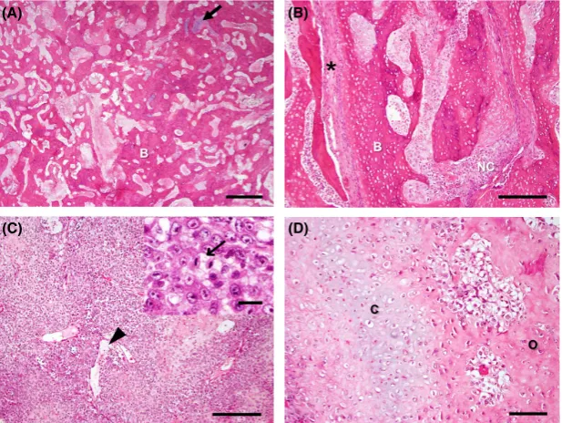
bilaterally absent menace responses the day after surgery. He gradually improved, was discharged from hospitaliza-tion 5 days after surgery and was, according to the own-ers, neurologically normal 7 days after surgery. Histopathological examination of cruciate sections repre-sentative of the mass and surgical margins revealed a neo-plasm composed of multiple islands and anastomosing trabeculae of well-differentiated woven bone and occa-sional cartilage surrounded by few polygonal to spindloid cells, separated by thin, irregular fibrovascular septa. There was mild anisocytosis and anisokaryosis, and five mitoses were observed in 10 high-power fields (Fig. 3). Neoplastic cells extended to the surgical margins. These findings were consistent with a multilobular tumor of bone (grade II) [1]. Eight days after surgery, the dog developed hypermetria of the left thoracic limb, which rapidly progressed to nonambulatory ataxia and a right-sided head tilt. Neurological examination revealed non-ambulatory vestibular ataxia, with a right-sided head tilt and a tendency for rolling and falling to the right side, a miotic right pupil, and disconjugate nystagmus, which changed directions with changes in position of the head.
(A) (B)
(C) (D)
These findings were suggestive for a central vestibular syndrome. General anesthesia was induced and main-tained with the aforementioned protocol. Repeat MRI examination of the head revealed a large, poorly demar-cated, extra-axial mass lesion at the site of the previous surgery. The lesion had a heterogeneous, predominantly hypointense intensity on all sequences compared to sur-rounding brain parenchyma (Fig. 1D). No IV contrast medium was administered during this study. Hematoma formation at the site of surgery and/or tumor regrowth was considered the most likely causes for this postopera-tive neurological deterioration. The next day, the dog was anesthetized again as described above and underwent sur-gical exploration of the previous surgery site. Surgery revealed a large firm mass lesion at the same site of previ-ous tumor resection (Fig. 2C), suggesting tumor regrowth. At this time, the owners of the dog elected euthanasia during surgery. Postmortem examination revealed a roughly spherical, 20915915 mm bony mass that compressed the left occipital lobe and left cere-bellar hemisphere (Fig. 2D). Gross postmortem examina-tion of the thoracic cavity, abdominal cavity, appendicular, and axial skeleton did not reveal any evi-dence of tumor metastasis. Histopathological examination of the mass revealed disorganized sheets of polygonal to
spindloid cells, with occasional fibrous septa and blood vessels. There were disorganized islands and trabeculae of osteoid, woven bone, chondroid matrix and cartilage, and extensive necrosis. Anisocytosis and anisokaryosis were marked, and 12 mitoses were counted per 10 high-power fields (Fig. 3). The underlying compressed cerebellum was partially malacic (necrotic). These findings were consis-tent with a multilobular tumor of bone with osteosarco-matous differentiation (grade III) [1].
Discussion
Multilobular tumors of bone (MTBs) (synonyms; multi-lobular osteochondrosarcoma, chondroma rodens, carti-lage analogue of fibromatosis, calcifying aponeurotic fibroma, juvenile aponeurotic fibroma and multilobular chondroma or osteoma) are uncommon, but not rare, neoplasms in dogs, which most often affect the flat bones of the skull [2]. Multiple additional sites including the axilla, hard palate, os penis, ribs and pelvis have also been reported [2–8]. Although the most common clinical pre-sentation is that of a solid and firm mass of the skull, neurological signs can result if compression of the brain occurs. The nature of neurological signs is then reflected by the specific location of the neoplasm. MTB is typically
(C) (D)
a slow-growing and locally invasive neoplasm with mod-erate potential for metastasis [2]. The relatively slow growth rate of this tumor allows it to grow to a relatively large size prior to causing clinical signs [2].
Multilobular tumors of bone are locally invasive, and recurrence after surgical resection is therefore common. Recurrence occurs typically only after a prolonged period of time and affected dogs therefore have a long disease-free interval and long survival times after surgery [1, 2]. In dogs undergoing surgical excision, with or without adjunctive radio- and/or chemotherapy, median time to recurrence was 14 months, while the median survival time was 22 months [1]. Even longer survival times were reported in another study, indicating median survival times and times to recurrence of 797 days after surgery [2]. In dogs receiving chemotherapy alone a median sur-vival of 144 days is reported [9]. Surgery therefore remains the treatment of choice and predictably, time to local recurrence has been reported to be shorter in dogs with a higher tumor grade (grade I to III, in which grade III represents the most malignant grade) and incomplete surgical margins (disease-free interval 330 days vs. 1332 days with complete surgical margins) [2]. Despite the presence of incomplete surgical margins in the dog described in this report, local recurrence in 8 days was still considered an unusually short disease-free interval.
Histopathological examination of both the originally excised tumor and the recurrent tumor revealed substan-tial morphological differences. The tissue obtained during the first surgery demonstrated relatively benign character-istics including a well-organized lobular or trabecular structure, low cellularity and mild cellular pleomorphism, typical of MTB. The tumor regrowth, on the other hand, displayed a markedly disorganized structure, high cellular-ity, extensive necrosis, marked cellular pleomorphism, and a higher mitotic count. These histopathological find-ings were considered consistent with osteosarcomatous transformation [10].
Although MTB with more malignant characteristics has occasionally been reported, the authors of this report are unaware of previous cases that experienced such rapid tumor regrowth [1, 2, 7]. Little information is therefore available on the frequency and risk factors for malignant transformation of MTB in dogs. It is possible that the com-bination of pre-existing hypoxia within malignant tissue, tumor-derived immunosuppression, a local inflammatory environment, and borderline immunosuppressive doses of dexamethasone postprocedurally, all created an environment conducive for malignant transformation [11–13]. Although the tumor was resected en bloc, microscopic examination indicated that tumor-free surgical margins had not been achieved. It can therefore also not be excluded that the most
(A) (B)
(C) (D)
typical features of malignancy including histologic grade and mitotic index [14]. Given the high grade and mitotic index of the second sample, a high p53 index would be expected. Although MTBs typically occur in middle to old-aged (median 8 years), medium to large-breed dogs, the dog in this report was less than 3 years old and con-sidered a small-breed dog as is rarely reported [2, 7, 10]. The dog in this report also experienced a short duration of observed clinical signs before he was admitted for ini-tial diagnostic investigations. It is, however, unclear if the atypical breed, the young age of the dog, and the short duration of clinical signs presented here could indeed be associated with a more malignant character of the diag-nosed MTB. Further studies are therefore necessary to evaluate if signalment and clinical presentation can be used as prognostic indicators for dogs with MTB.
This case report indicates that although resection of MTBs is typically associated with a good prognosis, long disease-free intervals, and prolonged survival, malignant transformation of residual tumor should be considered a potential cause for rapid recurrence of clinical signs. Fur-ther studies are necessary to evaluate risk factors for transformation for this type of neoplasia.
Acknowledgments
None.
Conflicts of Interest
None declared.
Authorship
SC: involved in case management, manuscript construc-tion, submission, and response to reviewers. AC: carried out histopathology, manuscript drafting, figure prepara-tion, and response to reviewers. RL: performed imaging interpretation, figure preparation, manuscript drafting, and revision. JF: involved in case management, revision surgery, manuscript drafting and revision. DN: involved in case anesthesia, manuscript drafting, and revision. SP:
the canine skull: 16 cases (1978–1988). J. Am. Vet. Med. Assoc. 195:1764–1769.
2. Dernell, W. S., R. C. Straw, M. F. Cooper, B. E. Powers, S. M. LaRue, and S. J. Withrow. 1998. Multilobular osteochondrosarcoma in 39 dogs: 1979–1993. J. Am. Anim. Hosp. Assoc. 34:11–18.
3. Hay, C. W., R. Roberts, and K. Latimer. 1994.
Multilobular tumour of bone at an unusual location in the axilla of a dog. J. Small Anim. Pract. 35:636.
4. Banks, T. A., and R. C. Straw. 2004. Multilobular osteochondrosarcoma of the hard palate in a dog. Aust. Vet. J. 82:409–412.
5. Liptak, J. M., and N. Ehrhart. 2005. Bone and joint tumours. Pp. 761–773inS. J. Ettinger and E. C. Feldman, eds. Textbook of veterinary internal medicine, 6th ed. W.B. Saunders, St. Louis.
6. Webb, J. A., J. M. Liptak, S. A. Hewitt, and A. R. Vince. 2009. Multilobular osteochondrosarcoma of the os penis in a dog. Can. Vet. J. 50:81–84.
7. Diamond, S. S., C. P. Raflo, and M. P. Anderson. 1980. Multilobular osteosarcoma in the dog. Vet Path 17:759– 780.
8. Fukui, K., and Y. Takamori. 1986. Multilobular osteoma (chondroma rodens) in a Pekingese. Vet Rec 118:483. 9. Vancil, J. M., C. J. Henry, R. J. Milner, A. M. McCoig,
J. C. Lattimer, J. A. Villamil, et al. 2012. Use of samarium Sm 153 lexidronam for the treatment of dogs with primary tumors of the skull: 20 cases (1986–2006). J. Am. Vet. Med. Assoc. 240:1310–1315.
10. Thompson, K. G., and R. R. Pool. 2002. Tumors of bone. Pp. 245–318inD. J. Meuten, ed. Tumors in domestic animals. 4th ed. Iowa State Press, Ames, IA.
11. Kim, R., M. Emi, K. Tanabe, and K. Arihiro. 2006. Tumor-driven evolution of immunosuppressive networks during malignant progression. Cancer Res. 66:5527–5536. 12. Karin, M. 2006. Nuclear factor-jB in cancer development
and progression. Nature 441:431–436.
13. Morrison, W. B. 2012. Inflammation and cancer: a comparative view. J. Vet. Intern. Med. 26:18–31. 14. Loukopoulos, P., J. R. Thornton, and W. F. Robinson.