Pilot study of the impact that bilateral sacroiliac
joint manipulation using a drop table technique
has on gait parameters in asymptomatic
individuals with a leg length inequality.
John Ward,
DC, MA, MS1Ken Sorrels,
DC, BA2Jesse Coats,
DC, BS, DAAPM, CCSP3Amir Pourmoghaddam,
PhD4Carlos DeLeon,
BS5Paige Daigneault,
BS51 Associate Professor/Research Fellow, Department of Physiology and Chemistry, Texas Chiropractic College 2 Professor, Department of Technique, Principles and Therapeutics, Texas Chiropractic College
3 Professor, Chairman, Department of Clinical Specialties, Texas Chiropractic College 4 Memorial Bone & Joint Clinic Researcher
5 TCC graduate student assistant
Corresponding author: e-mail: [email protected]
Texas Chiropractic College, 5912 Spencer Highway, Pasadena, TX 77505 Campus phone (281) 998-5704, Fax: (281) 487-0581
Acknowledgments: This study was supported with an internal grant from TCC ©JCCA 2014
Purpose: The purpose of this study was to pilot test our
study procedures and estimate parameters for sample size calculations for a randomized controlled trial to determine if bilateral sacroiliac (SI) joint manipulation affects specific gait parameters in asymptomatic individuals with a leg length inequality (LLI). Methods: Twenty-one asymptomatic chiropractic students engaged in a baseline 90-second walking kinematic analysis using infrared Vicon® cameras. Following this, participants underwent a functional LLI test. Upon examination participants were classified as: left short leg, right short leg, or no short leg. Half of the participants in each short leg group were
Objectif : Le but de cette étude était de mettre à l’essai
un projet pilote concernant nos procédures d’étude et d’estimer les paramètres pour le calcul de la taille de l’échantillon d’un essai contrôlé randomisé afin de déterminer si la manipulation de l’articulation sacro-iliaque bilatérale affecte les paramètres spécifiques de marche chez les personnes asymptomatiques ayant un problème d’inégalité de longueur des membres inférieurs (ILMI).
Méthodologie : Vingt et un étudiants en chiropratique
asymptomatiques ont pris part à une analyse cinématique de base de la marche de 90 secondes à l’aide de caméras infrarouges ViconMD, à la suite de
Introduction
Two types of leg length inequality (LLI) exist, anatomical and functional LLI.1,2 It has been suggested that a
conse-quence of possessing a short lower limb is that it places abnormal mechanical stress on both lower limbs.1-3 The
longer limb may develop greater foot pronation, and the shorter limb may be predisposed to degenerative joint changes.2,3
An anatomically short lower limb occurs when some-one is born or in some way develops a lower limb weight-bearing bone that is smaller than its contralateral counter-part.2 This can occur when individuals are born with a
shorter than normal femur or tibia.
A consequence of an anatomically short lower limb is that the pelvis will undergo torsion to biomechanically adapt.1 Depending on the degree of short LLI back pain,
knee pain, lower limb stress fractures, and increased rates of lower limb osteoarthritis have been reported.1,4-9 If a
LLI is untreated the body quite obviously would have to adapt to the difference in limb length and that could lead to the development of a functional adaptive scoliosis.1
Another form of LLI is a functional LLI. This form
of short lower limb is believed to be due to malposition of one or both innominate bones in relation to the sac-rum, resulting in a limb that is shorter than normal.10 One
theoretical mechanism for this occurring is greater than normal suprapelvic muscle hypertonicity which leads to an alteration in pelvic rotation.10-13 Pelvic girdle
malpos-ition like this is thought to occur when one innominate bone rotates anteriorly or posteriorly, resulting in the de-velopment of a functional LLI.10
The long-term consequence of a functionally short
then randomized to receive bilateral corrective SI joint chiropractic manipulative therapy (CMT). All participants then underwent another 90-second gait analysis. Pre- versus post-intervention gait data were then analyzed within treatment groups by an individual who was blinded to participant group status. For the primary analysis, all p-values were corrected for multiple comparisons using the Bonferroni method. Results: Within groups, no differences in measured gait parameters were statistically significant after correcting for multiple comparisons.
Conclusions: The protocol of this study was acceptable to all subjects who were invited to participate. No participants refused randomization. Based on the data collected, we estimated that a larger main study would require 34 participants in each comparison group to detect a moderate effect size.
(JCCA 2014;58(1):85-95)
k e y w o r d s: chiropractic, manipulation, gait,
biomechanics, locomotion, drop table technique, randomization
hasard un traitement chiropratique de manipulation de l’articulation sacro-iliaque bilatérale. Tous les participants ont ensuite pris part à une autre analyse de 90 secondes de la marche. Les données de marche avant et après l’intervention ont ensuite été analysées pour les groupes par une personne qui ne connaissait pas l’état des groupes de participants. Pour l’analyse principale, toutes les valeurs p ont été corrigées pour tenir compte des comparaisons multiples en utilisant la méthode de Bonferroni.
Résultats : Au sein des groupes, aucune différence dans lesparamètres mesurés de marche n’était
statistiquement significative après la correction pour les comparaisons multiples.
Conclusions : Le protocole de cette étude était
acceptable pour tous les sujets invités à y participer. Aucun des participants n’a refusé la randomisation. En fonction des données recueillies, nous avons estimé qu’il faudrait, pour une étude principale plus importante, 34 participants dans chaque groupe de comparaison afin de détecter un effet d’une ampleur modeste.
(JCCA 2014;58(1):85-95)
m o t s c l é s : chiropratique, manipulation, démarche,
lower limb is not clearly known.10 Theoretically,
individ-uals with a significant functional LLI could experience similar symptoms as a person with an anatomical LLI. A literature review using PubMed, Index to Chiro-practic Literature, and Alt Health Watch databases using keywords “chiropractic”, “biomechanics”, and “gait” yielded only two applicable gait-related chiropractic ma-nipulative therapy (CMT) articles. Sandell et al.14 found
that following CMT to the sacroiliac (SI) joint runners improved their hip extension capabilities. This interesting change, however, did not materialize into any improve-ment in running velocity post-CMT on a 30-meter sprint (p=0.572).14 Herzog, in his CMT gait biomechanics
arti-cle described how corrective SI joint CMT resulted in increased gait support time and improved gait symmetry based on ground reaction force analysis over the course of a multi-week study.15 These findings add credibility to
the belief that SI joint CMT may marginally alter the bio-mechanics of the lower limbs. Due to the limited research in this field more studies are warranted, particularly stud-ies utilizing state-of-the-art motion analysis technology. Chiropractors treat patients with functional LLI.16-18
The impact that SI joint CMT has on gait kinematics
should be studied further to help chiropractors better understand how they may impact gait when treating pa-tients with LLI. The overall purpose of this study was to pilot test a protocol that will be the basis for a series of larger studies aimed at measuring the impact of bilateral SI joint manipulation on gait parameters in asymptomatic individuals with a LLI. In the current study, our specific aims were to: 1) determine the feasibility of administering advanced motion analysis technology in a chiropractic re-search setting; and 2) generate point and range estimates to inform sample size estimates for a larger study of the potential effect of SI joint manipulation on improvement of gait symmetry.
Methods
This study received ethics approval from the Texas Chiro-practic College (TCC) Human Subjects Committee.
Study Design and Setting
This was a single-blind, randomized, controlled pilot study of the immediate impact that SI joint CMT had on walking kinematics in asymptomatic individuals with a LLI. As shown in Figures 1 and 2, participants initially en-Figure 1.
Experimental design. LLI = leg length inequality; SI = sacroiliac; CMT = chiropractic manipulative therapy; LSLM = left short leg manipulation; LSLN = left short leg no manipulation; RSLM = right short leg manipulation; RSLN =
gaged in a 90-second baseline gait analysis utilizing a Vicon® infrared camera imaging system (Vicon, Centen-nial, CO, USA). Next, they underwent a prone heel com-parison test to observe for a functionally short lower limb (Fig. 3). Study participants who possessed a short lower limb were then randomized into two groups: 1) Posterior Superior Iliac Spine (PSIS) CMT to the short leg side and ischial tuberosity CMT to the long leg side, or 2) no CMT. Next study participants underwent another 90-second gait trial. At the conclusion of that time the following
five study subgroups existed: left short leg-manipulation (LSLM), left short leg-no manipulation (LSLN), right short leg-manipulation (RSLM), right short leg-no ma-nipulation (RSLN), or no short leg (NSL) (Table 1). The LSLN, RSLN, and NSL groups were intended to serve as controls for comparison purposes.
Participants
Asymptomatic student volunteers were recruited with on-campus flyers and via word-of-mouth. All study ap-Figure 2.
plicants provided an informed written consent on college-approved documents. They were then screened against inclusion and exclusion criteria. Those that met inclusion/ exclusion criteria attended a single twenty-minute visit specifically for the study. Participants were given a study preparation handout of the exclusion criteria and were re-minded to avoid consuming caffeine, alcohol and receiv-ing CMT durreceiv-ing the day of study participation.
Inclusion/exclusion criteria
Inclusion criteria were: 1) completion of TCC student physical examination and absence of self-reported contra-indications to SI joint CMT; 2) age 18-45 years; 3) a “no” response to all exercise contraindication sections on a Physical Activity Readiness-Questionnaire (PAR-Q); 4) no engagement in strenuous exercise on the day of the study; and 5) willingness to provide informed written consent. Study participants with any of the following cri-teria were excluded from the study: 1) diagnosis of any lumbar, sacral, hip, or lower limb pathology that would prevent them from walking; 2) severe neurological con-ditions which would impact gait (e.g., type II diabetes, Parkinson’s disease, traumatic brain injury, dementia,
stroke, epilepsy, multiple sclerosis, myasthenia gravis, Huntington’s disease, etc.); 3) a history of alcohol abuse; 4) any health condition that would impair their ability to walk up to 3 mph; 5) visual impairment that would ren-der walking on a treadmill dangerous; 6) hypertonia; 7) reliance on a cane or similar assistive walking device; 8) taking medications that could alter motor function (e.g., acetylcholine-esterase inhibitors, L-dopa agonists, dopa-antagonists, or neuroleptics); 9) botulinum injection in their lower limb muscles within the past six months; 10) presence of severe pain in their lower limbs of greater in-tensity than 3 on a 10 cm Visual Analog Scale (VAS); 11) vertigo or history of falls within the past 60 days; or 12) any prior bone or muscle-related surgeries.
Baseline Preparation and Kinematic Recording
Participants were all given a verbal description of the walking study and LLI analysis prior to testing to reduce anxiety during the test. Upon arrival to the session they changed into standardized black spandex shorts and dark shoes. All males wore new MX409 New Balance® shoes and Women wore new WL574 New Balance® shoes (New Balance, Brighton, MA, USA). Any reflective Figure 3.
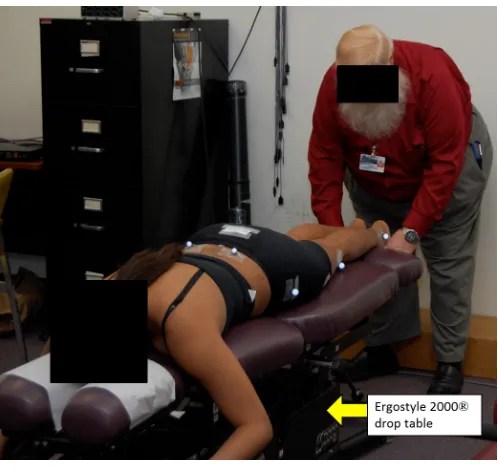
Illustration of the LLI test.
Table 1.
Baseline study participant attributes.
Group 1
LSLM Group 2 LSLN (control group #1)
Group 3
RSLM Group 4 RSLN (control group #2)
Group 5 NSL (control group #3) Sex
Males
Females 3 3 2 4 1 3 1 1 2 1
Age (y) 26.0 ± 4.7 29.7 ± 7.2 25.5 ± 5.1 24.7 ± 2.1 28.3 ± 4.0
Body Mass
(kg) 76.6 ± 12.7 77.4 ± 18.4 78.4 ± 40.0 74.8 ± 10.4 73.5 ± 5.2
Height (m) 1.74 ± 0.06 1.68 ± 0.06 1.70 ± 0.12 1.75 ± 0.04 1.74 ± 0.04
Body Mass
logos on the shoes were spray-painted with non-reflective paint. Prior to this study researchers purchased five pairs of male (sizes 8-12) and female (sizes 5-9) shoes in com-mon sizes. Standardized shoes were chosen as opposed to having participants walk barefoot to most closely emulate a real-world scenario. Next, trained research assistants placed 18 silver reflective markers on the participant’s lower body using double-sided marker fixing tape and surgical tape. Reflective markers were placed on the fol-lowing anatomic landmarks during this study bilaterally: ASIS, PSIS, greater trochanter, lateral epicondyle of the femur, tibial tuberosity, lateral malleolus, posterior calca-neus, top of the fifth metatarsal head, and top of the first metatarsal head (Fig. 2). Sixteen of the MoCap solutions reflective markers were 19 mm (MoCap solutions, Hun-tington Beach, CA, USA). The two PSIS markers used in this study were slightly smaller, at 14 mm. This was done in an attempt to gain better resolution by reducing the likelihood that those markers would be merged together by the Vicon® cameras considering how close the PSISs were on smaller participants with narrow hips.
Prior to a participant arriving at the lab each day the Vicon® system was calibrated as suggested by the manu-facturer. Once the participant was dressed properly and all of the reflective markers were in place they stood on top of the 400 Pro series Keys® treadmill (Keys Fitness Prod-ucts, Inc., Dallas, TX, USA) for their baseline 10-second model generation. Next the participant was instructed that they would be walking as they normally would at a velocity of 1.5 mph. A research assistant started the treadmill at the same time as another researcher began recording data with the Vicon® system. The lab’s Vicon® MX system consisted of 8 infrared Bonita 0.3 megapixel cameras. Kinematic data was recorded at 100 Hz. The displacement of the 18 reflective markers over time was recorded. At the conclusion of 100 seconds the researcher operating the Vicon® computer stopped the recording and then the treadmill was stopped. The study partici-pant was not given any indication of when the treadmill would be stopped prior to the examiner finishing his com-puter data recording. Immediately after the 100 second recording was made the initial 10 seconds was clipped from the data to remove any initial steps as the partici-pant became acclimated to the treadmill upon beginning the test. Following the baseline 90 seconds of data col-lection the participant then carefully stepped off of the
treadmill. After this the research assistants removed the participant’s shoes for them. They then removed the re-flective markers on the two tibial tuberosity and two ASIS points. Prior to removing those four reflective markers the research assistants made circular pen tracings around the markers on the participant’s skin. This was done in an at-tempt to leave a guide which would aid in placing the markers back as closely as possible where they were for the 90-second post-kinematic analysis.
LLI assessment
Study participants positioned themselves prone on a drop table with their shoes off. During positioning for the LLI test, an effort was made to ensure the participant’s whole body was in a neutral position on the table without spinal or pelvic frontal plane distortion. The treating doctor then held the participant’s ankles in a neutral position to pre-vent foot inversion or eversion. The leg length was visual-ized by comparing the inferior aspect of both compressed heels exclusively (Fig. 3). This technique was chosen as a previous study suggested that this form of LLI meas-urement demonstrated better inter-examiner reliability,19
although LLI tests in general do not have particularly high inter-rater reliability.20-23 If the examining
Randomization and blinding
Participants were subdivided into the three following groups based on the LLI test: left short leg, right short leg, and no short leg (as shown in Fig. 1). A computer-generated randomized intervention list was created be-fore the study began. That list determined if a participant with a short lower limb would undergo CMT or not. The doctor performing the SI joint CMT was aware of which group the study participant belonged to. The researcher who analyzed the motion capture data was blinded as to group designation. He was only told that he would be pro-vided with walking data from five distinct study groups and that he needed to determine gait kinematics and if any statistically significant differences existed within groups in terms of their pre versus post gait data.
Intervention
The intervention phase of the study was performed by a chiropractor with 35 years of experience. The interven-tion involved either: 1) a hypothenar ilium apex push to the PSIS on the short leg side24 in an attempt to rotate
the superior ilium anteriorly to elongate that lower limb and a hypothenar ischial tuberosity push on the long leg side, or 2) no manipulation. Bilateral SI joint CMT was chosen over unilateral short leg PSIS manipulation based on preliminary data by our lab on the lack of effectiveness of unilateral corrective CMT to improve gait symmetry of our student participants (unpublished data). All CMT con-sisted of a high-velocity low-amplitude force delivered three times in a row using a drop table (Ergostyle 2000, Chattanooga Group Inc., Hixson, TX, USA). The intent of the drop table was to try to keep the amount of force reasonably standardized. This prone form of CMT was selected to decrease the likelihood of making researchers remove more reflective surface markers than the five that were absolutely necessary to remove. One minute after receiving SI joint CMT or no CMT the study participant engaged in their walking post-kinematic analysis.
Kinematic Post-data Processing
The data was processed using a customized Matlab script (Mathworks, USA R2007a). The kinematic data was ana-lyzed to calculate characteristics of movement for each participant. Data for the dependent variables was aver-aged for each participant over all of their strides within each gait trial. In the current study we investigated the
changes in the functional active range of motion (in the sagittal plane) of the hip angle, knee angle, and ankle an-gle as a result of the intervention. This was performed by subtracting the minimum joint angle from the maximum joint angle for each of the aforementioned joints. In addi-tion, the double support time, percent double support time (duration both feet were on the ground in relation to the gait cycle), stance time, percent stance time (duration one foot was on the ground in relation to the gait cycle), step length, and stride length bilaterally were calculated. Approximate Entropy, a measure of gait variability, was additionally determined for each joint. In healthy in-dividuals there is a certain amount of acceptable variabil-ity that represents a normal (healthy) gait pattern. How-ever, highly variable gait patterns are typically indicative of some type of pathology or loss of coordination,25 which
may render a person at risk for falling.26 Gait variability
has been identified by the application of a mathematical technique called approximate entropy (ApnEn) that may reveal small changes in the gait pattern.25,27,28 Values near
“0” represent a stable gait, while values near “2” repre-sent a very unstable gait.
Statistical Analysis
Data was analyzed in SPSS version 19 (Release Version 19.0.1). Pre- and post-intervention gait parameter meas-urements were summarized as mean + standard devia-tions (SD) unless otherwise specified. Parametric with-in-groups, dependent variables were compared using a paired-samples t-test. Since we intended to utilize a series of t-tests we engaged in a Bonferroni adjustment to avoid type I statistical error. As a result, the alpha level of p < 0.002 was considered statistically significant for all an-alyses. For data analysis purposes both short leg groups had their data merged into one group based on ipsilat-eral short leg effects (e.g., the R lower limb data for the RSLM group and L lower limb data for the LSLM group) and contralateral long leg effects (e.g., the L lower limb data for the RSLM group and R lower limb data for the LSLM group) pre- and post-intervention. Similarly, the RSLN and LSLN groups had their data merged for com-parison purposes. The NSL group had its bilateral data values averaged together.
Results
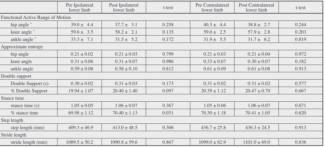
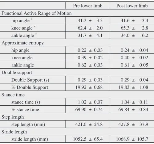
par-ticipants. Just over half of our participants were female (n = 12). The mean age was = 27.2 (sd = 5.1) years, mean height was = 1.72 (sd = 0.07) m, and mean body mass was = 76.6 (sd = 18.2) kg. Only two interested subjects were excluded from this study based on exclusion criteria (one with a history of lower limb surgery, another due to existing foot drop). CMT to the SI joint resulted in no statistically significant change in functional active range of motion and other parameters of gait (Table 2a-2c). This, however, was a pilot study with only twenty-one participants and did not follow a power analysis. Few at-tributes in this study even approached statistical signifi-cance when adjusted for multiple comparisons. However, from an exploratory analysis perspective, the change in pre ipsilateral lower limb % stance time was significant at an unadjusted alpha level (p=0.031) for RSLM. In the RSLN and LSLN, our analysis demonstrated a change in the knee joint angle on the long limb side of almost 1˚ (p=0.011) and an increase in percentage of stance time by 0.4% (p=0.010) on the short limb side. This, along with the normal gait data demonstrated in Table 2c illustrates the amount of variability in gait without CMT.
Addi-tionally, there was no discernible pattern changes in the RSLM and LSLM groups.
Discussion
The changes in walking kinematics in response to CMT in this pilot study were small. This study intentionally was a pilot study with a small sample size within each group. Subsequent larger studies should follow a power analysis. Using G*Power version 3.1.3 (Universität Kiel, Germany)29,30 we determined post-hoc that future studies
should have 34 participants per study group to compare 2 groups (experimental and control). This analysis was in accordance with a desired f effect size of 0.5, α of 0.05, and power of 0.8 through a 2-group ANOVA using [stride length] as the primary outcome of interest. We do feel that we would be capable of recruiting this number of student participants with future studies if we merge right and left short leg group data together (e.g., RSLM and LSLM into one group). Our rate of eligibility for this study was 91.3% out of all applicants from our college. Recruiting outside non-student participants (general public) would also be possible as an alternative, but that would require Table 2a.
Summary of RSLM and LSLM merged group data (n=10).
Pre Ipsilateral
lower limb Post Ipsilateral lower limb t-test Pre Contralateral lower limb Post Contralateral lower limb t-test Functional Active Range of Motion
hip angle ˚ 39.0 ± 4.4 37.7 ± 3.1 0.258 40.3 ± 4.4 38.8 ± 2.7 0.244
knee angle ˚ 59.6 ± 3.5 58.2 ± 2.1 0.135 59.0 ± 2.5 57.9 ± 2.8 0.203
ankle angle ˚ 33.3 ± 7.1 31.5 ± 5.2 0.172 31.9 ± 5.5 31.7 ± 6.2 0.819
Approximate entropy
hip angle 0.21 ± 0.02 0.21 ± 0.03 0.799 0.21 ± 0.03 0.21 ± 0.04 0.972
knee angle 0.31 ± 0.06 0.31 ± 0.07 0.986 0.33 ± 0.07 0.30 ± 0.07 0.182
ankle angle 0.59 ± 0.08 0.58 ± 0.10 0.812 0.61 ± 0.09 0.61 ± 0.08 0.913
Double support
Double Support (s) 0.30 ± 0.02 0.31 ± 0.03 0.173 0.31 ± 0.02 0.31 ± 0.02 0.577
% Double Support 19.94 ± 1.07 20.40 ± 1.40 0.097 20.39 ± 1.12 20.47 ± 0.79 0.667
Stance time
stance time (s) 1.05 ± 0.05 1.06 ± 0.07 0.367 1.05 ± 0.06 1.06 ± 0.07 0.671
% stance time 69.98 ± 1.12 70.40 ± 1.13 0.031 70.30 ± 1.18 70.41 ± 1.05 0.620
Step length
step length (mm) 409.3 ± 46.9 413.0 ± 48.5 0.306 436.7 ± 25.8 436.3 ± 24.5 0.913
Stride length
stride length (mm) 1089.5 ± 50.2 1090.8 ± 59.6 0.867 1099.0 ± 62.9 1101.0 ± 69.0 0.836
further resources. Additionally, a future definitive study could involve randomizing NSL participants to manipula-tion or no manipulamanipula-tion groups in order to constitute an additional comparison group.
Our eight camera Vicon® motion analysis system worked consistently in our study. No participants were in-jured at any point. For safety reasons following the com-pletion of this experiment we have installed thin side rails on the treadmill that provide a safety bar participants can grab onto in case they lose their balance on the treadmill. We are also in the process of installing an overhead safety harness system to prevent falling.
Our method of using a blinded biomechanist appeared to be effective in this pilot study. One issue we did have was the transport of the large kinematic data files we were generating (approximately 82 MB per participant). As a result, it took our offsite biomechanist many hours to transfer all of the files from our lab computer to his com-puter for analysis using a trial version of TeamViewer 8 ® software (TeamViewer Inc., Tampa, FL). For future stud-ies we will need to purchase a more robust file transfer system to handle transferring large quantities of data.
Table 2c.
Summary of NSL bilateral mean data (n=3).
Pre lower limb Post lower limb Functional Active Range of Motion
hip angle ˚ 41.2 ± 3.3 41.6 ± 3.4
knee angle ˚ 62.4 ± 2.0 65.3 ± 2.8
ankle angle ˚ 31.7 ± 4.1 34.0 ± 6.2
Approximate entropy
hip angle 0.22 ± 0.03 0.24 ± 0.04
knee angle 0.39 ± 0.02 0.40 ± 0.02
ankle angle 0.62 ± 0.03 0.61 ± 0.05
Double support
Double Support (s) 0.29 ± 0.03 0.29 ± 0.04 % Double Support 19.92 ± 0.68 19.83 ± 1.08 Stance time
stance time (s) 1.02 ± 0.07 1.04 ± 0.11
% stance time 69.90 ± 0.74 69.84 ± 0.84
Step length
step length (mm) 421.0 ± 24.8 427.8 ± 37.9 Stride length
stride length (mm) 1052.5 ± 65.4 1068.9 ± 105.7 Data listed as mean ± SD for group dependent variable data.
Table 2b.
Summary of RSLN and LSLN merged group data (n=8).
Pre Ipsilateral
lower limb Post Ipsilateral lower limb t-test Pre Contralateral lower limb Post Contralateral lower limb t-test Functional Active Range of Motion
hip angle ˚ 38.7 ± 3.0 39.3 ± 3.5 0.534 38.9 ± 2.9 39.9 ± 3.8 0.207
knee angle ˚ 59.5 ± 3.7 60.0 ± 4.8 0.535 59.7 ± 3.9 60.6 ± 3.7 0.011
ankle angle ˚ 33.0 ± 6.1 32.0 ± 5.0 0.456 32.5 ± 5.5 32.5 ± 4.6 0.984
Approximate entropy
hip angle 0.21 ± 0.03 0.22 ± 0.03 0.258 0.21 ± 0.02 0.23 ± 0.04 0.095
knee angle 0.33 ± 0.07 0.34 ± 0.07 0.303 0.33 ± 0.07 0.33 ± 0.06 0.951
ankle angle 0.61 ± 0.10 0.60 ± 0.10 0.761 0.62 ± 0.08 0.59 ± 0.08 0.239
Double support
Double Support (s) 0.30 ± 0.04 0.31 ± 0.04 0.183 0.30 ± 0.04 0.31 ± 0.04 0.499
% Double Support 19.99 ± 1.51 20.41 ± 1.72 0.032 20.05 ± 1.55 20.24 ± 1.38 0.361 Stance time
stance time (s) 1.05 ± 0.10 1.06 ± 0.09 0.677 1.06 ± 0.09 1.06 ± 0.08 0.857
% stance time 69.69 ± 1.40 70.09 ± 1.53 0.010 70.33 ± 1.33 70.51 ± 1.37 0.117 Step length
step length (mm) 417.2 ± 50.8 416.6 ± 47.7 0.906 427.2 ± 41.8 425.7 ± 39.5 0.728 Stride length
stride length (mm) 1086.9 ± 113.3 1088.0 ± 104.5 0.924 1098.9 ± 110.7 1099.1 ± 101.4 0.985
Manual determination of LLI by chiropractors is oc-casionally performed, but it has not been shown to be as accurate as more expensive imaging methods.8,19 When
choosing an ideal method of measuring LLI we, similar to field practitioners, had to consider reliability, accur-acy, magnification, radiation dose, cost, need for special equipment, convenience, and the opportunity to image an entire extremity.9 In our study design we opted to utilize
a technique that we theorized would be more common amongst chiropractic general practitioners. Our original hypothesis was that we expected participants with a LLI to have a slightly asymmetric gait. Then after corrective SI joint CMT we hypothesized that the participant’s gait would be more symmetrical. This belief did not material-ize.
The experience we gained through this pilot study was invaluable for the generation of subsequent larger stud-ies following a power analysis. If SI joint CMT improves gait symmetry that may directly have implications on bal-ance while walking. Further study to determine the true physiological impact of SI joint manipulation on gait is required.
The main intent of this study was to determine if our design would be feasible to engage in a series of larger studies using advanced motion analysis technology. This was shown to be possible. This lab now intends to de-velop the three following walking studies involving SI joint CMT over the course of September 2012-September 2015 using large study groups: 1) normative data with a combination of healthy non-chiropractic student partici-pants (mainstream public) versus chiropractic students, 2) adult SI joint pain patient data (mainstream public), and 3) geriatric at-risk-for-fall data.
As this field is developed further additional direc-tions that should be explored would be running kinematic changes in response to SI joint and/or lumbar spine CMT and changes in gait that may be induced in ataxic special populations. Also surface EMG should be used to explore if any changes found in gait are induced by alterations in motor recruitment patterns of the lower limb muscles in response to CMT.
Limitations
This study only informs us as to the potential immediate impact SI joint CMT may have on specific gait param-eters in young, asymptomatic individuals. It is possible
that a CMT dose-response relationship may exist related to improvements in gait performance, similar to what Herzog discovered.15
One issue this study must accept is the limitation on ex-ternal validity. The population we sampled was composed of chiropractic students who regularly receive CMT. It is possible that the general public may react differently than individuals who receive CMT often.
Another issue this study faced is that LLI tests do not have high levels of validity and reliability.13,19,20-24 In our
study design we opted to use a test that was a modified form of a Derifield Pelvic Leg Check LLI test that only involved comparison of heel length. It is possible that our test was not ideal since a reliable and valid test does not clearly exist. No X-ray or similar imaging lower limb procedure was used to ensure that participants truly had a functionally short lower limb and not an anatomically short lower limb.2,31-35 Despite this, the primary goal of
this study was to observe raw marginal change in kin-ematics pre versus post SI joint CMT (e.g., can SI joint CMT truly induce a subtle change in gait kinematics or not).
Conclusions
There is minimal research into how spinal manipulation may augment gait. The focus of this experiment was to determine if developing a series of larger SI joint CMT biomechanics studies using motion analysis technology was possible through our study design. The findings of our study suggest larger studies are feasible and we will proceed accordingly.
Funding sources and potential conflicts of interest
This study was supported by a grant from Dr. S. M. Elliot, TCC President Emeritus. The authors report no conflicts of interest.
References
1. Knutson G. Anatomic and functional leg-length inequality: a review and recommendation for clinical decision-making. Part I, anatomic leg-length inequality: prevalence, magnitude, effects and clinical significance. Chiropr Osteop. 2005;13:11.
2. McCaw S, Bates B. Biomechanical implications of mild leg length inequality. Br J Sp Med. 1991;25(1):10-3. 3. Langer S. Structural leg shortage: a case report. I Am
4. Friberg O. Clinical symptoms and biomechanics of lumbar spine and hip joint in leg length inequality. Spine. 1983;8(6):643-51.
5. Sabharwal S, Kumar A. Methods for assessing leg length discrepancy. Clin Orthop Relat Res. 2008;466:2910-22. 6. Golightly Y, Allen K, Helmick C, Renner J, Salazar A,
Jordan J. Relationship of limb length inequality with radiographic knee and hip osteoarthritis. Osteoarthritis Cartilage. 2007;15(7):824-9.
7. Harvey W, Yang M, Cooke T, Segal N, Lane N, Lewis C, Felson D. Associations of leg length inequality with prevalent, incident, and progressive knee osteoarthritis: a cohort study. Ann Intern Med. 2010;152(5):287-95. 8. Anderson R, Hayak R, Foggerty M. Leg length
inequality and the side of low back pain. Comsig Review. 1995;4(2):33-6.
9. Reid D, Smith B. Leg length inequality: a review of etiology and management. Physio Can. 1984;36:177-82. 10. Knutson G. Anatomic and functional leg-length inequality:
a review and recommendation for clinical decision-making. Part II, the functional or unloaded leg-length asymmetry. Chiropr Osteop. 2005;13:12.
11. Cooperstein R, Lisi A. Pelvic torsion: anatomic considerations, construct validity, and chiropractic examination procedures. Top Clin Chiro. 2000;7(3):38-49. 12. Giles L, Taylor J. Lumbar spine structural changes
associated with leg length inequality. Spine. 1982;7(2):159-62.
13. Mannello D. Leg Length Inequality. J Manipulative Physiol Ther. 1992;15(9):576-90.
14. Sandell J, Palmgren P, Björndahl L. Effect of chiropractic treatment of hip extension ability and running velocity among young male running athletes. JCM. 2008;7:39-47. 15. Herzog W. Biomechanical studies of spinal manipulative
therapy. JCCA. 1991;91:156-64.
16. Haas M, Peterson D, Panzer D, Rothman E, Solomon S, Krein R, Johansen R. Reactivity of leg alignment to articular pressure testing: evaluation of a diagnostic test using a randomized crossover clinical trial approach. J Manipulative Physiol Ther. 1993;16(4):220-7. 17. Nguyen H, Resnick D, Caldwell S, Elston E, Bishop B,
Steinhouser J, et al. Interexaminer reliability of activator methods relative leg-length evaluation in the prone extended position. J Manipulative Physiol Ther. 1999;22(9):565-9.
18. Thompson J. Thompson technique reference manual, Elgin, IL 1984. Thompson Educational Workshops SM and Williams Manufacturing.
19. Walker B, Buchbinder R. Most commonly used methods of detecting subluxation and the preferred term for
its description: a survey of chiropractors in Victoria, Australia. J Manipulative Physiol Ther. 1997;20(9):583-8. 20. Fuhr A, Osterbauer P. Interexaminer reliability of relative
leg-length evaluations in the prone, extended position. Chiropractic Technique. 1989;1(1):13-8.
21. Haas M, Peterson D. Interexaminer reliability of relative leg-length evaluations in the prone, extended position [letter, comment]. Chiropractic Technique. 1989;1(4):150-1.
22. DeBoer K, Harmon R, Savoie S, Tuttle C. Inter- and intra-examiner reliability of the leg-length differential measurement: a preliminary study. J Manipulative Physiol Ther. 1983;10(3):61-6.
23. Danelius D. Letter to the editor: inter- and intra-examiner reliability of leg-length measurement: a preliminary study. J Manipulative Physiol Ther. 1987;10(3):132.
24. Bergmann T, Peterson D. Chiropractic Technique: principles and procedures. 3rd ed. St. Louis: Elsevier-Mosby; 2011.
25. Myers S, Stergiou N, Pipinos I, Johanning J. Gait
variability patterns are altered in healthy young individuals during the acute reperfusion phase of
ischemia-reperfusion. J Surg Res. 2010;164(1):6-12.
26. Maki B. Gait changes in older adults: predictors of falls or indicators of fear. J Am Geriatr Soc. 1997;45(3):313-20. 27. Buzzi U, Stergiou N, Kurz M, Hageman P, Heidel J.
Nonlinear dynamics indicates aging affects variability during gait. Clin Biomech. 2003;18:435-43.
28. Pincus S. Approximate entropy as a measure of system complexity. Proc Natl Acad Sci USA. 1991;88:2297-301. 29. Buchner A, Erdfelder E, Faul F. (1997). How to Use
G*Power [WWW document]. URL http://www.psycho. uni-duesseldorf.de/aap/projects/gpower/how_to_use_ gpower.html.
30. Erdfelder E, Faul F, Buchner A. GPOWER: A general power analysis program. Behav Res Meth Ins. 1996;28:1-11.
31. Clarke G. Unequal leg length: an accurate method of detection and some clinical results. Rheum Phys Med. 1972;11:385-90.
32. Beal M. The short leg problem. J Am Osteopath Assoc. 1977;76:745-51.
33. Gofton J, Trueman G. Studies in osteoarthritis of the hip. Part II: Osteoarthritis of the hip and leg-length inequality. Can Med Assoc J. 1971;104:791-9.
34. Friberg 0, Koivisto E, Wegelius C. A radiographic method for measurement of leg length inequality. Diagn Imag Clin Med. 1985;54:54-81.