Epidemiologic and Histopathologic Feature of Lung
Cancer in Central Iran (2012-2018)
Mohammad Sadegh Khalilian1,2, Sina Narrei2, Mahdi Hadian1,2, Mehrdad
Zeinalian2,3*
1. Student Research Committee, Isfahan University of Medical Sciences, Isfahan, Iran
2. Ala Cancer Prevention and Control Center, Isfahan, Iran
3. Department of Genetics and Molecular medicine, School of Medicine, Isfahan University of
Medical Science, Isfahan, Iran (Corresponding author): zeinalianmehrdad@gmail.com
Abstract
Background: Lung cancer is one of the common causes of death worldwide.
Although the incidence rate of lung cancer in Western countries is decreasing, it
presents a growing trend in developed countries. Since there is no accurate enough
information about the epidemiological and Histopathologic features of lung cancer in
central Iran, Isfahan, we were motivated to conduct this research.
Materials and Methods: This was a descriptive, cross-sectional study carried out in
central Iran, Isfahan. All demographic, histopathological and clinical data of the lung
cancer patients registered in MACSA, a referral charity-based cancer center in
central Iran, was analyzed within 2012-2018 using SPSS v.22 software.
Results: Altogether 260 patients with lung cancer were included in this study from
6127 cancer patients registered within 2012-2018 (4.2%). Out of them, 66.2% were
men, and 18.8 % of the patients were alive at the time of the study. The mean age of
the patients at diagnosis was 61.56 (SD=14.11, range: 9-93). Altogether, 63.1% of
patients had metastasis of whom 57.6% were in stage IV at diagnosis. The Frequency
of different types of lung cancer was 36.9% adenocarcinomas, 14.2% squamous cell
carcinoma, 9.6% bronchogenic carcinoma and 8.1% small cell lung cancer,
respectively. Altogether, 128 cases were smokers with an average 35.45 ± 14 pack-
years. Only in 36.2% of the patients, the diagnostic and therapeutic biomarkers had
been checked, and CK7 was positive in 88.9% of the cases in which the biomarker
had been checked.
Conclusion: Despite to similar Iranian studies, the most common histopathologic
type of lung cancer among the patients was adenocarcinoma that it may be attributed
to the lower consumption of smoking in our population and their different genetic
context. Molecular biomarkers had been checked in a small portion of the patients.
More education of the clinicians along with the development of cancer molecular
testing may lead to promote the personalized-based approach.
Key word: lung cancer, Epidemiology, Histopathology, Central Iran, Isfahan
Introduction
Cancer is the second leading cause of death in developed countries and the third one in
developing countries like Iran(1,2). The incidence of cancer in developing countries is
increasing due to the population aging and the raising of some lifestyle-related cancer
risk factors like smoking, physical inactivity, obesity, stress, etc. Cancer is the third most
common cause of mortality after cardiovascular disease and car accidents in Iran(3).
Lung cancer is one of the common causes of death worldwide (4). Lung cancer affects
more than a million people a year, according to the International Agency for Research on
Cancer (IARC). Because of the high mortality rate of lung cancer, scientists pay more
attention to its diagnosis, treatment, and prognosis (5). In the United States, lung cancer is
the second most common cancer among men and women (6). Moreover, lung cancer is
the third most common cancer in both genders with the highest rate of death in Europe
(7). Although the incidence rate of lung cancer is estimated low in Iran, its survival rate is
Lung cancer is usually divided into two groups: small cell lung cancer (SCLC) and
non-small cell lung cancer (NSCLC). NSCLCs accounts for about 85% of all lung cancers
and are classified into squamous cell carcinoma (SCC), adenocarcinoma (AC), and large
cell carcinoma (9). The epidemiology of lung cancer is changing in many parts of the
world, as we enter the 21st century. The incidence trend of lung cancer is not promising,
however, it moves from developed to less developed countries (10). However, the
relationship between smoking and various types of histological findings of lung cancer is
not similar, so there is a significant relationship between smoking and the occurrence of
SCLC and SCC, but its association with adenocarcinoma is lower (11).
There are Limited epidemiologic studies on lung cancer in Iran. Some studies show that
the incidence of lung cancer may be increased by changes in the smoking pattern. The
incidence of lung cancer in the North West and West provinces of Iran has been
estimated higher than other regions (12). Since there is no accurate enough information
about the epidemiological and Histopathologic feature of the lung cancer in central Iran,
Isfahan, we were motivated to conduct this research.
Materials and Methods
This was a descriptive, cross-sectional study carried out in Ala cancer control and
prevention center (MACSA), a charity-based institute for supportive and palliative care
of cancer in central Iran, Isfahan. All the patients registered in MACSA have an
electronic database including clinical and para-clinical information. In this study,
demographic, histopathological and clinicopathological information of all patients with
lung cancer admitted to MACSA was obtained from 2012 to 2018. Demographic
variables included age at diagnosis, age at death, gender, and location of living. Moreover,
some cancer-related risk factors such as smoking (cigarette, opium or Hookah), alcohol
addiction, and chronic contact with chemical substance were also included.
Histopathological variables including pathologic feature, metastasis state, the tumor stage,
clinicopathological factors containing the first main symptom, past medical history, the
family history of cancer and the type of treatment were included. The collected data was
analyzed to determine the statistical information and correlations by SPSS v.22 software.
Results
Altogether among 6127 cancer patients registered in MACSA within 2012-2018, 260
patients with lung cancer (4.2%) were included in this study. Out of this population, 172
(66.2%) were men, and 49 (18.8 %) patients were alive at the time of the study. The male
to female ratio is 1.95. Overall, 242 patients (93.1%) were urban residents, and the others
(6.9%) were rural. The average age of the patients at diagnosis was 60.30 (SD=14.31,
9-93), and at the time of death 62.59 (SD=13.71). Moreover, the mean period time from
diagnosis to death was 13.36 (SD=12.31) months.
Out of total lung cancer patients, 135 cases (51.9%) had a positive family history of
cancer of whom 92 (68.1%) cases have at-least a first-degree relative with cancer. The
most common malignancies among the affected family members were in lung (32,
23.7%), breast (17, 12.6%) and stomach (17, 12.6%), respectively. (Table 1)
Altogether, 164 (63.1%) and 150 (57.6%) patients had metastasis at the time of study and
diagnosis, respectively (Table 2). The common sites of metastasis were bone (73, 44.5%),
brain (60, 36.6%) and liver (31, 18.9%), respectively.
According to the pathological reports, the Frequency of different types of lung cancer
were 96 (36.9%) cases of adenocarcinomas, 37 (14.2%) cases of squamous cell
carcinoma, 25 (9.6%) cases of bronchogenic carcinoma and 21 (8.1%) cases of small cell
lung cancer, respectively.
The histopathological feature of the studied malignancies were categorized in four types
include: 21 SCLC (8.1%), 162 NSCLC (62.3%), 51 cases other types of lung cancer
(19.6%) and 26 cases (10%) unknown. In the unknown category the pathological feature
of the patients’ tumors had not been identified (Table3).Overall, 41% of the tumors were
Out of 260 patients, 128 cases (49.2%) were smokers with an average (35.45 ± 14) pack
per year.
The exposure rate of the patients with some of the environmental factors investigated in
this study listed in Table 4. According to our study, 125 (72.6%) of men and 3 (3.4%) of
women were active cigarette smoker.
The most common early symptoms in the patients were cough (209, 80.4%), dyspnea (31,
11.9%), hoarseness (11, 4.2%) and hemoptysis (9, 3.5%), respectively. Moreover, 158
cases (60.8%) had a history of a chronic disease, including 40 (25.3%) patients with
ischemic heart disease, 37 (23.4%) with diabetes and 49 (31%) with hypertension.
Also, a history of COPD (Chronic Obstructive Pulmonary Disease), Asthma, and TB
(Tuberculosis) has been reported for 4 (1.5 %), 2 (0.8%) and 1(0.4 %) patients,
respectively.
Radiation or chemo-procedures for therapeutic or palliative intention had been done for
175 (67.3%) patients of whom 95 patients (54.3%) had tolerated palliative chemotherapy,
and 80 patients (45.7%) had received therapeutic chemotherapy. Radiation therapy had
been done for 160 patients of whom 99 cases (61.9%) were therapeutic and the others
were palliative.
Altogether, 53 molecular biomarkers had been checked by Immunohistochemical (IHC)
staining of tumor samples. The most common biomarkers have been listed in Table 5.
Discussion
Epidemiology
Although the incidence rate of lung cancer in Western countries is decreasing, it presents
a growing trend in developed countries (13). According to the some previous studies, it
seems the incidence rate of lung cancer in Iran is lower than Western countries (6). The
national reports have showed that the prevalence of lung cancer is ranked as seventh or
eighth among men and above the tenth among women, while on the global scale, it is the
patients with lung cancer out of 6127 cancer patients registered in MACSA within
2012-2018. The aim of this study was to investigate the epidemiological and
histoclinicopathological features among the lung cancer patients in central Iran.
According to the previous studies in Iran, male to female ratio has been reported 2.85
(12), 3.22 (15) and 5.09 (16), while in our study it was 1.95. In other countries like Spain
and India the ratio was 8.1 (17) and 4.1 (18) respectively. The lower male to female ratio
with the lung cancer compared with other countries can be due to difference in
environmental exposures of Iranian women, also, some probable genetics variations.
Further investigations based on the risk assessment are highly recommended for this
population.
In our study, the mean age of the patients at the time of study was 61.5 (SD=14).
According to the results of other studies in Iran the mean age of the patients were 65.7
(SD= 11.2) (16), 59.9 (SD= 13) (19) and 64.09 (SD= 9.44) (20). In studies at other
countries like India (21) and Canada (22) the mean age of the patients were 56 and
75years respectively .This can be attributed to the different genetic context and
environmental and lifestyle-related factors (15). The mean age at time of study among
men (61.69, SD=12.84) and women (61.30, SD= 16.39) (P=0.041) were similar to the
studies at Tehran (P=0.001, P=0.004)(15,23) and different with study at
Qazvin(P=0.171)(16).
Altogether, 51.9% of the patients had a positive family history of cancer (the presence of
different cancers among the first or second-degree relatives of the patient) of whom 68.1%
were in the first-degree relatives. Studies in Ardebil (west- north of Iran)(20) and Tehran
(23)showed the 17.3% and 11.6 % of patients had positive family history of cancer.
Additionally, the most common cancers among the family members were lung (23.7%),
breast (12.6%) and stomach (12.6%), respectively. In a similar Turkish study,38% of 213
patients had a positive family history of cancer, including 41.9% lung, 19%
gastrointestinal, and 7.6% breast cancers(24). Although MACSA is considered as a
referral center for cancer patients in central Iran, Isfahan, apparently more evaluations in
future studies, with emphasis on the genetic context of the disease could be helpful.
Residence of the patients was 93.1% in urban and 6.9% in rural areas (13.49 times). In
Ardebil(20), 54.1% were urban. Since according to the last study of Statistical Center of
Iran, the 85.42% and 14.57% of total population of Isfahan Province were urban and rural,
respectively(25), we expect this proportion would be also among the patients with lung
cancer if there is no difference between urban and rural populations regarding to the
incidence rate of the disease. With comparison the results, it is revealed that the
proportion of the urban patients to the rural ones is 13.49, while it is 5.86 in Isfahan
general population. It means the incidence rate of the disease in the urban population is
estimated 2.3 times more than the rural populations. It may be attributed to the air
pollution and other environmental cancer-related risk factors which have been
accumulated in the urban regions more than rural areas. More population-based studies
can evaluate this hypothesis.
Histopathological features
Overall, 63.1% of the patients had metastasis at the time of study of which 57.6% had
presented metastasis at diagnosis. Common metastatic sites were bone, brain, and liver.
According to one study in India(26), the rate of lung cancer metastasis was 32.5%. In the
study at Tehran(15) 67.3% of patients had metastasis in which the sites included
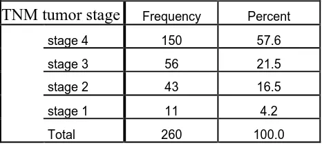
contralateral lung, bone, liver, brain. According to table 2, 57.6% patients were diagnosed
in stage 4 and 21.5% in stage 3. In the studies at Tehran(15), 85.3% were in stages 3 to 4,
in other study at Tehran(22), 48.5% were in Stage 4, and in the study at Sari( north of
Iran) (27), 85.1% of patients were in stage 4.
In most cases of lung cancer, the disease is diagnosed in advanced stages (28). Based on
this information the screening program in Iran should be paid more attention to early
detection of lung cancers and making biological clues for different sites of metastasis.
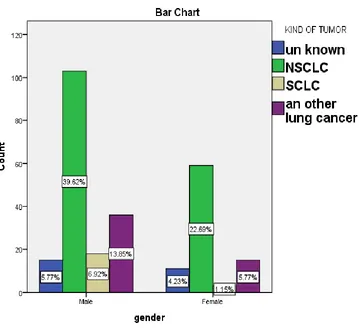
According to the pathological reports (Table 3), the frequency of different types of lung
cancer were 36.9% cases of adenocarcinomas, 14.2% cases of squamous cell carcinoma,
9.6% cases of bronchogenic carcinoma and 8.1% cases small cell lung cancer,
respectively. There was significant relationship between different types of lung cancer
and sex (p<0.001). (Figure 1)
In the study at Yazd( Iran)(8) 34.9% of patients had SCC and In study at Tehran (23), 19%
of patients were SCC (Table 6).
According to the results, totally 93 patients (35.7%) had IHC assay reports in which 53
different diagnostic, prognostic, and predictive biomarkers had been measured (Table 5,
7).
Although all of the measured biomarkers have a diagnostic significance (29–34), the
most relevant biomarkers which have been recently identified, CK20 and CK7, have an
important prognostic value in lung cancer patients(35). Moreover, predictive markers like
LCA (31) has been checked in a few patients. It seems, currently, IHC tests for cancer in
Iranian patients are requested for diagnostic indications than predictive ones. It may be
due to the limitation of cancer molecular and genetic testing facilities in Iran. Apparently,
development of new advanced cancer genetic labs along with the education of clinicians
towards personalized medicine-associated approaches on cancer could promote the
clinical applications of molecular cancer biomarkers in Iran.
Environmental risk factors
Smoking, is the most important lung cancer-related risk factor, so 90-85% of all lung
cancers are directly caused by exposure to cigarettes (36) . In our study, 49.2% of the
patients were cigarette smokers with an average 35.45 (SD= 14) pack per year. In other
studies at Tehran, 57.2% (15) and 72.9%(23) and in study at Ardebil(north of Iran) (20),
90.8% were cigarette smokers with an average 25.7 (SD= 38.67) pack per year. It seems
other environmental factors are suspected as the underlying cause of catching lung cancer
in the patients. So, we investigated other factors according to table 4, hookah smoking,
opium addiction and alcohol consumption, as the other potential reasons for catching
lung cancer in the studied population. On the other hand, 125 (72.6%) of men and 3
(3.4%) of women were active cigarette smokers and there is a significant difference
between sexes and cigarette smoking, hookah smoking, opium addiction and alcohol
consumption (p<0.001). In the study at Tehran(15), 70.3% of male and 5% of female
lung cancer patients were cigarette smoker. This difference is likely due to the less
consumption of cigarettes among Iranian women because of the cultural reason when we
proportion among the Iranian patients with lung cancer be significantly more than other
countries with high prevalence of smoking among their women. This proportion,
however, is less than other countries, according to our results and their comparison with
other foreign studies(17,18). It could be referred to the different genetic background in
our populations in comparison to the other countries and more effect of the inheritable
genetic factors in pathogenesis of the disease. This conclusion may be approved by the
high prevalence of familial lung cancer among the patients compared to other populations,
an issue which needs more evaluations in the next studies.
In other studies of Iranian population like Teaching Hospital of Qazvin (Iran)(16), 17.2%
of the patients had opium addiction, 47.4% baking bread history, 2.4 % contact with a
chemical substance. In one study at Ardebil(20), 34.7% had opium addiction and 11.3%
had a history of baking bread. Totally our study suggests, instead of concentrating on
cigarette consumption between lung cancer patients, we should pay more attention to the
other ways of smoking (hookah and opium) and environmental exposures in Iranian
patients to achieve comprehensive risk factors underlying lung cancer.
According to other studies, smoking is associated with a higher proportion of SCC. The
lower prevalence of SCC in our study may be due to the lower consumption of cigarettes
in the population than other populations studied in Iran. There was, also, a significant
difference in the histopathologic type of lung cancer (p<0.001) between sexes in this
study, similar to other Iranian studies (15,16,23).
Due to the limitations of our study (inaccessibility to the other risk factors and
comprehensive molecular study), we could not conclude any other items.
Clinical aspects
According to the results, the most common early symptoms of lung cancer among the
patients were cough and dyspnea. In the Qazvin study (16), 76.5% patients had cough as
an early symptom but in the Ardebil study (20) the most common symptom was
hemoptysis (32.7%). This controversy in studies needs more comprehensive data to make
an accurate decision about the early symptoms of lung cancer in Iranian patients.
Altogether, 67.3% patients had a history of chemotherapy and 38.1% had a history of
tolerated chemotherapy. These results may be correlated to the stage of diagnosis in our
patients. Curative chemotherapy and radiotherapy are more acceptable for the patients
who are not in the advanced stage.
Conclusion
This study was performed on 260 patients with lung cancer who had been referred to
MACSA-Isfahan within 2012-2018. The prevalence of disease was significantly more in
men and in urban patients.
There was a significant difference between the mean age of women and men with lung
cancer. Most of the patients had metastasis at the time of referral and bones were the
most common site of metastasis. There was a significant relationship between sex and
smoking. The most common histopathologic feature of lung cancer among the patients
was adenocarcinoma, probably due to the lower consumption of smoking in our
population. In some patients, the diagnostic and therapeutic biomarkers had been checked,
and CK7 was positive in most of the cases in which the biomarker was checked. Given
the limitations of this study regarding the accessibility of the patients’ data and sample
size, more investigations are recommended to reveal a more accurate epidemiologic and
clinicohistopathologic feature of the disease and its risk factors in central Iran.
ACKNOWLEDGEMENTS
The authors declare the full gratitude of the health-workers in MACSA center, as
well as the Student Research Committee at the Isfahan University of medical
References
1. Ferlay J, Shin H-R, Bray F, Forman D, Mathers C, Parkin DM. Estimates of worldwide
burden of cancer in 2008: GLOBOCAN 2008. Int J Cancer. 2010 Dec 15;127(12):2893–
917.
2. Saadat S, Yousefifard M, Asady H, Moghadas Jafari A, Fayaz M, Hosseini M. The Most
Important Causes of Death in Iranian Population; a Retrospective Cohort Study. Emergency.
2015;3(1):16–21.
3. FARHOOD B, GERAILY G, ALIZADEH A. Incidence and Mortality of Various Cancers
in Iran and Compare to Other Countries: A Review Article. Iran J Public Health. 2018
Mar;47(3):309–16.
4. Zhang M, Wu C-H, Zhu X-L, Wang Y-J. Loss of imprinting of insulin-like growth factor 2
is associated with increased risk of primary lung cancer in the central China region. Asian
Pac J Cancer Prev APJCP. 2014;15(18):7799–803.
5. Jemal A, Bray F, Center MM, Ferlay J, Ward E, Forman D. Global cancer statistics. CA
Cancer J Clin. 2011 Mar 1;61(2):69–90.
6. Cancer Statistics Review, 1975-2014 - SEER Statistics [Internet]. SEER. [cited 2018 Nov
29]. Available from: https://seer.cancer.gov/archive/csr/1975_2014/
7. Bosetti C, Bertuccio P, Levi F, Lucchini F, Negri E, La Vecchia C. Cancer mortality in the
European Union, 1970-2003, with a joinpoint analysis. Ann Oncol Off J Eur Soc Med
Oncol. 2008 Apr;19(4):631–40.
8. Zahir ST, Mirtalebi M. Survival of patients with lung cancer, Yazd, Iran. Asian Pac J
Cancer Prev APJCP. 2012;13(9):4387–91.
9. Types of Lung Cancer: Small Cell and Non-Small Cell Lung Cancer Types [Internet]. [cited
2018 Nov 29]. Available from: https://www.webmd.com/lung-cancer/lung-cancer-types#1
10. de Groot P, Munden RF. Lung cancer epidemiology, risk factors, and prevention. Radiol
Clin North Am. 2012 Sep;50(5):863–76.
11. Rosai and Ackerman’s Surgical Pathology - 2 Volume Set - 10th Edition [Internet]. [cited
2018 Sep 22]. Available from:
https://www.elsevier.com/books/rosai-and-ackermans-surgical-pathology-2-volume-set/rosai/978-0-323-06969-4
12. Almasi Z, Salehiniya H, Amoori N, Enayatrad M. Epidemiology Characteristics and Trends
13. Siegel R, Naishadham D, Jemal A. Cancer statistics, 2013. CA Cancer J Clin. 2013
Jan;63(1):11–30.
14. Mousavi SM, Gouya MM, Ramazani R, Davanlou M, Hajsadeghi N, Seddighi Z. Cancer
incidence and mortality in Iran. Ann Oncol Off J Eur Soc Med Oncol. 2009 Mar;20(3):556–
63.
15. Adnan K, Zahra E-M, Sharareh S, Shirin K, Habib E, Kian K. Clinicopathological
Characteristics of Iranian Patients with Lung Cancer: a Single Institute Experience. :6.
16. Hajmanoochehri F, Mohammadi N, Zohal MA, Sodagar A, Ebtehaj M. Epidemiological
and clinicopathological characteristics of lung cancer in a teaching hospital in Iran. Asian
Pac J Cancer Prev APJCP. 2014;15(6):2495–500.
17. Santos-Martínez MJ, Curull V, Blanco ML, Macià F, Mojal S, Vila J, et al. Lung Cancer at
a University Hospital: Epidemiological and Histological Characteristics of a Recent and a
Historical Series. Arch Bronconeumol Engl Ed. 2005 Jun;41(6):307–12.
18. Dey A, Biswas D, Saha SK, Kundu S, Kundu S, Sengupta A. Comparison study of
clinicoradiological profile of primary lung cancer cases: An Eastern India experience.
Indian J Cancer. 2012 Jan 1;49(1):89.
19. Hosseini M, Naghan PA, Karimi S, SeyedAlinaghi S, Bahadori M, Khodadad K, et al.
Environmental risk factors for lung cancer in Iran: a case–control study. Int J Epidemiol.
2009 Aug 1;38(4):989–96.
20. Ghobadi H, Sharghi A, Sadat-Kermani Zh. Epidemiology and Risk Factors for Lung Cancer
in Ardabil, Iran. Journal of Ardabil University of Medical Sciences.2013 Jun 15;13(2):220–
8.
21. Noronha V, Dikshit R, Raut N, Joshi A, Pramesh CS, George K, et al. Epidemiology of
lung cancer in India: focus on the differences between non-smokers and smokers: a
single-centre experience. Indian J Cancer. 2012 Mar;49(1):74–81.
22. Cancer in Canada: Focus on Lung, Colorectal, Breast and Prostate [Internet]. [cited 2019
Apr 1]. Available from:
https://www150.statcan.gc.ca/n1/pub/82-624-x/2011001/article/11596-eng.htm
23. Hosseini M, Naghan PA, Karimi S, SeyedAlinaghi S, Bahadori M, Khodadad K, et al.
Environmental risk factors for lung cancer in Iran: a case–control study. Int J Epidemiol.
2009 Aug 1;38(4):989–96.
24. (PDF) The changing epidemiological trends for carcinoma of the lung in Turkey [Internet].
https://www.researchgate.net/publication/5398954_The_changing_epidemiological_trends_
for_carcinoma_of_the_lung_in_Turkey
25. WebCite query result [Internet]. [cited 2019 May 6]. Available from:
http://www.webcitation.org/6CvbN9BL9
26. al MS et. Clinico-pathology of lung cancer in a regional cancer center in Northeastern India.
- PubMed - NCBI [Internet]. [cited 2019 Mar 7]. Available from:
https://www.ncbi.nlm.nih.gov/pubmed/24460288
27. Abedi S, Janbabaee G, Moosazadeh M, Alashti MR, Alizadeh-Navaei R,
Hedayatizadeh-Omran A. Epidemiology of Lung Cancer Patients Attending Tooba Clinic and Imam
Khomeini Hospital, Sari, Iran 2010-2014. :8.
28. Silvestri GA, Alberg AJ, Ravenel J. The changing epidemiology of lung cancer with a focus
on screening. BMJ. 2009 Aug 17;339:b3053.
29. Cowan ML, Li QK, Illei PB. CDX-2 Expression in Primary Lung Adenocarcinoma. Appl
Immunohistochem Mol Morphol AIMM. 2016 Jan;24(1):16–9.
30. He P, Kuhara H, Tachibana I, Jin Y, Takeda Y, Tetsumoto S, et al. Calretinin mediates
apoptosis in small cell lung cancer cells expressing tetraspanin CD9. FEBS Open Bio.
2013;3:225–30.
31. Majem M, Rudin CM. Small-cell lung cancer in the era of immunotherapy. Transl Lung
Cancer Res. 2017 Dec;6(Suppl 1):S67–70.
32. Argon A, Nart D, Veral A. The Value of Cytokeratin 5/6, p63 and Thyroid Transcription
Factor-1 in Adenocarcinoma, Squamous Cell Carcinoma and Non-Small-Cell Lung Cancer
of the Lung. Turk Patoloji Derg. 2015;31(2):81–8.
33. Inamura K. Update on Immunohistochemistry for the Diagnosis of Lung Cancer. Cancers.
2018 Mar 14;10(3).
34. Woo JS, Reddy OL, Koo M, Xiong Y, Li F, Xu H. Application of Immunohistochemistry in
the Diagnosis of Pulmonary and Pleural Neoplasms. Arch Pathol Lab Med. 2017
Sep;141(9):1195–213.
35. Luo H-T, Liang C-X, Luo R-C, Gu W-G. Identification of relevant prognostic values of
cytokeratin 20 and cytokeratin 7 expressions in lung cancer. Biosci Rep. 2017 Dec 22;37(6).
36. Kazemi-Lomedasht F, Rami A, Zarghami N. Comparison of inhibitory effect of curcumin
nanoparticles and free curcumin in human telomerase reverse transcriptase gene expression
Tables and Figures
Table 1: The site of cancers among the family members of the patients with lung cancer
registered in MACSA-Isfahan within 2012-2018
The site of cancer Frequency of
affected members
Percent
(percent in total 260 patients)
Valid percent
(percent in total 135 patients)
Lung 32 12.3 23.7
Breast 17 6.5 12.6
Stomach 17 6.5 12.6
Colorectal 13 5.2 9.1
Brain 12 4.6 8.9
Hematopoietic tract 12 4.8 8.4
Liver 10 3.8 7.4
Prostate 10 3.8 7.4
Small Intestine 9 3.5 6.7
Uterine 7 2.7 5.2
Skin 6 2.4 4.2
Larynx 4 1.5 3.0
Pancreas 4 1.5 3.0
Esophagus 2 0.8 1.5
MUO 1 0.4 0.7
Kidney 1 0.4 0.7
Testis 1 0.4 0.7
Table 2: The Frequency of histopathological TNM tumor stage at the time of diagnosis
in the patients with lung cancer registered in MACSA-Isfahan within 2012-2018
TNM tumor stage Frequency Percent
stage 4 150 57.6
stage 3 56 21.5
stage 2 43 16.5
stage 1 11 4.2
Total 260 100.0
Table 3: The frequency of different histopathological types of lung cancer in the patients with lung cancer registered in MACSA-Isfahan within 2012-2018
SCC: Squamous Cell Carcinoma
SCLC: Small cell lung cancer
NSCLC: Non-small cell lung cancer
histopathological type Frequency Percent
Adenocarcinoma1 96 36.9
SCC1 37 14.2
unknown4 26 10.0
Bronchogenic carcinoma3 25 9.6
SCLC2 21 8.1
Sarcomatoid carcinoma1 18 6.9
Neuroendocrine
carcinoma3 12 4.6
Mesotheliuma3 11 4.2
NSCLC1 9 3.5
Lymphoma3 3 1.2
Large cell carcinoma1 2 .8
Total 260 100.0
Table 4: The exposure rate with some of the environmental factors in the patients with
lung cancer registered in MACSA-Isfahan within 2012-2018
SCLC: Small cell lung cancer NSCLC: Non-small cell lung cancer
Risk Factor NSCLC SCLC Other Un known Total
number (%) Cigarette
smoking
68 (52.7%) 14 (73.6%) 40 (46.5%) 6 (23.0%) 128(49.2%)
Passive smoker
23 (17.8%) 2 (10.52%) 19 (22.09%) 7 (26.92%) 51(19.61%)
Hookah smoking
52 (40.3%) 12 (63.1%) 33 (38.37%) 4 (15.38%) 101(38.8%)
Opium addiction
40 (31%) 8 (42.1%) 17 (19.76%) 1 (3.84%) 66(25.38%)
Alcoholic
drinker
9 (6.97%) 0 3 (3.48%) 1 (3.84%) 13(5%)
Cooking
bread
11 (8.52%) 0 7 (8.1%) 3 (11.53%) 21(8%)
Carpet
weaving
12 (9.30%) 0 9 (10.4%) 2 (7.69%) 23(8.84%)
Chemical substance
23 (17.82 %) 3 (15.7%) 16 (18.6%) 2 (7.69%) 44(16.92%)
Asbestosis 1 (0.77%) 0 1 (1.16%) 0 2(0.7%)
Table 5: The frequency distribution of the lung cancer patients according to the
molecular biomarkers checked on their tumor samples
valid Frequency Biomarker
frequency
unavailable
data
Percent
of
positive
Valid
percent of
positive positive negative
CK7 48 6 206 18.5 88.9
TTF1 42 17 201 16.2 71.2
PR 0 3 257 0 0
CK5 4 3 253 1.5 57.1
CK20 11 37 212 4.2 22.9
CD56 0 8 252 0 0
CD19 0 1 259 0 0
CD20 3 9 248 1.2 25.0
CD3 2 4 254 0.8 33.3
CD7 0 1 259 0 0
CD33 1 0 259 0.4 100.0
CD68 1 0 259 0.4 100.0
AE1/AE3 1 0 259 0.4 100.0
TG 0 1 259 0 0
CD99 5 1 254 1.9 83.3
CALRETININ 4 14 242 1.5 22.2
CD34 2 1 257 0.8 66.7
S100 2 4 254 0.8 33.3
DESMIN 2 4 254 0.8 33.3
BCL2 2 1 257 0.8 66.7
CD117 1 3 256 0.4 25.0
CYTOCERATIN7/20 0 1 259 0 0
EMA 3 2 255 1.2 60.0
CYTOCIN20 1 0 259 0.4 100.0
GCDFP15 0 2 258 0 0
CD10 1 1 258 0.4 50.0
CK6 4 4 252 1.5 50.0
CA125 0 4 256 0 0
ALK1 0 1 259 0 0
EGFR 2 2 256 0.8 50.0
CD5 1 3 256 0.4 25.0
TDT 1 1 258 0.4 50.0
WT1 2 10 248 0.8 16.7
CEA 5 6 249 1.9 45.5
GATA3 1 4 255 0.4 20.0
VIMENTIN 6 6 248 2.3 50.0
CHOROMOGRANIN 13 7 240 5.0 65.0
SYNAPTOPHYSIN 15 4 241 5.8 78.9
P63 9 7 244 3.5 56.3
CDX2 1 18 241 0.4 5.3
CK 21 7 232 8.1 75.0
CK8 1 0 259 0.4 100.0
CK18 1 0 259 0.4 100.0
ER 1 7 252 0.4 12.5
CD56 2 0 258 0.8 100.0
GFAP 0 1 259 0 0
LCA 4 11 245 1.5 26.7
NAPSINE 5 1 254 1.9 83.3
PSA 0 3 257 0 0
SMA 1 0 259 0.4 100.0
Figure 1: The frequency distribution of different types of lung cancer according to the
sex, in the patients registered in MACSA-Isfahan within 2012-2018
Table 6: The frequency of histopathologic types of lung cancer in the current study and
other similar studies in Iran.
histopathologic type Current study
Tehran study(15)
Ardebil study(20)
Tehran study(23)
Qazvin study(16)
Yazd study(8)
Adenocarcinoma (AC)
36.9% 45.01% 14.5% 28.9% 14.8% 23.5%
Squamous cell carcinoma(SCC)
14.2% 23.28% 61.3% 19% 52.7% 34.9%
Small cell lung cancer(SCLC)
Table 7: The most relevant biomarkers that have been checked in the patients with lung
cancer registered in MACSA-Isfahan within 2012-2018
Biomarker
Total number of checked
biomarker (percent in
total 93 Patients)
Number of Positive
(percent)
Number of
Negative(percent)
CK20*&** 60(64.5%) 14(23.3%) 46(76.6%)
TTF1* 59(63.4%) 42(71.1%) 17(28.8%)
CK7*&** 54(58%) 48(88.8%) 6(11.1%)
CK5/6* 28(30.1%) 21(75%) 7(25%)
Chromogranin* 20(21.5%) 13(65%) 7(35%)
Synaptophysin* 19(20.4%) 15(78.9%) 4(21%)
CDX2* 19(20.4%) 1(5.2%) 18(94.7%)
Calretinin* 18(19.3%) 4(22.2%) 14(77.7%)
P63* 16(17.2%) 9(56.2%) 7(43.7%)
LCA*** 15(16.1%) 4(26.6%) 11(73.3%)
VIMENTIN** 12(12.9%) 6(50%) 6(50%)
WT1** 12(12.9%) 2(16.6%) 10(83.3%)
CEA** 11(11.8%) 5(45.4%) 6(54.5%)