Serial Serum C-Reactive Protein Levels in the Diagnosis of Neonatal
Infection
William E. Benitz, MD; Michael Y. Han, BS; Ashima Madan, MD; and Pramela Ramachandra, MD
ABSTRACT. Objective. To evaluate serial serum C-reactive protein (CRP) levels for diagnosis of neonatal infection.
Setting. A regional intensive care nursery and two community intensive care nurseries.
Methods. All neonates treated for suspected bacterial infection were prospectively evaluated using a standard-ized clinical pathway. Infants were categorstandard-ized as having proven sepsis (bacteria isolated from blood, cerebrospi-nal fluid, or urine culture), probable sepsis (clinical and laboratory findings consistent with bacterial infection without a positive culture), or no sepsis (findings not consistent with sepsis), without consideration of CRP levels. Infants whose blood cultures yielded skin flora but who demonstrated no other signs of bacterial infec-tion were not considered to have sepsis. CRP levels were determined at the initial evaluation and on each of the next two mornings. Sensitivity, specificity, predictive values, and likelihood ratios were calculated for the first (CRP #1), second (CRP #2), higher of the second and third (CRP #2 and #3), or highest of all three CRP levels (CRP33).
Results. Sepsis was suspected within the first 3 days after birth in 1002 infants (early-onset) and on 184 occa-sions in 134 older infants (late-onset). There were 20 early-onset and 53 late-onset episodes of proven sepsis, and 74 early-onset and 12 late-onset episodes of probable sepsis. CRP #1 had sensitivities of 39.4% and 64.6% for proven or probable sepsis and 35.0% and 61.5% for proven sepsis in early-onset and late-onset episodes, re-spectively. CRP levels on the morning after the initial evaluation (CRP #2) had higher sensitivities (92.9% and 85.0% for proven or probable sepsis and 78.9% and 84.4% for proven sepsis in early-onset and late-onset episodes, respectively), and normal results were associated with lower likelihoods of infection (likelihood ratios for nor-mal results of 0.10 and 0.19 for proven or probable sepsis and 0.27 and 0.21 for proven sepsis, in early-onset and late-onset episodes, respectively). Three serial serum CRP levels had sensitivities of 97.8% and 98.1% for proven or probable sepsis and 88.9% and 97.5% for proven sepsis in early-onset and late-onset episodes, re-spectively. The negative predictive values for CRP 3 3 were 99.7% and 98.7% for both proven or probable sepsis and for proven sepsis in early-onset and late-onset epi-sodes, respectively. A CRP level obtained at the time of the initial evaluation can be omitted without significant loss of sensitivity or negative predictive value: the
sen-sitivities of CRP #2 and #3 were 97.6% and 94.4% for proven or probable sepsis and 88.9% and 96.4% for proven sepsis in early-onset and late-onset episodes, re-spectively; negative predictive values were 99.7% both for proven and for proven or probable early-onset sepsis, 97.6% for proven or probable late-onset infection, and 98.8% for proven late-onset infection. Serial normal CRP levels were associated with a markedly reduced likeli-hood of infection as compared with that in the entire population before testing, with likelihood ratios ranging from 0.03 to 0.16 for the various subgroups. Maximum CRP levels >3 mg/dL had positive predictive values >20% for proven or probable early-onset infections and for proven or probable and proven late-onset infections, but only those>6 mg/dL had such a high positive pre-dictive value for proven early-onset sepsis.
Conclusions. Serial CRP levels are useful in the diag-nostic evaluation of neonates with suspected infection. Two CRP levels<1 mg/dL obtained 24 hours apart, 8 to 48 hours after presentation, indicate that bacterial infec-tion is unlikely. The sensitivity of a normal CRP at the initial evaluation is not sufficient to justify withholding antibiotic therapy. The positive predictive value of ele-vated CRP levels is low, especially for culture-proven early-onset infections.Pediatrics1998;102(4). URL: http:// www.pediatrics.org/cgi/content/full/102/4/e41; bacterial infection, C-reactive protein, human, infant-newborn, sen-sitivity and specificity, acute-phase proteins, neonatal sepsis.
ABBREVIATIONS. CSF, cerebrospinal fluid; CRP, C-reactive pro-tein; ROC, receiver-operator characteristic; AUC, area under the curve; CI, confidence interval; NPV, negative predictive value; PPV, positive predictive value.
B
acterial sepsis is one of the most common di-agnostic challenges in newborn medicine. A definitive diagnosis— based on culture of blood, cerebrospinal fluid (CSF), or urine—is usually reached only after a delay of a day or two, yet rapid progression of untreated infection may greatly in-crease morbidity or mortality. Initiation of antibiotic therapy before diagnostic results are available is rec-ommended for neonates with clinical signs or epide-miologic factors associated with neonatal sepsis.1 These findings are diverse, often subtle, and nonspe-cific,2–5so empiric therapy may result in treatment of as many as 30 uninfected infants for every 1 who is ultimately determined to have been infected.6 – 8 There have been many attempts to develop screening tests or scoring systems that can identify infected infants at the time of initial assessment, sparing oth-ers from invasive diagnostic procedures, intravenous antibiotic therapy, mother-infant separation, andFrom the Division of Neonatal and Developmental Medicine, Department of Pediatrics, Stanford University School of Medicine, Stanford, California. Received for publication Sep 11, 1997; accepted Apr 29, 1998.
Reprint requests to (W.E.B.) Division of Neonatal and Developmental Med-icine, Department of Pediatrics, Stanford University School of MedMed-icine, 750 Welch Rd, Suite 315, Palo Alto, CA 94304.
heightened parental anxiety. Various laboratory studies have been evaluated, but no single test or combination of findings is sufficiently sensitive to permit withholding treatment from at-risk infants on the basis of negative results, or sufficiently specific to avoid unnecessary treatment of uninfected infants with abnormal results. The sensitivity of laboratory methods is limited at the time of presentation, but may substantially improve within 12 to 24 hours. Recent reports indicate that serial C-reactive protein (CRP) levels during this interval may be useful for early identification of infants for whom antibiotic therapy can be safely discontinued.9
Prompted by reports of high negative predictive values (NPVs) of serial normal CRP levels, we adopted a standardized approach to evaluation of infants with suspected sepsis in June of 1993. CRP levels were obtained for all such infants, both with the initial diagnostic evaluation and on each of the next two mornings. To determine if the sensitivity of serial normal CRP levels is sufficient to justify dis-continuation of therapy before final culture results are available, we reviewed our cumulative experi-ence in.1000 infants. The sensitivity of CRP at the time of presentation was also reevaluated, and four different strategies for use of CRP measurements were compared. Measurement of CRP contempora-neously with initiation of empiric therapy is not suf-ficiently sensitive to permit withholding treatment, but levels determined on each of the next two morn-ings are sufficient to guide discontinuation of antibi-otic treatment.
METHODS Patient Population
Neonates who were treated for sepsis in the regional intensive care nursery at Lucile Packard Children’s Hospital (Palo Alto, CA) and two affiliated community intensive care nurseries (El Camino Hospital, Mountain View, CA, and Washington Hospital, Fre-mont, CA) between June, 1993 and March, 1996 were assessed using a standard clinical pathway. Indications for initiation of antibiotic therapy included obstetrical factors (intrapartum fever, chorioamnionitis, prolonged rupture of membranes, premature preterm rupture of membranes, premature onset of labor, preterm labor refractory to tocolysis, fetal tachycardia, meconium staining of amniotic fluid) and neonatal clinical signs (meconium aspira-tion, lethargy, poor feeding, feeding intolerance, hypothermia, respiratory distress, increased or new apnea or bradycardia epi-sodes, feeding intolerance, hematochezia, bilious emesis or other signs of necrotizing enterocolitis, impaired cardiac output, con-gestive heart failure, shock, abnormal glucose homeostasis or hyperlipidemia, metabolic acidosis, nonphysiologic jaundice, and signs of localized infection). Complete blood counts with differ-entials, platelet counts, and blood cultures were performed before antibiotic treatment. Other diagnostic studies, including cultures of CSF or urine obtained by suprapubic aspirate, and decisions regarding initiation or duration of therapy were at the discretion of the attending neonatologist. Radiographic findings consistent with sepsis included pulmonary infiltrates, pleural effusions, pneumatosis intestinalis, or intraperitoneal free air. Abnormal hematologic findings were defined as described by Manroe et al10
and Rodwell et al.11Pleocytosis, an elevated protein concentration,
or a decreased glucose level in the CSF were considered to indicate probable bacterial meningitis.12Serum CRP levels were obtained
at the initial evaluation and on each of the next two mornings, with at least 8 hours between the first two measurements. Repeat CRP measurements were not required after an elevated CRP or positive culture was reported.
Birth weight, gestational age, postnatal age, discharge diag-noses, and final outcome were recorded for each infant. Infants
.44-weeks’ postconceptional age (estimated gestational age at birth plus postnatal age) were not included. For each episode of suspected infection, clinical and laboratory data, including white blood cell and platelet counts, CSF analyses, results of cultures, and CRP levels, were recorded. All laboratory studies were con-ducted in the clinical laboratory at each hospital. CRP levels were determined by rate immunonephelometry (Beckman Instruments, Fullerton, CA).
Patient Classification
The diagnosis at each evaluation was categorized without con-sideration of CRP levels. Infants were considered to have proven sepsis if a culture yielded pathogenic bacteria, probable sepsis if: clinical, radiographic, and laboratory findings were consistent with this diagnosis but cultures were negative; or no sepsis if there were no clinical, radiographic, or laboratory findings attributable to sepsis. Clinical signs were attributed to infection and the patient was considered to have probable sepsis unless these findings were fully explained by another diagnosis or resolved completely within 8 hours. Bacteria recovered in cultures were considered to be pathogenic unless they were normal skin or upper respiratory flora, all other laboratory studies were normal, and the infant either had no clinical signs of infection or such signs resolved without antimicrobial therapy.
Statistical Methods
Sensitivity, specificity, positive predictive value (PPV), and NPV were calculated as defined by Feinstein.13Differences in the
preva-lence of proven or probable sepsis between groups were analyzed using the G-test of independence.14Receiver-operator characteristic
(ROC) curves were constructed to permit selection of threshold val-ues for test results and comparison of different testing strategies.15
Areas under ROC curves and their standard errors were determined using the method of Centor,16and compared using the normal
dis-tribution, with correction for correlation of observations derived from the same cases.17A larger area under a ROC curve (AUC) indicates
superior test performance, with 1 representing 100% sensitivity and specificity and 0.5 representing no discriminatory utility. The cutoff limit for an abnormal test result that produces the point nearest the upper left corner on the ROC graph is optimal if false-positive and false-negative results are equally undesirable.18However, it is
pref-erable to treat several infants with false-positive diagnoses rather than fail to adequately treat a single infant with sepsis because of a false-negative result, so we used ROC curves to identify threshold values with the maximum sensitivity consistent with specificity of at least 50%. Confidence intervals (CIs) for sensitivity, specificity, PPV, and NPV were calculated using the normal or binomial distribu-tions.19Likelihood ratios and CIs were calculated as described by
Armitage and Berry20 and Fleiss,21 respectively. AllP values are
two-tailed. Statistical significance was assumed atP,.05.
RESULTS Patient Ascertainment and Classification
findings were abnormal in 15, the blood culture yielded an additional organism (Enterococcus faecalis
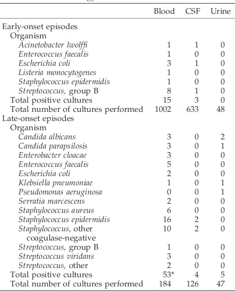
or yeast) in 3, the CSF yielded the same organism in 2, signs of local infection (necrotizing enterocolitis, omphalitis, pneumonia) were noted in 3, and treat-ment was continued for 7 days or more at the dis-cretion of the attending physician because of clinical instability along with the positive culture in 2 others. The most frequently isolated organisms were Strep-tococcus agalactiaein early-onset episodes and coagu-lase-negative staphylococci in late-onset episodes. Proven sepsis was less frequent in early-onset (attack rate 2%) than in late-onset episodes (attack rate 29%), but probable sepsis was diagnosed in;7% of episodes in each group.
Thirteen blood cultures yielded only skin or upper respiratory normal flora, including 6 with multiple organisms. Two positive blood cultures were
ob-tained from infants who never became ill and recov-ered without specific therapy. Fifteen episodes with positive CSF cultures were classified as no sepsis; those cultures yielded only normal skin flora or had scant bacterial growth in only 1 of 3 culture media, and all concomitant CSF cell counts, glucose, and protein levels were normal. These episodes were classified as having no sepsis.
Correlation of Diagnoses and Serum CRP Levels CRP levels were obtained in 1002 episodes of sus-pected early-onset infection and 184 of sussus-pected late-onset infection. The relationship between CRP levels and diagnoses is shown in Table 3. Four dif-ferent uses of these measurements were considered: the initial level alone (CRP #1), the second level alone (CRP #2), the higher of the second and third levels (CRP #2 and #3), or the highest of all three levels (CRP33). For each test, the sensitivity was highest at a cutoff of 1 mg/dL; this value was used in all evaluations. Proven or probable sepsis was strongly correlated with elevated CRP levels ($1.0 mg/dL) in all of these testing strategies, for both early- and late-onset episodes (P,10-5by 23 3 G-tests,,10-6 by 232 G-tests for no sepsis versus either proven or probable sepsis), supporting the diagnostic utility of CRP levels. The calculated sensitivities and specific-ities of each testing strategy are shown in Table 4.
TABLE 1. Demographic Characteristics of Study Population
Early-onset Episodes
Late-onset Episodes
Total patients 1002 134
Total episodes 1002 184
Birth weight (g)* 273461029 187461123 (550–6759) (385–4920) Gestational age (weeks)* 36.264.4 32.265.8
(23–43) (23–42) Age at evaluation (days)* 0.260.6 22.8620.2
(0–3) (4–118)
Male:female 553:449 80:54
Proven sepsis 20 53
Probable sepsis 74 12
Sepsis excluded 908 119
* Mean6standard deviation (range).
TABLE 2. Bacteriology of Positive Cultures
Blood CSF Urine
Early-onset episodes Organism
Acinetobacter lwolffi 1 1 0
Enterococcus faecalis 1 0 0
Escherichia coli 3 1 0
Listeria monocytogenes 1 0 0
Staphylococcus epidermidis 1 0 0
Streptococcus,group B 8 1 0
Total positive cultures 15 3 0
Total number of cultures performed 1002 633 48 Late-onset episodes
Organism
Candida albicans 3 0 2
Candida parapsilosis 3 0 1
Enterobacter cloacae 3 0 0
Enterococcus faecalis 5 0 0
Escherichia coli 2 0 0
Klebsiella pneumoniae 1 0 1
Pseudomonas aeruginosa 0 0 1
Serratia marcescens 2 0 0
Staphylococcus aureus 6 0 0
Staphylococcus epidermidis 16 2 0 Staphylococcus,other
coagulase-negative
10 2 0
Streptococcus,group B 1 0 0
Streptococcus viridans 3 0 0
Streptococcus,other 2 0 0
Total positive cultures 53* 4 5
Total number of cultures performed 184 126 47
* Some cultures yielded more than one organism.
TABLE 3. C-Reactive Protein Levels in Suspected Neonatal Infection
CRP #1 CRP #2 CRP #3 No Sepsis
Probable Sepsis
Proven Sepsis
Early-onset episodes
,1 Not done Not done 1* 0 1
,1 Not done 0 0 1
,1 692 0 2
1 42 2 1
1 Not done 20 12 5
,1 38 7 1
1 47 23 2
1 Not done Not done 10 8 0
,1 Not done 12 0 0
,1 7 0 0
1 0 0 0
1 Not done 10 13 3
,1 10 1 0
1 19 8 4
Total 908 74 20
Late-onset episodes
,1 Not done Not done 1† 0 9
,1 Not done 1‡ 0 4
,1 78 0 1
1 3 0 0
1 Not done 3 0 2
,1 1 0 1
1 1 3 3
1 Not done Not done 8 4 12
,1 Not done 3 0 0
,1 2 1 0
1 0 0 0
1 Not done 11 2 14
,1 2 0 1
1 5 2 6
Total 109 12 53
* Blood culture positive forStreptococcus salivarius.
† Endotracheal aspirate culture positive forCandida albicansand Staphylococcus aureus.
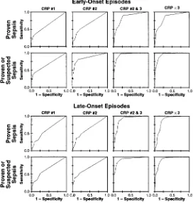
The sensitivity of a single measurement with the initial evaluation (CRP #1) for early-onset infection was low, both for proven sepsis (35.0%) and for proven or prob-able sepsis (39.4%). Although the sensitivity of CRP #1 was higher for late-onset infections, initial CRP levels were normal in more than one-third of all sepsis epi-sodes. The sensitivity of a delayed (8–24 hours after presentation) CRP level (CRP #2) was substantially higher, but maximum sensitivities were achieved only by combination of the second and third (CRP #2 and #3) or all three (CRP33) CRP levels. The discrimina-tory power (AUC, Fig 1) of CRP #2 and #3 or CRP33
was significantly greater than that of either CRP #1 or CRP #2 for both proven sepsis and proven or probable sepsis in both early- and late-onset episodes (P,.005). The AUC for CRP #1 differed from that for CRP #2 only for proven or probable sepsis in early-onset episodes (P,.001). The AUC for CRP #2 and #3 and CRP33 ROC curves were not significantly different.
Predictive Values and Bayesian Analysis
To assess the ability of abnormal and normal CRP levels to identify the presence or absence of infection, respectively, the positive and NPV for each testing
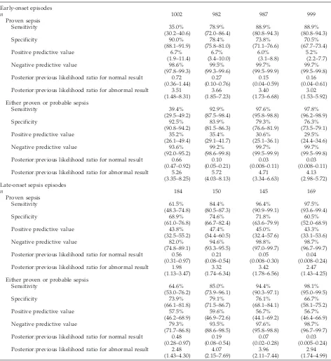
TABLE 4. Performance of C-Reactive Protein Measurements in Diagnosis of Neonatal Bacterial or Fungal Infection*
CRP #1 CRP #2 CRP #2 and #3 CRP33
Early-onset episodes
n 1002 982 987 999
Proven sepsis
Sensitivity 35.0% 78.9% 88.9% 88.9%
(30.2–40.6) (72.0–86.4) (80.8–94.3) (80.8–94.3)
Specificity 90.0% 78.4% 73.8% 70.5%
(88.1–91.9) (75.8–81.0) (71.1–76.6) (67.7–73.4)
Positive predictive value 6.7% 6.7% 6.0% 5.2%
(1.9–11.4) (3.4–10.0) (3.1–8.8) (2.2–7.7)
Negative predictive value 98.6% 99.5% 99.7% 99.7%
(97.8–99.3) (99.3–99.6) (99.5–99.9) (99.5–99.8) Posterior:previous likelihood ratio for normal result 0.72 0.27 0.15 0.16
(0.36–1.44) (0.10–0.76) (0.04–0.59) (0.04–0.61) Posterior:previous likelihood ratio for abnormal result 3.51 3.66 3.40 3.02
(1.48–8.31) (1.85–7.23) (1.73–6.68) (1.53–5.92) Either proven or probable sepsis
Sensitivity 39.4% 92.9% 97.6% 97.8%
(29.5–49.2) (87.5–98.4) (95.8–98.8) (96.2–98.9)
Specificity 92.5% 83.9% 79.3% 76.3%
(90.8–94.2) (81.5–86.3) (76.6–81.9) (73.5–79.1)
Positive predictive value 35.2% 35.4% 30.6% 29.5%
(26.1–49.4) (29.1–41.7) (25.1–36.1) (24.4–34.6)
Negative predictive value 93.6% 99.2% 99.7% 99.7%
(92.0–95.2) (98.6–99.8) (99.5–99.9) (99.5–99.8) Posterior:previous likelihood ratio for normal result 0.66 0.10 0.03 0.03
(0.47–0.92) (0.05–0.21) (0.008–0.11) (0.008–0.11) Posterior:previous likelihood ratio for abnormal result 5.26 5.72 4.71 4.13
(3.35–8.25) (4.03–8.13) (3.34–6.63) (2.98–5.72) Late-onset sepsis episodes
n 184 150 145 169
Proven sepsis
Sensitivity 61.5% 84.4% 96.4% 97.5%
(48.3–74.8) (80.5–87.3) (90.9–99.1) (93.6–99.4)
Specificity 68.9% 74.6% 71.8% 60.5%
(61.0–76.8) (66.7–82.4) (63.6–79.9) (52.0–68.9)
Positive predictive value 43.8% 47.4% 45.0% 43.3%
(32.5–55.2) (34.4–60.5) (32.4–57.6) (33.1–53.6)
Negative predictive value 82.0% 94.6% 98.8% 98.7%
(74.8–89.1) (93.3–95.5) (97.0–99.7) (96.7–99.7) Posterior:previous likelihood ratio for normal result 0.56 0.21 0.05 0.04
(0.31–0.97) (0.08–0.54) (0.008–0.30) (0.008–0.24) Posterior:previous likelihood ratio for abnormal result 1.98 3.32 3.42 2.47
(1.13–3.47) (1.74–6.34) (1.78–6.56) (1.43–4.25) Either proven or probable sepsis
Sensitivity 64.6% 85.0% 94.4% 98.1%
(53.0–76.2) (73.9–96.1) (90.3–97.1) (95.0–99.5)
Specificity 73.9% 79.1% 76.1% 66.7%
(66.1–81.8) (71.5–86.7) (68.1–84.1) (58.1–75.2)
Positive predictive value 57.5% 59.6% 56.7% 56.7%
(46.2–68.9) (46.9–72.6) (44.1–69.2) (46.4–66.9)
Negative predictive value 79.3% 93.5% 97.6% 98.7%
(71.7–86.8) (88.6–98.5) (95.8–98.8) (96.7–99.7) Posterior:previous likelihood ratio for normal result 0.48 0.19 0.07 0.03
(0.28–0.97) (0.08–0.54) (0.02–0.28) (0.005–0.24) Posterior:previous likelihood ratio for abnormal result 2.48 4.07 3.96 2.94
(1.43–4.30) (2.15–7.69) (2.11–7.44) (1.74–4.99)
strategy were calculated (Table 4). The PPV were low: among episodes in which a CRP level was ele-vated, proven sepsis was diagnosed in fewer than 10% of early-onset and fewer than 50% of late-onset evaluations, and proven or probable sepsis was iden-tified in only 30% to 35% of early-onset and 55% to 60% of late-onset evaluations. The NPV were.90% for all groups except those evaluated with a single CRP level at the time of presentation (CRP #1) with suspected late-onset disease, reflecting the low prev-alence of sepsis in these populations. To quantify the extent to which CRP measurements added diagnos-tic information, the Bayesian ratios of the likelihoods of infection before and after obtaining each test result were also calculated (Table 4). Normal initial CRP levels (CRP #1) were associated with modest (28%– 52%) reductions in the likelihood of infection (P ,
.05 except for proven sepsis in early-onset episodes). Normal CRP levels 8 to 24 hours later (CRP #2) were associated with more substantial (75%–90%) reduc-tions in the likelihood of both proven and probable infection (P,.05). If the last 2 (CRP #2 and #3) or all 3 (CRP 3 3) levels were normal, the likelihood of infection was only 3% to 15% of that in the entire population. Abnormal CRP results were associated with increases in the likelihood of infection of;3- to 6-fold for early-onset episodes and 1.5- to 2.5-fold for late-onset episodes.
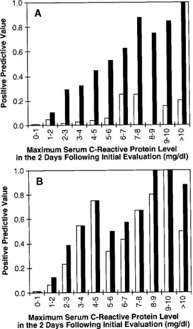
To determine whether greater elevations in CRP levels were associated with a higher probability of infection, the PPV were calculated for maximum CRP levels obtained 8 to 48 hours after presentation (CRP #2 and #3) for each successive interval of 1 mg/dL up to 10 mg/dL, for both early- and late-onset and proven or probable infection (Fig 2). In the
first 3 days of life, the PPV for mildly elevated CRP levels (1–2 mg/dL) were only 5% and 12% for proven and either proven or probable sepsis, respec-tively. All infected infants with such mildly elevated CRP levels were symptomatic. CRP levels.2 mg/dL were associated with a probability of either proven or probable infection .25%, but the probability of proven infection was .10% only of episodes in which CRP levels exceeded 5 mg/dL (Fig 2A). In contrast, episodes with CRP levels .2 mg/dL after the first 3 days of life had at least a 12% risk of proven infection and a 20% chance of proven or probable infection (Fig 2B).
Potential False-negative Results
Elevated CRP levels were not observed in 18 epi-sodes in which blood cultures yielded pathogenic organisms. In 11 of these episodes, CRP levels were obtained only with the initial evaluation, and CRP levels were obtained only in the first 16 hours after presentation in 4 additional episodes. Subsequent levels were not measured because positive culture results had already been reported. An infant deliv-ered by cesarean section at 32-weeks’ gestation for maternal indications was evaluated because of mild respiratory distress; she had neither a left-shifted differential blood count nor other clinical findings suggestive of Gram-negative sepsis, butAcinetobacter lwolffi was recovered from her blood culture and three CRP levels were ,1 mg/dL. One infant had
.105 colonies of Klebsiella pneumoniae per mL in a urine sample obtained by suprapubic aspiration; he had an increased proportion of immature granulo-cytes but CRP levels were persistently normal. One premature infant born after prolonged rupture of Fig 1. Receiver-operator characteristic curves for
membranes for.10 days died withStreptococcus viri-dans sepsis and progressive granulocytopenia, but serial CRP levels were not elevated.
Discontinuation of Antibiotics
Three normal CRP levels were obtained in 694 of the 1002 infants evaluated for early-onset infection. Of the 499 such infants for whom antibiotics were discontinued within 3 days, 13 required reevaluation for suspected infection within 14 days of the initial evaluation. Five of these infants were infected: 2 with
Staphylococcus epidermidissepsis, 1 withS aureus sep-sis, 1 with combined S aureus and E faecalis sepsis, and 1 withK pneumoniaeurinary tract infection. An-tibiotics were administered for .3 days in 195 in-fants, but only 85 infants were treated for 7 days or more. Indications for prolongation of antibiotic ther-apy included positive blood cultures in 2 patients (Acinetobacter lwolffiandS viridans, see above), abnor-mal hematologic findings alone in 40, a history of chorioamnionitis in 1, radiographic findings consis-tent with pneumonia in 3, and persisconsis-tent clinical instability without other explanation in the remain-der.
Three normal CRP levels were obtained in 79 of the 184 episodes of suspected late-onset infection. Of the 43 such episodes in which antibiotics were
dis-continued within 3 days, reevaluation for suspected infection within 14 days of the initial evaluation was required in 9, and 2 (5%) were associated with bac-teremia (S epidermidis, E faecalis). In retrospect, among the 36 episodes in which multiple CRP levels were normal but treatment was given for .3 days, only 1 had evidence of bacterial infection (K pneu-moniae urinary tract infection). Twenty of these epi-sodes were treated for 7 days or more. Treatment was continued for 3 to 7 days in 5 episodes in which blood cultures were positive for coagulase-negative staphylococci and three CRP levels were normal; only 1 was evaluated for suspected sepsis within 14 days and was not infected.
DISCUSSION
Serum concentrations of CRP increase several hun-dredfold in response to bacterial infection, making it an attractive diagnostic test for neonatal sepsis. Be-cause many of the more than 70 publications on this subject that have appeared during the past 30 years were flawed by imprecise diagnostic criteria, absent or inappropriate controls (eg, healthy neonates), in-complete description of results, or inadequate sam-ple sizes, the role of this test in evaluation of neo-nates remains controversial. Early reports described a high prevalence of elevated CRP levels in infected infants, but levels are elevated in only 35% to 65% of neonates with bacterial infection at the onset of ill-ness. Recognition that a delay of at least several hours is intrinsic to the cascade of events leading to elevation of serum CRP levels (including activation of neutrophils, elaboration of interleuk6, and in-duction of hepatic synthesis of CRP) led to appropri-ate criticism of this test as having insufficient sensi-tivity to guide therapy either by reliably diagnosing or excluding bacterial infection. Noting that CRP levels are consistently elevated 24 to 48 hours after the onset of infection, Philip22 and later others23–26 suggested that serial normal levels may be useful for identification of infants who do not have bacterial infection. In a series of 218 infants (13 with sepsis), Gerdes and Polin8 reported high sensitivity (93%) and NPV (99%) for CRP levels determined by a latex agglutination method at the time of evaluation and 12 to 24 hours later. Pourcyrous et al9found that two levels measured over the first 3 days in 140 neonates (36 with infection) had a high NPV, and suggested that the utility of CRP levels might be optimized by obtaining serial levels at 12-hour intervals in the first 24 to 36 hours of illness. In 292 evaluations, Krediet et al27found that two levels in the first 24 hours had only modest sensitivity (53% to 88%) and NPV (80% to 97%), depending on the criteria for diagnosis (proven or probable sepsis) and the time of onset of the infection (early or late). In the largest series pub-lished to date, including 689 evaluations in 491 in-fants and 187 episodes of culture-proven or clinically definite sepsis, Pourcyrous et al9 described similar results when CRP levels were obtained at the initial evaluation and twice at 12 hour intervals, emphasiz-ing the importance of serial levels for achievement of optimum sensitivity. Philip and Hewitt7reported no recurrence of infection within 7 days of discontinu-Fig 2. Positive predictive values for the highest of two serum
ation of antibiotics based on three normal CRP de-terminations within 48 hours and negative cultures in 147 low birth infants at risk for early-onset infec-tion. Based on these data, determination of serial CRP levels was incorporated into our diagnostic ap-proach to suspected neonatal sepsis. Levels were obtained at the initial evaluation and with routine morning laboratory studies on each of the next 2 days. This analysis was undertaken to determine whether our experience was similar to previous re-ports.
Evaluation of the performance of a diagnostic test requires an objective and reliable reference method for identifying patients with and without the disease of interest, but there are no simple criteria for diag-nosis of sepsis in the neonate. The most objective evidence of invasive infection is usually considered to be recovery of an organism from body fluids ob-tained using sterile technique: blood, CSF, or urine obtained by suprapubic aspirate, in particular. These studies may yield false-positive results, particularly for organisms that may represent either skin flora or potential pathogens in low birth weight or debili-tated neonates, such as coagulase-negative staphylo-cocci. To minimize false-positive diagnoses, patients whose cultures were positive for skin flora only and who had no other clinical findings consistent with infection were not considered to have infection. The remaining patients with positive culture results con-stituted the group with proven sepsis. This category is most useful for assessment of the sensitivity of CRP levels—that is, the probability of an abnormal CRP level in those patients who are unequivocally septic. False-negative results may result from sub-mission of small aliquots of blood for culture, inter-mittent or low-density bacteremia, or suppression of bacterial growth by earlier (eg, intrapartum) antibi-otic administration. To minimize errors resulting from false-negative culture results, infants with neg-ative cultures but whose clinical and laboratory find-ings were suggestive of infection were assigned to a group with probable sepsis. Patients who lacked findings suggestive of infection were assigned to a group with no sepsis, in which infection could be excluded with confidence. This group is most useful for evaluation of the specificity and NPV of one or more normal CRP levels. The possibility that infants may have been assigned an incorrect diagnosis re-mains, however, and is intrinsic to all studies of this nature.
This analysis of the relationship between serum CRP levels and neonatal infection, which includes 1186 diagnostic evaluations in 1066 neonates, is the largest such study yet published. In these popula-tions, proven infection was much less likely in early-onset (2%) than in late-early-onset (40%) episodes, reflect-ing early evaluation of many infants without clinical signs of infection because of obstetrical factors (eg, prolonged rupture of membranes) alone. The preva-lence of probable sepsis was similar in the early- and late-onset groups. The higher rate of confirmed in-fection in the late-onset patients is similar to that previously reported in other comparisons of the per-formance of CRP in early- and late-onset disease,27–29
and probably results from selection of infants for late-onset evaluations only if they had clinical signs of possible infection. As in previous series, elevated CRP levels strongly correlated with infection for both early- and late-onset episodes, whether single or se-rial levels were considered, and independent of whether probable cases were grouped with proven cases, with noninfected infants, or considered sepa-rately. There were few differences in test perfor-mance between early- and late-onset groups. Other than the higher PPV of elevated CRP levels in late-onset episodes, which reflects the much higher prev-alence of sepsis in this group, only the higher sensi-tivity of the initial CRP in the late-onset group (for both proven and proven or probable sepsis) was statistically significant. The higher prevalence of el-evated CRP levels in these patients is not unex-pected, because their infections had necessarily been present for sufficient time to produce persistent clin-ical signs that led to diagnostic assessment. ROC curve analysis (Fig 1) established a serum level of 1 mg/dL, an appropriate threshold above which re-sults should be considered abnormal, provided the first statistical confirmation that serial levels are di-agnostically superior to single measurements, and confirmed previous observations that a single CRP level at the beginning of an evaluation lacks sensi-tivity and negative predictive power. The sensisensi-tivity and NPV are improved by delaying testing for 8 to 24 hours, as previously reported,18,30 but the highest levels of these performance parameters were achieved only with multiple serial levels. These ob-servations, as well as more recent reports,31–33 sup-port the previously resup-ported clinical superiority of serial CRP determinations.8,27,34
Traditional descriptors—sensitivity, specificity, PPV, and NPV—may not accurately represent test performance, because they are heavily influenced by the prevalence of disease in the sample population. For example, the NPV of a nondiscriminant test will be high in a population in which the disease of interest is rare. Diagnostic test performance is better reflected in Bayesian ratios of the likelihood of infec-tion in each of the subgroups defined by the test results (posterior probability) to that in the entire population (previous probability).35 Such posterior: previous probability ratios are an effective indication of the information added by the test result. In earlier studies in which CRP measurements were per-formed only during the first 24 hours after presenta-tion, the Bayesian ratios associated with normal se-rial CRP levels were 0.09 (95% CI, 0.02– 0.55; n 5
the Bayesian ratio for infection was 0.16 or less for early-onset cases and 0.08 or less for late-onset cases (Table 4). These data, based on nearly twice the num-ber of episodes included in all previous reports, in-dicate that serial normal CRP levels during the first 2 to 3 days of suspected infection are associated with a substantial reduction in the probability that such a neonate is infected.
Our data confirm previous reports of poor sensi-tivity of single CRP levels on presentation of an infant with possible infection. In isolation, a normal initial CRP is associated with a risk reduction of only
;50% or less (Table 4), which will rarely affect treat-ment. If combined with two subsequent normal lev-els, the initial CRP had little impact on the NPV and Bayesian likelihood ratios, as the differences between these values for CRP #2 and #3 and those for CRP3 3 were small and neither clinically nor statistically significant. Exclusion of episodes in which an ele-vated initial CRP level precluded performance of subsequent levels could artifactually skew these re-sults, but the data in Table 3 indicates that this does not lead to diagnostic errors. Of the 37 infants in the early-onset group in whom only the initial level was elevated, none had proven infection, and none of the 8 with probable sepsis had a second CRP level. Of the 30 late-onset episodes in which only the initial level was elevated, 12 had proven infection (a posi-tive culture), and only 1 of the 5 with probable sepsis had more than one CRP. In that patient, the initial level was 1.0 mg/dL and two subsequent levels were
#0.5 mg/dL; infection was probable because the proportion of immature granulocytes was increased. The second or third CRP level was elevated in all of the 66 infants with early-onset probable sepsis and in 7 of the 8 episodes of late-onset probable sepsis in which more than one CRP was obtained, making it unlikely that subsequent levels would have been normal had they been done. Exclusion of these pa-tients from the CRP #2 and #3 group did not com-promise the NPV of that test. Omission of subse-quent levels in episodes with positive cultures reduced the prevalence of proven infection in the sample populations and may have reduced the cal-culated specificity and PPV of CRP #2, CRP #2 and #3, and CRP33. CRP levels at the initial evaluation can be omitted without compromising the diagnostic utility of serial levels obtained during the next 48 hours. These data do not address the possibility that early levels may have utility as a component of a multifactorial screening panel.
In this experience, false-negative results (positive cultures with normal CRP levels) were most com-mon in infants who had serum samples obtained only early in the evaluation. In these instances, pos-itive culture results were available before CRP levels became elevated, and additional levels were not ob-tained. As noted by Buck and Pohlandt,37the timing of CRP levels is critical to achievement of optimal sensitivity, and it is possible that abnormal levels did develop, but were not measured, within 48 hours of diagnosis. Others have described inconsistent re-sponses to infections caused by mildly pathogenic organisms (particularly coagulase-negative
staphylo-cocci),38,39which may also produce false-negative re-sults. In the 2 cases in which pathogenic organisms (Acinetobacter lwoffiandE faecalis) were isolated from blood but no other clinical or laboratory findings consistent with sepsis were apparent, the positive culture results may have been factitious. Lack of a demonstrable acute phase response in the patient with urinary tract infection is consistent with previ-ous reports that CRP levels are often not elevated in infants and children whose infection is limited to the lower urinary tract.40,41In addition, infants with over-whelming bacterial sepsis may exhibit little or no increment in serum CRP levels when the infection is associated with severe granulocytopenia.42 Thus, whereas serial measurements increase sensitivity substantially, perfect sensitivity is not achievable.
The outcome of at-risk infants in whom antibiotic therapy is discontinued on the basis of serial normal CRP levels may be the best indicator of their utility for exclusion of infection. Because discontinuation of antibiotic therapy was left to the judgment of the attending neonatologist, the data reported here are not conclusive. In episodes in which antibiotics were discontinued after three normal CRP levels, the nos-ocomial infection rate (1 per 1000 patient days) was much less than the prevalent rate in our nurseries during that period (6 –9 per 1000 patient days). The organisms associated with subsequent infectious ep-isodes after early discontinuation of therapy for sus-pected early-onset sepsis were typical of late-onset nosocomial infections, and these episodes occurred 7 to 11 days after the initial evaluation. Of those epi-sodes in which serial CRP levels were normal and treatment was given for.3 days, antibiotic therapy was discontinued before completion of a 7 day course in more than half, reflecting the belief of the attending neonatologists that these infants were not infected. Among the 105 episodes in which a full course ($7 days) of antibiotic therapy was provided, only 2 infants would have been at serious risk had antibiotics been discontinued prematurely. Because these infants had Klebsiella urinary tract infection and S viridans sepsis with severe granulocytopenia, the diagnosis was not obscure, and a basis for dis-counting the normal CRP levels was readily evident. These data suggest that the risk of recurrence of an inadequately treated infection is extremely small when normal serial CRP levels are the basis for dis-continuation of antibiotics. However, the latter 2 cases emphasize the need for continued application of this test within the context of other clinical data, and additional experience is needed to demonstrate that discontinuation of antibiotics based on serial normal CRP levels is safe in infants whose blood cultures are positive for coagulase-negative staphy-lococci.
study populations. Elevated CRP levels occur in as-sociation with many other conditions in addition to bacterial or fungal infection. Interleukin-6 of mater-nal origin43in cord blood may produce elevated CRP levels in uninfected infants born to women with chorioamnionitis. CRP levels may be elevated in ne-onates with thermal burns,44,45pneumothoraces,46 in-traventricular hemorrhage,46 meconium aspiration syndrome with negative cultures,23,34necrotizing en-terocolitis,47– 49 and after surgical procedures,50,51 car-diopulmonary bypass,52–54 or immunizations55 but not in those with uncomplicated respiratory distress syndrome,46,56,57perinatal asphyxia,56prolonged rup-ture of membranes,56or jaundice.56 Most acute viral infections are not associated with abnormal CRP lev-els.58,59 Severe viral infections, such as herpes sim-plex,60 may be associated with elevated CRP levels, which may be an indication of bacterial superinfec-tion.58 We identified 4 patients with positive viral cultures and others with acute tissue injuries, hema-tologic disorders, or during extracorporeal life sup-port, who did not have apparent bacterial infection but did have increased CRP levels (data not shown). In .80% of the cases in which CRP levels were abnormal in the absence of bacterial or fungal infec-tion, the levels did not exceed 5 mg/dL, suggesting that markedly elevated levels might have a higher PPV. The additional analysis summarized in Figure 2 demonstrated that CRP levels.2 mg/dL were asso-ciated with a risk of either proven or probable sepsis of.25%, and levels.3 mg/dL in infants evaluated for late-onset infection were associated with proven bacterial or fungal infection in ;50%. Infants with modest elevations (up to 2 mg/dL) may not require antimicrobial therapy, but those with greater eleva-tions probably should be treated until infection can be excluded using other data.
Measurements of serial CRP concentrations in se-rum may be useful in treatment of suspected neona-tal sepsis. The greatest utility is in the ability of serial levels within the first 48 hours of suspected illness to distinguish infants with bacterial infection and neg-ative nonpermissive cultures (false-negneg-ative cul-tures), who may require continuation of antibiotic treatment, from those whose clinical findings are not related to bacterial or fungal infection (true-negative cultures), for whom antibiotic therapy can be safely discontinued. Persistent normal CRP levels may also identify infants with false-positive cultures, espe-cially if other data (clinical course and blood counts) are also inconsistent with infection. An elevated CRP level may help guide initiation or adjustment of an-timicrobial therapy, as in routine postoperative screening for intercurrent infection36 or monitoring for acquisition of resistance to antibiotic treatment. In most cases, however, information from CRP levels obtained at that juncture will not alter a decision to start antibiotic therapy. The role of CRP levels taken at the time of admission as part of a multicomponent sepsis screen was not addressed by this study, and the utility of such a sepsis screen may be demon-strated by future prospective studies.
ACKNOWLEDGMENTS
This work was supported by General Clinical Research Center grant RR-00070 and by the Medical Scholars Program of the Stan-ford Medical Alumni Association.
We thank Deirdre Umbenhower and Rose Machie for assis-tance with the Lucile Packard Children’s Hospital information systems, Sunita Shastry and Tropty Singh for abstracting data from El Camino Hospital medical records, and Patricia Cross and Linda Hanna for their assistance with funding applications.
REFERENCES
1. Centers for Disease Control and Prevention. Prevention of perinatal group B streptococcal disease: a public health perspective.MMWR Morb Mortal Wkly Rep. 1996;45:1–24
2. McCracken GH Jr, Shinefield HR. Changes in the pattern of neonatal septicemia and meningitis.Am J Dis Child. 1966;112:33–39
3. Gluck L, Wood HF, Fousek MD. Septicemia of the newborn.Pediatr Clin North Am. 1966;13:1131–1148
4. Spector SA, Ticknor W, Grossman M. Study of the usefulness of clinical and hematologic findings in the diagnosis of neonatal bacterial infec-tions.Clin Pediatr (Phila). 1981;20:385–392
5. Yancey MK, Duff P, Kubilis P, Clark P, Frentzen BH. Risk factors for neonatal sepsis.Obstet Gynecol. 1996;87:188 –194
6. Hammerschlag MR, Klein JO, Herschel M, Chen FC, Fermin R. Patterns of use of antibiotics in two newborn nurseries.N Engl J Med. 1977;296: 1268 –1269
7. Philip AG, Hewitt JR. Early diagnosis of neonatal sepsis.Pediatrics. 1980;65:1036 –1041
8. Gerdes JS, Polin RA. Sepsis screen in neonates with evaluation of plasma fibronectin.Pediatr Infect Dis J. 1987;6:443– 446
9. Pourcyrous M, Bada HS, Korones SB, Baselski V, Wong SP. Significance of serial C-reactive protein responses in neonatal infection and other disorders.Pediatrics. 1993;92:431– 435
10. Manroe BL, Weinberg AG, Rosenfeld CR, Browne R. The neonatal blood count in health and disease. I. Reference values for neutrophilic cells.
J Pediatr. 1979;95:89 –98
11. Rodwell RL, Leslie AL, Tudehope DI. Early diagnosis of neonatal sepsis using a hematologic scoring system.J Pediatr. 1988;112:761–767 12. Klein J, Marcy S. Bacterial sepsis and meningitis. In: Remington J, Klein
J, eds.Infectious Diseases of the Fetus and Newborn Infant. Philadelphia, PA: WB Saunders; 1995:866 – 868
13. Feinstein AR. Clinical biostatistics XXXI. On the sensitivity, specificity, and discrimination of diagnostic tests.Clin Pharmacol Ther. 1975;17: 104 –116
14. Sokol R, Rohlf F.Biometry. New York, NY: WH Freeman; 1995:724 –760 15. Zweig MH, Campbell G. Receiver-operating characteristic (ROC) plots: a fundamental evaluation tool in clinical medicine.Clin Chem. 1993;39: 561–577
16. Centor RM. A Visicalc program for estimating the area under a receiver operating characteristic (ROC) curve.Med Decis Making. 1985;5:139 –148 17. Hanley JA, McNeil BJ. A method of comparing the areas under receiver operating characteristic curves derived from the same cases.Radiology. 1983;148:839 – 843
18. Messer J, Eyer D, Donato L, Gallati H, Matis J, Simeoni U. Evaluation of interleukin-6 and soluble receptors of tumor necrosis factor for early diagnosis of neonatal infection.J Pediatr. 1996;129:574 –580
19. Rosner B.Fundamentals of Biostatistics. Belmont, CA: Duxbury Press; 1994;173–178
20. Armitage P, Berry G.Statistical Methods in Medical Research. London, England: Blackwell Science; 1994:71–76
21. Fleiss JL.Statistical Methods for Rates and Proportions. New York, NY: John Wiley and Sons; 1981:71–75
22. Philip AGS. Commentary. In: Oski FA, Stockman JA, eds.Year Book of Pediatrics. Chicago, IL: Year Book Medical Publishers; 1981:17 23. Ainbender E, Cabatu EE, Guzman DM, Sweet AY. Serum C-reactive
protein and problems of newborn infants.J Pediatr. 1982;101:438 – 440 24. Squire EN Jr, Reich HM, Merenstein GB, Favara BE, Todd JK. Criteria
for the discontinuation of antibiotic therapy during presumptive treat-ment of suspected neonatal infection.Pediatr Infect Dis. 1982;1:85–90 25. Speer C, Bruns A, Gahr M. Sequential determination of CRP, alpha
1-antitrypsin and haptoglobin in neonatal septicaemia.Acta Paediatr Scand. 1983;72:679 – 683
27. Krediet T, Gerards L, Fleer A, van Stekelenburg G. The predictive value of CRP and I/T-ratio in neonatal infection.J Perinat Med. 1992;20: 479 – 485
28. Seibert K, Yu VY, Doery JC, Embury D. The value of C-reactive protein measurement in the diagnosis of neonatal infection.J Paediatr Child Health. 1990;26:267–270
29. Russell GA, Smyth A, Cooke RW. Receiver operating characteristic curves for comparison of serial neutrophil band forms and C reactive protein in neonates at risk of infection.Arch Dis Child. 1992;67:808 – 812 30. Mathers NJ, Pohlandt F. Diagnostic audit of C-reactive protein in
neo-natal infection.Eur J Pediatr. 1987;146:147–151
31. Wagle S, Grauaug A, Kohan R, Evans SF. C-reactive protein as a diagnostic tool of sepsis in very immature babies.J Paediatr Child Health. 1994;30:40 – 44
32. Berger C, Uehlinger J, Ghelfi D, Blau N, Fanconi S. Comparison of C-reactive protein and white blood cell count with differential in neo-nates at risk for septicaemia.Eur J Pediatr. 1995;154:138 –144
33. Kawamura M, Nishida H. The usefulness of serial C-reactive protein measurement in managing neonatal infection.Acta Paediatr. 1995;84: 10 –13
34. Pourcyrous M, Bada HS, Korones SB, Barrett FF, Jennings W, Lockey T. Acute phase reactants in neonatal bacterial infection.J Perinatol. 1991; 11:319 –325
35. Philip AG. Diagnosis of neonatal bacteraemia [letter].Arch Dis Child. 1989;64:1514
36. Chwals WJ, Fernandez ME, Jamie AC, Charles BJ, Rushing JT. Detection of postoperative sepsis in infants with the use of metabolic stress monitoring.Arch Surg. 1994;129:437– 442
37. Buck C, Pohlandt F. Accuracy of leukocyte indices and C-reactive protein for diagnosis of neonatal infection [letter].Pediatr Infect Dis J. 1995;14:1119 –1120
38. Schmidt BK, Kirpalani HM, Corey M, Low DE, Philip AG, Ford-Jones EL. Coagulase-negative staphylococci as true pathogens in newborn infants: a cohort study.Pediatr Infect Dis J. 1987;6:1026 –1031
39. Tegtmeyer FK, Horn C, Richter A, van Wees J. Elastase alpha 1 protein-ase inhibitor complex, granulocyte count, ratio of immature to total granulocyte count, and C-reactive protein in neonatal septicaemia.Eur J Pediatr. 1992;151:353–356
40. Wientzen RL, McCracken GH Jr, Petruska ML, Swinson SG, Kaijser B, Hanson LA. Localization and therapy of urinary tract infections of childhood.Pediatrics. 1979;63:467– 474
41. Johnson CE, Shurin PA, Marchant CD, et al. Identification of children requiring radiologic evaluation for urinary infection.Pediatr Infect Dis. 1985;4:656 – 663
42. Sabel KG, Hanson LA. The clinical usefulness of C-reactive protein (CRP) determinations in bacterial meningitis and septicemia in infancy.
Acta Paediatr Scand. 1974;63:381–388
43. Singh B, Merchant P, Walker CR, Kryworuchko M, Diaz-Mitoma F.
Interleukin-6 expression in cord blood of patients with clinical chorio-amnionitis.Pediatr Res. 1996;39:976 –979
44. Daniels JC, Larson DL, Abston S, Ritzmann SE. Serum protein profiles in thermal burns. II. Protease inhibitors, complement factors, and C-reactive protein.J Trauma. 1974;14:153–162
45. Pruchniewski D, Pawlowski T, Morkowski J, Mackiewicz S. C-reactive protein in management of children’s burns.Ann Clin Res. 1987;19: 334 –338
46. Dyck RF, Bingham W, Tan L, Rogers SL. Serum levels of C-reactive protein in neonatal respiratory distress syndrome.Clin Pediatr (Phila). 1984;23:381–383
47. Philip AG, Sann L, Bienvenu F. Acute phase proteins in neonatal necrotizing enterocolitis.Acta Paediatr Scand. 1986;75:1032–1033 48. Isaacs D, North J, Lindsell D, Wilkinson AR. Serum acute phase
reac-tants in necrotizing enterocolitis.Acta Paediatr Scand. 1987;76:923–927 49. Schober PH, Nassiri J. Risk factors and severity indices in necrotizing
enterocolitis.Acta Paediatr Suppl. 1994;396:49 –52
50. Ito H, Kishikawa T, Yamakawa Y, et al. Serum acute phase reactants in pediatric patients; especially in neonates.Jpn J Surg. 1983;13:506 –511 51. Chwals WJ, Letton RW, Jamie A, Charles B. Stratification of injury
severity using energy expenditure response in surgical infants.J Pediatr Surg. 1995;30:1161–1164
52. Aronen M, Leijala M, Meri S. Value of C-reactive protein in reflecting the magnitude of complement activation in children undergoing open heart surgery.Intensive Care Med. 1990;16:128 –132
53. Mitchell IM, Pollock JC, Jamieson MP, Donaghey SF, Paton RD, Logan RW. C-reactive protein and cardiopulmonary bypass in infants under 5 kilograms.Ann Clin Biochem. 1991;28:109 –110
54. Butler J, Pathi VL, Paton RD, et al. Acute-phase responses to cardiopul-monary bypass in children weighing less than 10 kilograms.Ann Thorac Surg. 1996;62:538 –542
55. Balkundi DR, Nycyk JA, Cooke RW. Immunisation and C reactive protein in infants on neonatal intensive care units [letter].Arch Dis Child. 1994;71:F149
56. Schouten-Van Meeteren NY, Rietveld A, Moolenaar AJ, Van Bel F. Influence of perinatal conditions on C-reactive protein production.J Pe-diatr. 1992;120:621– 624
57. Senses DA, Toppare MF, Kaya IS, Dilmen U, Kitapci F. C-reactive protein and respiratory distress syndrome [letter].J Pediatr. 1993;122:164 58. Peltola H, Jaakkola M. C-reactive protein in early detection of bactere-mic versus viral infections in immunocompetent and compromised children.J Pediatr. 1988;113:641– 646
59. Borgnolo G, Barbone F, Guidobaldi G, Olivo G. C-reactive protein in viral and bacterial gastroenteritis in childhood.Acta Paediatr. 1996;85: 670 – 674
DOI: 10.1542/peds.102.4.e41
1998;102;e41
Pediatrics
William E. Benitz, Michael Y. Han, Ashima Madan and Pramela Ramachandra
Serial Serum C-Reactive Protein Levels in the Diagnosis of Neonatal Infection
Services
Updated Information &
http://pediatrics.aappublications.org/content/102/4/e41
including high resolution figures, can be found at:
References
http://pediatrics.aappublications.org/content/102/4/e41#BIBL
This article cites 54 articles, 6 of which you can access for free at:
Permissions & Licensing
http://www.aappublications.org/site/misc/Permissions.xhtml
in its entirety can be found online at:
Information about reproducing this article in parts (figures, tables) or
Reprints
http://www.aappublications.org/site/misc/reprints.xhtml
DOI: 10.1542/peds.102.4.e41
1998;102;e41
Pediatrics
William E. Benitz, Michael Y. Han, Ashima Madan and Pramela Ramachandra
Serial Serum C-Reactive Protein Levels in the Diagnosis of Neonatal Infection
http://pediatrics.aappublications.org/content/102/4/e41
located on the World Wide Web at:
The online version of this article, along with updated information and services, is
by the American Academy of Pediatrics. All rights reserved. Print ISSN: 1073-0397.