ORIGINAL ARTICLE
IJPHY
ABSTRACT
Background: Providing new objective valid and reliable methods of assessment of a range of motion is always a per-sistent need for clinical practitioners and researchers in physical therapy for obtaining précised and realistic diagnostic and treatment decisions. So this study was carried out to test the validity and intra-rater reliability of the laser goni-ometer via comparing repeated measures of laser and electro-gonigoni-ometers in measuring a range of motion of shoulder movements considering the electro-goniometer as the reference standard.
Methods: one hundred healthy males with ages ranging between 20-30 years shared in this study. Three consecutive measures of bilateral shoulder flexion, abduction, internal and external rotation range of motion were performed by the same examiner on each subject by each of the laser and electro-goniometer, with standardized measurement proce-dures, subjects’ positions, and stabilizations.
Results: Pearson (r), paired T-test, and intra-class (ICC) correlation coefficients were used to test the validity and intra-rater reliability of the laser goniometer in comparison to the electro-goniometer. And the results of the validity testing showed very strong relationship between readings by both devices (r=0.84 to 0.93) and also no significant dif-ferences between means of readings of both devices with the p-value ranging between 0.13 and 0.97. Also, ICC revealed high intra-rater reliability of laser goniometer on repeated measures of shoulder range of motions (ICC=0.98-0.99). Conclusion: laser goniometer can be used as a new valid, reliable digital objective method of measurement of shoulder range of motion.
Keywords: laser goniometer; electro-goniometer; range of motion; assessment; validity; reliability, shoulder, objective measurement, physical therapy.
Received 05th June 2019, accepted 27th September 2019, published 09th October 2019
www.ijphy.org
10.15621/ijphy/2019/v6i5/186838
CORRESPONDING AUTHOR
Int J Physiother. Vol 6(5), 169-176, October (2019) ISSN: 2348 - 8336
VALIDITY AND INTRA-RATER RELIABILITY OF LASER
GONIOMETER VERSUS ELECTRO-GONIOMETER IN
MEASURING SHOULDER RANGE OF MOTION
¹Mohammed H. Elgendy
*2Wael O. A. Abd El-khalek
*2Wael O. A. Abd El-khalek
Lecturer, Department of Basic Sciences, Faculty of Physical Therapy,
Badr University of Cairo (BUC), Egypt. email: [email protected] ¹Professor, Department of Basic Sciences,
Faculty of Physical Therapy, Cairo University, Egypt.
INTRODUCTION
Clinical range of motion (ROM) measurement is an essen-tial evaluation procedure used in physical therapy, and it is known as goniometry which when dealing with muscu-loskeletal conditions is considered a vital evaluation skill, whose resulting measures can be interpreted to prove the absence or presence of functional affection in the normal mobility of body joints, that is to develop and generate evi-dence of the effect of treatment strategies [1].
Unfortunately, -till nowadays- the most common way for measuring joint range of motion is the universal plastic goniometer (UG) which is considered as a subjective inac-curate method that suffers from the factor of human error and inconsistency, including the reading and interpretation of the readings [2], improper goniometric application, er-roneous specification of the bony landmarks’ location, the axis of joint rotation and inability to keep the goniometer’s fulcrum on this axis, which affect its validity and reliability. So due to the increased demand of health practitioners to-ward providing recent qualified services for human health, and the increased orientation about clinical practice based on evidence, efforts are made and continuously done to find reliable and valid clinical objective evaluation tools can be used to evaluate ROM of different joints as it has become important demand for physical therapists for valu-able and effective clinical practice [1, 3].
One of the modalities that has become well known as a standardized, valid and reliable method of measuring joint range of motion is the electro-goniometer “ELGON” [1, 4], which converts the angular displacement of the joint into an electric signal readout [3], produced in a simple digital form with measurement’s accuracy ±2◦ in range of ±90◦ [2].
So it is used primarily in research studies considering be-ing expensive, time-consumbe-ing for calibration [4, 5] and the required skill for application [3], which makes it lim-iting factor for being used in clinical and practical settings [3-5].
The validity and reliability of electrogoniometer had been evaluated and proven by several studies on different body regions, one of those is the study carried out by Da Silva et al. (2015) to compare the electrogoniometer and the universal goniometer regarding intra-examiner and inter-examiner reliability as well as the inter-device reliability while test-ing wrist range of motion, and they concluded that electro goniometer has shown to be a reliable measurement tool for clinical usage when compared to universal goniometer [6]. Another study carried out by Rowe et al. (2001) to as-sess the validity and reliability of electrogoniometer com-pared to motion analysis system in measuring knee motion during gait, and they concluded that the electrogoniometer is a valid, reliable instrument providing accurate, precise and stable readings on repeated measures [7].
The laser goniometer is a new modality that has the advan-tage of the ease and speed measurement of range of mo-tion, it has two laser beams that compensate for the fixed and movable arms of the traditional goniometer configu-ration, the degree of change of the direction of the laser
beams represent the degree of change of angular displace-ment of the joint and the readings are shown on an LCD screen in digital form, it just requires to be applied over the region or segment whose range will be measured, and then pressing the zero reference button to determine the zero reference point. By holding the device in the same position during the movement with the laser beams pointing to the bony landmarks all over the measurement, the result can be displayed in the form of digital reading with high pre-cision [2].
This study aimed to test the laser goniometer’s validity and intra-examiner reliability in comparison with the elec-tro-goniometer considering the later as the reference stan-dard in providing accurate, consistent and precise mea-sures of range of movement of the different body regions that the physical therapists can rely on in clinical research and practical evaluation approaches.
MATERIAL AND METHODS Design
This study’s research design was a single group within-sub-ject validity and reliability test. Before sharing in this study, a written consent form was signed down by each subject.
Participants
One hundred healthy volunteer male physical therapy stu-dents and practitioners from faculty of physical therapy at Misr University for science and technology, with ages, ranging from 20-30 years, heights range between 160-180 cm and body mass indices (BMI) range between 18.5-29.9 kg/m2 have shared in this study. This study was carried in
the period from 11th June 2016 to 31st August 2016. Sampling
The sample shared in this study was sequentially selected through an announcement for who would like to share as a volunteer and then the inclusive and exclusive criteria were applied for all the volunteers thus only those that have met the criteria have shared and who not were excluded.
Materials
-Weight scale: A CAMRY mechanical personal weight scale Model BR5002 with serial no. C101100430, shown in Fig. 1; was calibrated before measuring each subject’s weight.
-An adhesive ruler and water bubble scale: for measuring height sticker ruler from IKEA adhered to the wall and a water bubble scale were used to measure the participants’ height as shown in Fig. 2.
Figure 2: IKEA adhesive sticker ruler and water bubble scale for measuring height
- Chair: standard 1100 back reclined chair
- Plinth.
- Straps for stabilization.
- Small sheets: for padding at humerus during measuring shoulder rotation movements.
- The electro-goniometer
The digital electro-goniometer - model 12-1027 – that works by an electrical signal induced by means of a battery was used as the reference standard in this study. The device converts the angular displacement of the joint (represented in the degree of change of the angle between the two arms of the device) into an electric signal readout [3], which is produced in a simple digital form [2] as shown in Fig. 3.
Figure 3: The Digital Electro-goniometer. -The laser goniometer
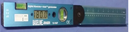
The laser goniometer HALO which is class 1 laser product with maximum out the power of 0.39 mW and wavelength of 635 nm – model HG1 - serial no. HG1XSH225G - de-signed in Australia, made in Malaysia, was used as the test-ed device in this study, it has 2 laser beams that compensate for the fixed and movable arms of the traditional goniom-eter configuration, the degree of change of the direction of the laser beams represent the degree of change of angular displacement of the joint and the readings are shown on an LCD screen in digital form, shown in Fig. 4.
Figure 4a: The digital laser goniometer HALO with the two laser beams emitting from it.
Figure 4b: HALO configurations.
Procedures
Weight and height and body mass index score of each of the participants who shared in this study were measured, then the inclusion and exclusion criteria were applied. Each participant of those who met the criteria was asked to sit for a while until he feels rested while the whole idea and procedures of the study were explained to him, and then after his approval to share in the study, he was asked to sign the consent form.
The sequence of movements’ measurement was randomly performed for each patient to avoid effect of sequencing on the examiner’s decisions or measurements. Also, the se-quence of devices used in measurement was randomized for each patient and each movement to avoid bias by the examiner.
Shoulder flexion, abduction, internal, and external rota-tion were measured bilaterally. Range of movement of each of these selected joint movements was measured for three consecutive times by the same examiner on the same sub-jects for each side, by each of the laser and electro-goniom-eters, one after another, taking into consideration the stan-dardization of all the measurement procedures, subjects’ positions, and stabilizations, considering the electro-goni-ometer as the reference standard. Each subject was asked to take off any clothes that may restrict movement of the measured region and to perform active range of motion for each of the tested movements for each joint with the assumption that each of the participants have followed the instructions strictly and acted freely, efficiently and in the same way while performing these movements during tak-ing the repeated measures by both devices.
For measuring the selected shoulder movements, the posi-tion used as the supine posiposi-tion, in which the participant was asked to lie down restfully on his back with his trunk bare skin to allow free movement of both arms during measurement of each side, stabilization with straps to the chest region was applied to avoid substitutions by trunk movement [3, 5].
and the thumb pointing upward. The axis of measurement was the lateral part of greater tubercle and the lateral epi-condyle as the reference point. The fixed arm was placed parallel to the surface of the floor, the movable arm was placed parallel to the lateral aspect of the upper arm (hu-merus) [3, 5], as shown in Fig. 5.
Figure 5a: Measuring shoulder flexionusing an electro-goniometer.
Figure 5b: Measuring shoulder flexion Laser goniometer. For shoulder abduction, the starting position for the par-ticipant’s arm was laterally rotated beside his body with the elbow extended and palm facing upward without flexion or extension of shoulder. The axis of measurement was the anterior aspect of the acromion process and the me-dial epicondyle as the reference point. The fixed arm was placed parallel to the surface of the floor, the movable arm was placed parallel to the anterior aspect of the upper arm (humerus) [3, 5], as shown in Fig. 6.
Figure 6a: Measuring shoulder abduction using electro-goniometer.
Figure 6b: Measuring shoulder abduction using a laser goniometer.
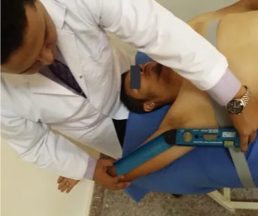
For shoulder internal and external rotation, the starting position for the participant’s arm was abducted 900 and
elbow flexed 900 with the forearm perpendicular on the
plinth, with his palm facing toward his feet without fore-arm pronation or supination and elbow outside plinth, a small pad was used under the humerus to be in level with the acromion. The distal end of humerus, thorax, and clav-icle was stabilized. The axis of measurement was the olec-ranon process, and the ulnar styloid process as the refer-ence point [3, 5], internal rotation shown in Fig. Seven and external rotation is shown in Fig. 8.
Figure 7a: Measuring shoulder internal rotation using electrogoniometer.
Figure 8a: Shoulder lateral rotation Using an electrogoni-ometer.
Figure 8b: Shoulder lateral rotation using a laser goni-ometer.
Statistical methods
Correlation between measures obtained by both devices was done by using Pearson’s correlation coefficient (r); also comparison using paired T-test was done, to test laser go-niometer’s validity about the electro-goniometer as a refer-ence standard.
Intra-rater reliability of the laser goniometer has been measured on repeated measures using intra-class correla-tion coefficient (ICC) value at confidence level (CI) 95%.
RESULTS
One hundred healthy males have shared in the study with mean ± SD ages, weights, heights, and Body mass indices as follows 21.53 ± 2.15 years, 75.03 ± 12.68 kg, 173.81 ± 5.58 cm, and 24.73 ± 3.41 kg/m² respectively, shown in ta-ble 1.
Table 1: Descriptive statistics for the mean age, weight, height and BMI of the study group.
Χ±SD Minimum Maximum Range Age (years) 21.53 ± 2.15 20 30 10 Weight (kg) 75.03 ± 12.68 50 96 46 Height (cm) 173.81 ± 5.58 160 180 20 BMI (kg/m²) 24.73 ± 3.41 18.69 29.65 10.96 Χ : Mean
SD: Standard Deviation
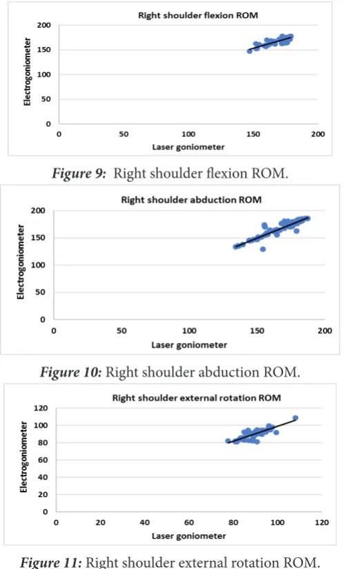
Corresponding to validity, the results of the correlation be-tween the measures of both devices in shoulder range of motions are presented in Figs. 9-16.
Figure 9: Right shoulder flexion ROM.
Figure 10:Right shoulder abduction ROM.
Figure 11: Right shoulder external rotation ROM.
Figure 12: Right shoulder internal rotation ROM.
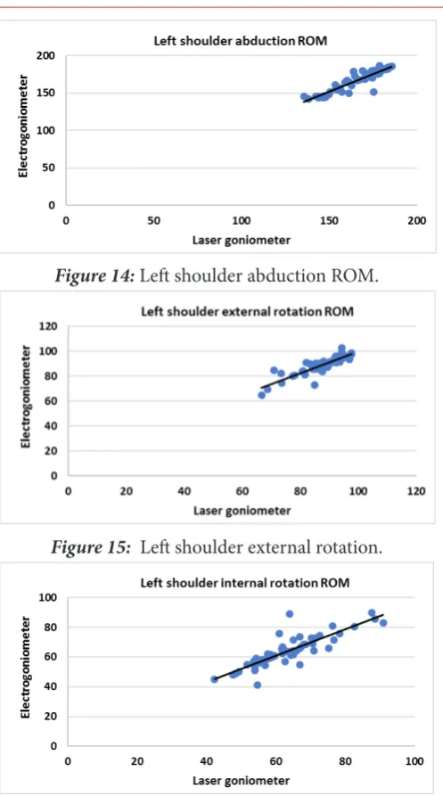
Figure 14:Left shoulder abduction ROM.
Figure 15: Left shoulder external rotation.
Figure 16: Right shoulder internal rotation ROM. The correlation and paired t-test between ROM of the right shoulder measured by laser goniometer and that measured by electro-goniometer were very strong positive significant correlation, with the r-value equals 0.86, 0.93, 0.89, 0.84 and the p values equals 0.36, 0.97, 0.72, and 0.35, for flex-ion, abductflex-ion, internal rotatflex-ion, external rotation respec-tively, with the level of significance set to 0.05, as shown in Table 2.
Table 2: Descriptive analysis, Correlation, and compari-son between shoulder ROM measured by laser
goniome-ter and that measured by electrogoniomegoniome-ter:
Shoulder ROM (degrees)
Laser
go-niometer Electro- go-niometer val-r ue
t- val-ue P-val-ue
Sig- nifi-cance
Χ±SD Χ±SD
Right shoul-der
Flexion 169.52 8.11± 168.26 7.05± 0.86 0.9 0.36 NS
Abduc-tion 166.74 14.77± 166.67 15.82± 0.93 0.02 0.97 NS External
rotation 90.56 5.13± 90.89 5.21± 0.84 -0.35 0.72 NS Internal
rotation 62.67 9.39± 64.36 10.62± 0.89 -0.92 0.35 NS
Left
shoul-der
Flexion 170.7 6.56± 168.86 6.64± 0.84 1.52 0.13 NS
Abduc-tion 167.88 14.15± 168.77 14.41± 0.93 -0.34 0.73 NS External
rotation 87.22 6.86± 88.62 6.83± 0.86 -1.11 0.26 NS Internal
rotation 63.19 9.96± 63.77 10.26± 0.85 -0.31 0.75 NS
Χ: Mean SD: Standard deviation r value: correlation coefficient value
P-value: Probability value NS: Non-significant
The correlation and paired t-test between ROM of the left shoulder measured by laser goniometer and that measured by electro-goniometer were very strong positive significant correlation with the r-value equals 0.84, 0.93, 0.85, 0.86 and the p values equals 0.13, 0.73, 0.26, and 0.75, for flex-ion, abductflex-ion, internal rotatflex-ion, external rotation respec-tively, with the level of significance set to 0.05, as shown in Table 2.
Corresponding to the reliability, laser goniometershowed high intra-examiner reliability in all shoulder ROM mea-surements with intra-class correlation coefficient (ICC) value of 0.98-0.99, as shown in Table 3.
Table 3: ICC for intra-rater reliability of laser goniometer in the measurement of shoulder ROM:
Shoulder ROM Lower ICC bound
(95% CI) Upper bound
Right shoul-der
Flexion 0.99 0.98 0.99
Abduction 0.99 0.99 0.99
External rotation 0.98 0.97 0.98
Internal rotation 0.99 0.99 0.99
Left shoulder
Flexion 0.98 0.98 0.99
Abduction 0.99 0.99 0.99
External rotation 0.98 0.98 0.99
Internal rotation 0.99 0.98 0.99
DISCUSSION
In order to develop and overcome the low validity and reliability of the traditional plastic universal goniometer, many strategies have been taken to develop other types of goniometers. A study carried out by Carey et al. (2010) was done to test reliability and validity of digital goniometer in comparison to universal goniometer and revealed that the digital goniometer could be used as a valid and reliable method of measuring range of motion [8]. Milanese et al. (2014) carried out a study to compare a smartphone ap-plication (the knee goniometer app (ockendon©)) (KGA)
error of measurement values [1].
Brosseau et al. (1997) carried out a study to test the cri-terion validity, intra and inter tater reliability of parallel-ogram goniometers in comparison to universal goniome-ters applied on sixty healthy subjects regarding active knee flexion, the results showed that the parallelogram was valid and reliable as well as the universal goniometer with the advantage of fast and few adjustments’ application of par-allelogram [9].
Valid, objective and reliable ROM measures can be ob-tained by using digital images, radiographs, photocopies, photographs, plumb line, electro-goniometer, and flexom-eter, but all those are not always available to be used in clin-ical or practclin-ical setting [4], other examples of goniometric devices include the inclinometer (also known as the bubble goniometer, gravity goniometer, and pendulum goniome-ter), and video recording equipment. Of these mentioned devices, the inclinometer is the most probably and widely used, since it is portable and is relative to low cost [3]. The validity and reliability of the electro-goniometer for measuring a range of motions of different regions have been studied, and the results of these studies had proved that the electro-goniometer could be used as an objective valid and reliable method in measuring range of movement of differ-ent body regions. One study was carried out by Bronner et al. (2010) comparing the electrogoniometer to digital pro-tractor and motion analysis in measuring range of motion of sagittal plane angular movements of the hip, knee and ankle, the intra-rater correlations and the correlations to the protractor and the correlations of concurrent validity in relation to the motion analysis were all high [10]. A study carried out by Mullaney et al. (2010) to compare a construction grade digital goniometer with the univer-sal goniometer in measuring shoulder joint active-assisted range of motion in 20 patients with unilateral pathology, the results showed that the digital goniometer is highly re-liable but cannot be used interchangeably with the univer-sal goniometer [11].
A study was carried out by Feipel et al. (1999) to establish clinical reference and normal database of active range of motion of cervical spine using electrogoniometer on 250 asymptomatic volunteers cervical and they stated that the results they had obtained agreed with previous observa-tions, which indicated the validity of the methodology used [12].
Another study carried out by Tajali et al. (2016) to test va-lidity, intra and inter tester reliability of two electrogoni-ometers in measuring active range of motion of hand and wrist and passive flexion of proximal interphalangeal joint of index finger in 44 patients with limited motion, the re-sults showed high Intra and inter tester reliability coeffi-cients in measuring active wrist and hand range of motion and passive flexion of proximal interphalangeal joint of index finger in patients with limited range of motion [13]. So in regard to validity, it is believed that the electro-go-niometer is nowadays used as a standardized, valid,
reli-able, and accurate, objective method of measuring range of motion of different joints [1, 4], so it can be used as a reference standard to test and compare the readings and results of measuring range of motion taken by another new measurement tool.
The laser goniometer HALO is a newly arising device de-signed for range of movement measurement, it is featured and characterized by being easy and not consuming time for obtaining the measurement, it just requires to be ap-plied over the region or segment whose range will be mea-sured, the emitting 2 safe low-level laser beams replace the stationary and movable arms of the universal goniometer and then pressing the zero reference button to determine the zero reference point. By holding the device in the same position during the movement, the result can be read from a digital display with a high degree of precision [2].
In our study the range of movement of flexion, abduction, lateral and medial rotation of both shoulders were assessed by the same examiner using the laser goniometer and the electro-goniometer - with standardization of all the mea-surement positions and stabilization in all measures for all subjects - that is to compare the results of measurements of both devices, using the electro-goniometer as the reference standard, to determine the validity and intra-rater reliabil-ity of the laser goniometer.
This study’s results showed no significant difference be-tween the measurements obtained by the laser goniome-ter and those obtained by the electro-goniomegoniome-ter in all the tested shoulder movements, and there was consistency of the readings taken by the laser goniometer. This may be due to the precision and the accuracy and sensitivity of the laser goniometer to any change of the angular displace-ment of the device represented in the shift in the direction of the laser beams.
With regards to shoulder range of motion, the results showed that there is strong to the excellent relationship be-tween the readings of both devices in measuring all shoul-der movements.
Concerning reliability, the laser goniometer showed that it has very high intra-class correlation coefficient values that mean that it has excellent intra-rater reliability on repeated measures for each movement on each side for each subject. This showed that the laser goniometer capable of providing high precision, accuracy, and consistency in measuring the involved movements repeatedly. The factor of long practi-cal experience and training of the examiner and the good standardization each procedure of each measurement help to leading for these high-reliability correlation coefficient values [14].
reliability goniometer with laser beam having the highest reliability scores corresponding to the 2 examiners, also the goniometer with laser beam showed to have high intra-ex-aminer reliability and moderate inter-exintra-ex-aminer reliability while the scores of other modalities were low. They re-ferred this to the advantage that the beam provided by the laser acts as a good reference for the vertical axis of femur that helps to ensure more accuracy of measurement[15].
CONCLUSION
Laser goniometer can be used as a valid and reliable digital objective method of measuring the shoulder range of mo-tions as a standardized alternative for the electro-goniom-eter that physical therapists can rely on in research studies and clinical practice.
REFERENCES
[1] Milanese, S., Gordon, S., Buettner, P., Flavell, C., Rus-ton, S., Coe, D., ... & McCormack, S. Reliability and concurrent validity of knee angle measurement: smart phone app versus universal goniometer used by experienced and novice clinicians. Manual thera-py. 2014;19(6): 569-574.
[2] Sobel, D., Kwiatkowski, J., Ryt, A., Domzal, M., Jedra-siak, K., Janik, L., & Nawrat, A.. Range of Motion Mea-surements Using Motion Capture Data and Augment-ed Reality Visualisation. In International Conference on Computer Vision and Graphics.2014 September; (pp. 594-601). Springer, Cham.
[3] Berryman Reese, N., & Bandy, W. D. Joint range of mo-tion and muscle length testing. Philadelphia, Pa: Saun-ders. 2002;p.11-12.
[4] Hazel, M. C. Musculoskeletal Assessment-Joint
Mo-tion and Muscle Testing. 2013;p.17.
[5] Norkin, C. C., & White, D. J. Measurement of joint motion: a guide to goniometry. FA Davis.2009;p.3-17, p.19-37, p.39-53.
[6] Da Silva Camassuti, P. A., Marcolino, A., Tamanini, G., Barbosa, R. I., Barbosa, A. M., & de Cássia Registro Fonseca, M. Inter-rater, intra-rater and inter-instru-ment reliability of an electrogoniometer to measure wrist range of motion. Hand Therapy. 2001;20(1): 3-10.
[7] Rowe, P. J., Myles, C. M., Hillmann, S. J., & Hazle-wood, M. E. Validation of flexible electrogoniome-try as a measure of joint kinematics. Physiotherapy. 2001;87(9): 479-488.
[8] Carey, M. A., Laird, D. E., Murray, K. A., & Stevenson, J. R. Reliability, validity, and clinical usability of a digi-tal goniometer. Work. 2009;36(1): 55-66.
[9] Brosseau, L., Balmer, S., Tousignant, M., O’Sullivan, J. P., Goudreault, C., Goudreault, M., & Gringras, S. In-tra-and intertester reliability and criterion validity of the parallelogram and universal goniometers for mea-suring maximum active knee flexion and extension of patients with knee restrictions. Archives of physical medicine and rehabilitation. 2001;82(3); 396-402.
[10] Bronner, S., Agraharasamakulam, S., & Ojofeitimi, S.
Reliability and validity of electrogoniometry measure-ment of lower extremity movemeasure-ment. Journal of
medi-cal engineering & technology. 2010;34(3): 232-242.
[11] Mullaney, M. J., McHugh, M. P., Johnson, C. P., &
Ty-ler, T. F. Reliability of shoulder range of motion com-paring a goniometer to a digital level. Physiotherapy theory and practice. 2010;26(5): 327-333.
[12] Feipel, V., Rondelet, B., Le Pallec, J. P., & Rooze,
M. Normal global motion of the cervical spine::
an electrogoniometric study. Clinical
Biomechan-ics. 1999;14(7): 462-470.
[13] Tajali, S. B., MacDermid, J. C., Grewal, R., & Young,
C. Reliability and validity of electro-goniometric range of motion measurements in patients with hand and wrist limitations. The open orthopaedics jour-nal. 2016;10: 190.
[14] Mohsin, F., McGarry, A., & Bowers, R. J. Factors
in-fluencing the reliability of the universal goniometer in measurement of lower-limb range of motion: a litera-ture review. JPO: Journal of Prosthetics and Orthotics. 2015;27(4):140-148.
[15] Choi, B. R., & Kang, S. Y. Intra-and