Comparative Evaluation of the Effect of Different Post and Core Materials on Stress
Distribution in Radicular Dentin by Three-Dimensional Finite Element Analysis
Saied Nokar 1, Mehran Bahrami 2, Azam Sadat Mostafavi 2
1
Associate Professor, Department of Prosthodontics, School of Dentistry, Tehran University of Medical Sciences, Tehran, Iran
2 Assistant Professor, Department of Prosthodontics, School of Dentistry, Tehran University of Medical Sciences, Tehran, Iran
Abstract
Objectives: The aim of this study was to investigate the stress distribution of different post and core materials in radicular dentin by three-dimensional finite element analysis (3D FEA).
Materialsand Methods: Twelve 3D models of a maxillary central incisor were simulated in the ANSYS 5.4 software program. The models were divided into three groups; the first group included: 1-Gold post and core and 2-Nickel-chromium (Ni-Cr) post and core restored with metal-ceramic restorations (MCRs). The second group included: 1-Stainless steel post, 2-Titanium post, 3-Carbon fiber post, 4-Glass fiber post, and 5-Quartz fiber post with composite cores and MCRs. The third group included: 1-Zirconia post and core, 2-Zirconia post, 3-Carbon fiber post, 4-Glass fiber post, and 5-Quartz fiber post; the last four models had composite cores restored with all-ceramic restorations (ACRs). Each specimen was subjected to a compressive load at a 45-degree angle relative to its longitudinal axis at a constant intensity of 100 N. The models were analyzed with regard to the stress distribution in dentin.
Results: Two stress concentration sites were detected in the models. The first group showed the lowest stress levels in the cervical region, while the stress levels detected in the second group were higher than those in the first group and lower than those found in the third group. Fiber-reinforced posts induced a higher stress concentration between the middle and cervical thirds of the root compared to other posts.
Conclusions:According to the results, since cast posts induce lower stresses in dentin, they are recommended for clinical use. Fiber-reinforced posts and ACRs caused the maximum stresses in dentin.
Key words: Finite Element Analysis; Post and Core Technique; Dental Stress Analysis
Journal of Dentistry, Tehran University of Medical Sciences, Tehran, Iran (2018; Vol. 15, No. 2
)
Corresponding author:
A. S. Mostafavi, Department of
Prosthodontics, School of
Dentistry, Tehran University of Medical Sciences, Tehran, Iran
Received: 16 September 2017 Accepted: 22 January 2018
INTRODUCTION
Restoration of endodontically treated teeth is
challenging. Since the time Pierre Fauchard used
gold, silver, or wooden dowels to retain crowns
[1], various types of post-and-core systems have
been introduced to dentistry. Endodontic posts
may be cast with the core, such as gold and
nickel-chromium (Ni-Cr) posts, or they may be
prefabricated, such as titanium and stainless steel
posts. Recently, non-metallic posts such as
fiber-reinforced composite (FRC) and ceramic posts
have been introduced as theoretically acceptable
Fig. 1: Classic tooth preparation for fabricating a cast post-and-core
In some studies, a high-modulus root canal dowel
has been recommended [9-13], while the others
have advocated that the Young’s modulus (E) of
a dowel should preferably be close to that of
dentin [14-16]. Different in-vitro studies have
determined the fracture resistance of the teeth
restored with a dowel under static loading;
however, their results are controversial. These
studies have expressed a lower [17-20], the same
[21-23], or a higher [2,24,25] strength in the teeth
restored with fiber dowels compared to those
restored with metal dowels. One reason for this
contradiction is that in-vitro studies are often
unable to control several clinical variables. In a
finite element analysis (FEA), Yaman et al [10]
expressed that cast gold posts and cores yielded
lower stress values than prefabricated stainless
steel and titanium posts. Some studies pointed to
a lower stress concentration in cast gold posts
compared to FRC posts [26,27], while some
others reported a lower stress concentration in
FRC posts compared to metallic posts [4,5,28].
Chen et al [16] expressed that polyethylene FRC
posts did not significantly change the stress
distribution compared to cast Ni-Cr posts.
Nonetheless, this is still a controversial subject.
The aim of this study was to evaluate common
post materials according to von Mises stress
(VMS) and to report their effect on the stress
distribution in radicular dentin by using
three-dimensional (3D) FEA. According to the null
hypothesis, there would be no significant
statistical differences among the studied post
materials with regard to the stress distribution in
radicular dentin.
MATERIALS AND METHODS
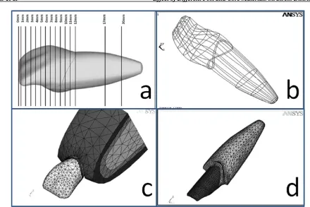
Fig. 2: (a) Twelve cross-sections in the crown, and two cross-sections in the root. (b) The lines, surfaces, and volumes were designed. (c) and (d) The mesh was generated
The maxillary central incisor was selected
because it is a single-rooted tooth with a
relatively simple anatomy, and it is highly
susceptible to fracture. The height of the
remaining dentin was 1.5 mm to create a ferrule
effect (Fig 1). In order to create a ferrule effect
by the core, a 45-degree contra bevel was
prepared around the vertical dentinal walls. In
addition, 4 mm of gutta-percha was retained to
preserve the apical seal.
The studied cores had a 9-mm length and a
4.7-mm diameter. The fabricated posts had a 1.7-4.7-mm
diameter and a 9-mm length. Panavia F 2.0 resin
cement (Kuraray America, Inc., New York, NY,
USA) was used for cementing the cast
post-and-core systems. The film thickness of the cement
was considered to be 67µm.
The models were divided into three groups:
The first group included gold post-and-core and
Ni-Cr post-and-core restored with metal-ceramic
restorations (MCRs). The second group included
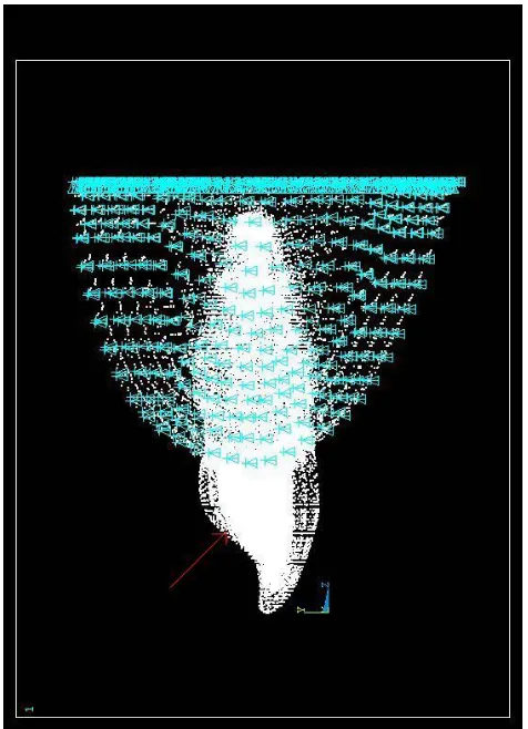
Fig. 3: A 100-N compressive load was applied to a load-bearing area of 1 mm2 on the lingual surface of the tooth
and a transparent acrylic resin was poured around
the tooth so that a polymeric hexahedral matrix
was produced with an artificial tooth in the
center. Twelve cross-sections were made in the
crown (due to the fine details), while two
cross-sections were made in the radicular part of the
model (Fig. 2a). The images of these 14
cross-sections were transferred to the Adobe
Photoshop software program (Adobe Systems
Inc., San Jose, CA, USA) by using a scanner, and
14 key points were chosen on each cross-section.
The position of each key point was determined
according to the three intersecting coordinate
planes of X, Y, and Z (Cartesian coordinates).
The data related to each key point were
transferred to the ANSYS software program, and
a 3D image of the tooth was generated. Next, the
lines, surfaces, and volumes were designed (Fig.
2b and 2c). The same method was used to create
the inner parts of the model such as the post,
cement, and gutta-percha. The gingiva, cancellous
bone, cortical bone, periodontal ligament (PDL),
lamina dura, and crown (metal-ceramic or
all-ceramic) were also simulated for each model. All the
materials, vital tissues, and continual interfaces
between the materials were presumed elastic,
homogenous, and isotropic. The mechanical
properties (Young’s modulus and Poisson’s ratio) of
each of the components used in this study are
summarized in Table 1 [9,11,27,29-32]. During the
meshing, the volumes were divided into smaller
parts named elements. Each element consisted of
eight nodes (a hexahedral element). The elements
were connected to each other at their nodes (Fig. 2c
and 2d). In this study, the finite element meshes
were composed of nearly 4300 elements and 6000
nodes.
Table 1. Young's modulus (E) and Poisson’s ratio (υ) of the materials in the present study
PDL=Periodontal ligament, Ni-Cr=Nickel-Chromium
Materials Young's modulus
(MN/m2)
Poisson’s ratio
Enamel 41E9 0.31
Dentin 18.6E9 0.30
PDL 68.9E6 0.45
Cortical bone 13.7E9 0.30
Cancellous bone 1.37E9 0.30
Gingiva 19.06E6 0.30
Gutta-percha 0.69E6 0.45
Porcelain 69E9 0.28
Stainless steel post 200E9 0.33
Gold post 88E9 0.35
Gold alloy coping 77E9 0.35
Quartz fiber 18.7E9 0.30
Carbon fiber 21E9 0.31
Glass fiber 40E9 0.26
Zirconia 200E9 0.33
Ni-Cr 200E9 0.33
Titanium 112E9 0.33
Composite core 12E9 0.30
IPS Empress II 96E9 0.25
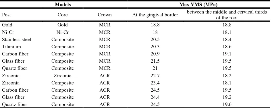
Table 2. Maximum von Mises stress (VMS) in the models
Ni-Cr=Nickel-Chromium, MCR=Metal-Ceramic Restoration, ACR=All-Ceramic Restoration
A compressive load with a constant intensity of
100 N was applied to a load-bearing area of
1 mm
2on the lingual surface of the tooth at an
angle of 45 degrees relative to the longitudinal
axis of the tooth in order to simulate a centric
occlusal contact with the opposite tooth (Fig 3).
Finally, the models were analyzed with regard to
the stress distribution in dentin.
RESULTS
In this FEA, VMS (equivalent stress) was
considered because it has a higher validity than
stress analysis. This parameter is shown by δe
and is obtained from the following formula:
δe = (1/2[(δ1 – δ2)2+ (δ2 - δ3)2+ (δ3 - δ1)2] )
½δ1, δ2, and δ3 are the principal stress components.
The VMS shows the location of the maximum stress
without determining its nature (either tensile or
compressive) [33]; therefore, it is useful in the
experiments which only determine the existence of
stress, similar to the current study.
In all the models, stress concentration was detected
at two areas of the root:
1- The junction of the middle and cervical thirds
of the root.
2- The cervical part of the root.
The results are presented as the maximum VMS
values in Table 2. Of course, The VMS is present
in all the components, but only radicular dentin
stresses are reported in this study. A convenient
way of reporting the VMS is a color representation
of the stress distribution.
DISCUSSION
In the current 3D FEA, the VMS of common post
materials was evaluated. The null hypothesis was
rejected. According to the results, in all the models,
two stress concentration regions were identified: 1)
the cervical region of the root, which was covered
with the cervical edges of the crown, and 2) between
the middle and cervical thirds of the root, where the
cortical bone comes to an end on the root. In both
regions, compressive stresses concentrated on the
buccal side, while tensile stresses concentrated on
the palatal side of the studied models. Several
studies have reported the cervical region of the root
as a stress concentration site [4,11,12,34,35]. Assif
and Gorfil [35] stated that this area is the interface
between materials with different Young's modulus
values.
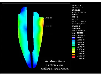
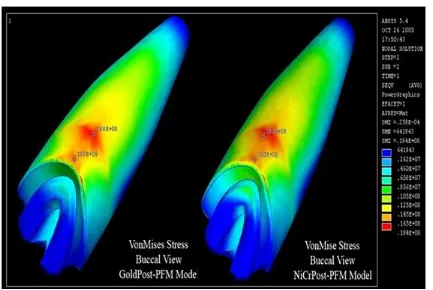
In the first group, stress levels in each model
were similar at both stress concentration regions.
However, Ni-Cr posts showed lower stress levels
compared to gold posts (Fig 4).
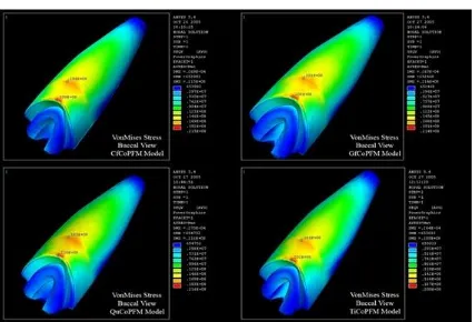
In the second group, stainless steel and titanium
posts showed lower stresses in dentin compared
to FRC posts. In all the five models of this group,
the VMS values in the cervical region of the root
Models Max VMS (MPa)
Post Core Crown At the gingival border between the middle and cervical thirds
of the root
Gold Gold MCR 18.8 18.8
Ni-Cr Ni-Cr MCR 18 18.1
Stainless steel Composite MCR 20.5 18.4
Titanium Composite MCR 20.3 18.6
Carbon fiber Composite MCR 20.9 19.1
Glass fiber Composite MCR 21.5 19.5
Quartz fiber Composite MCR 21 19.5
Zirconia Zirconia ACR 22.7 18.2
Zirconia Composite ACR 23.4 18.1
Carbon fiber Composite ACR 24.5 19.5
Glass fiber Composite ACR 24.4 19.2
Fig. 4: Maximum von Mises stress (VMS) in gold and-core (GoldPost-MCR) and nickel-chromium (Ni-Cr) post-and-core (NiCrPost-MCR) restored with metal-ceramic restorations (MCRs)
were higher than those between the middle and
cervical thirds of the root (Fig. 5). Yang et al [36]
reported the same results.
In the third group, cervical stresses were higher
compared to the other groups. The least amount of
stress was detected in the model with a zirconia
post-and-core (Fig. 6); this has also been confirmed by
similar studies [10,11,34]. Assmusen et al [11] and
Toksavul et al [34] reported lower stress levels for
zirconia post-and-core compared to titanium posts.
Therefore, zirconia post-and-core systems may be
an alternative to metallic posts.
According to the results of the present study, cast
post-and-core systems showed a more favorable
stress distribution pattern as they induced a lower
VMS in radicular dentin, especially in the cervical
region of the root. They also induced almost the
same stress levels in both stress concentration areas.
Among the prefabricated post models, stainless
steel, titanium, and zirconia posts demonstrated
nearly the same stress levels between the middle
and cervical thirds of the root. However, these
three posts showed lower levels of VMS between
the middle and cervical thirds of the root in
comparison with FRC posts (Fig. 6).
Cervical stresses in the models restored with
ACRs were significantly higher than those in the
models restored with MCRs because IPS
Empress II is stiffer than MCR [28,37]. This
finding was similar to the results of the studies by
Pegoretti et al [28] and Eskitaşcioğlu et al [37].
In the present research, the zirconia post-and-core
system showed a more favorable stress distribution
pattern than the zirconia post-composite core, which
confirms the results found by Heydecke et al [38]
and Butz et al [39].
Cast metal post-and-core systems caused lower
levels of stress compared to prefabricated metallic
posts, similar to the results of the study by Yaman et
al [40]. In addition, the findings of the current study
confirmed that an increase in the Young's modulus
of the dowels reduces dentinal stresses.
Some FEA
Fig. 5: Maximum von Mises stress (VMS) in carbon fiber post (CfCoMCR), glass fiber post (GfCoMCR), quartz fiber post (QuCoMCR), and titanium post (TiCoMCR) with composite cores and metal-ceramic restorations (MCRs)
Different mechanical properties (especially for
FRCs), different modeling techniques, use of 2D
or 3D FEA, different forces and directions of
load application are some of the factors which
may have affected the results of these studies.
Most mechanical experiments have recorded a
higher fracture threshold for metallic posts
compared to FRC posts; however, it has also
been explained that prefabricated FRC posts
show more favorable fractures in comparison
with metallic posts [9,17,19,40,41].
Ferrari et al [6] reported that the teeth restored
with carbon FRC posts had a significantly higher
survival rate after 4 years than the teeth restored
with metal posts. In a clinical trial designed by
King et al [42], the teeth restored with carbon
FRC posts did not perform as well as
conventional wrought precious alloy posts.
However, clinical studies comparing fiber
dowels with metal dowels are scarce. Heydecke
et al [38] and Butz et al [39] reported that zirconia
post-and-core systems could be used as an
alternative to metal posts; however, the survival
rate of zirconia posts/composite cores was lower
than that of cast posts. In the study by Dilmener
et al [43], cast metal post-and-core systems were
found to be more
fracture resistant
than zirconia
posts/composite cores. According to Fraga et al
[44], cast Ni-Cr post-and-core systems showed a
significantly higher resistance to fracture than
prefabricated stainless steel posts. Dilmener et al
[43] found the same results. In a research by
Barjau Escribano et al [29], a significantly lower
failure load was found for the teeth restored with
stainless steel posts compared to those restored
with glass fiber posts. According to Asmussen et
al [11] and Toksavul et al [34], zirconia ceramic
posts created less stress concentration in dentin
than glass FRC and titanium posts. In the present
study, cast metal posts showed a more favorable stress
distribution pattern than the other posts. Nevertheless,
supplementary clinical studies are required to further
evaluate the properties of FRC posts.
CONCLUSION
Within the limitations of this theoretical FEA, the
following conclusions were drawn:
1- In all the models, two sites of stress concentration
appeared: at the cervical edge of the root, and
between the middle and cervical thirds of the root.
2- Metallic cast posts showed the least amount of
stress concentration.
3- The models reconstructed with MCRs showed
higher stresses in the cervical region of the root.
These stresses increased in the models restored
with ACRs.
4- Stainless steel, titanium, and ceramic posts
induced a more favorable stress distribution
pattern in comparison with FRC posts.
5- Among the models, FRC posts showed higher
stress levels in the area between the middle and
cervical thirds of the root.
The findings of the current study may help the
clinicians to select the most suitable
post-and-core systems according to the clinical status of
each tooth. Of course, additional clinical
investigations are required to verify these
theoretical in-vitro results.
REFERENCES
1- Ingle JI, Taintor JFJ. Endodontics. 3rd ed. Lea &
Febiger, Philadelphia, 1985:46-52.
2- Akkayan B, Gülmez T. Resistance to fracture of endodontically treated teeth restored with different post systems. J Prosthet Dent. 2002 Apr;87(4):431-7. 3- Zhang Y, Lu Z, Wang K. [Fracture strength of custom-fabricated celay all-ceramic post and core]. [Article in Chinese]. Hua Xi Kou Qiang Yi Xue Za Zhi. 2002 Feb;20(1):39-41, 44.
4- de Castro Albuquerque R, Polleto LT, Fontana RH, Cimini CA. Stress analysis of an upper central incisor restored with different posts. J Oral Rehabil. 2003 Sep;30(9):936-43.
5- Lanza A, Aversa R, Rengo S, Apicella D, Apicella A. 3D FEA of cemented steel, glass and carbon posts in a maxillary incisor. Dent Mater. 2005 Aug;21(8):709-15. 6- Ferrari M, Vichi A, Garcia-Godoy F. Clinical evaluation of fiber-reinforced epoxy resin posts and cast post and cores. Am J Dent. 2000 May;13(Spec No):15B-18B.
7- Hassan-Ahangari A, Geramy A, Valian A. Ferrule Designs and Stress Distribution in Endodontically Treated Upper Central Incisors: 3D Finite Element Analysis. J Dent (Tehran). 2008 Feb;5(3):105-110. 8- Geramy A, Eghbal MJ, Ehsani S. Stress distribution changes after root canal therapy in canine model: a finite element study. Iran Endod J. 2008 Fall;3(4):113-8.
9- Asmussen E, Peutzfeldt A, Heitmann T. Stiffness, elastic limit, and strength of newer types of endodontic posts. J Dent. 1999 May;27(4):275-8. 10- Yaman SD, Karacaer O, Sahin M. Stress distribution of post-core applications in maxillary central incisors. J Biomater Appl. 2004 Jan;18(3):163-77.
11- Asmussen E, Peutzfeldt A, Sahafi A. Finite element analysis of stresses in endodontically treated, dowel-restored teeth. J Prosthet Dent. 2005 Oct;94(4):321-9.
12- Ho MH, Lee SY, Chen HH, Lee MC. Three-dimensional finite element analysis of the effects of posts on stress distribution in dentin. J Prosthet Dent. 1994 Oct;72(4):367-72.
14- Assif D, Oren E, Marshak BL, Aviv I.
Photoelastic analysis of stress transfer by
endodontically treated teeth to the supporting structure using different restorative techniques. J Prosthet Dent. 1989 May;61(5):535-43.
15- Isidor F, Odman P, Brøndum K. Intermittent loading of teeth restored using prefabricated carbon fiber posts. Int J Prosthodont. 1996 Mar-Apr;9(2):131-6.
16- Chen XT, Li XN, Guan ZQ, Liu XG, Gu YX. [Effects of post material on stress distribution in dentine]. [Article in Chinese]. Zhonghua Kou Qiang Yi Xue Za Zhi. 2004 Jul;39(4):302-5.
17- Fokkinga WA, Kreulen CM, Vallittu PK, Creugers NH. A structured analysis of in vitro failure loads and failure modes of fiber, metal and ceramic post-and-core systems. Int J Prosthodont. 2004 Jul-Aug;17(4):476-82.
18- Martínez-Insua A, da Silva L, Rilo B, Santana U. Comparison of the fracture resistances of pulpless teeth restored with a cast post and core or carbon fiber post with a composite core. J Prosthet Dent. 1998 Nov;80(5):527-32.
19- Sirimai S, Riis DN, Morgano SM. An in vitro study of the fracture resistance and the incidence of vertical root fracture of pulpless teeth restored with six post-and-core systems. J Prosthet Dent. 1999 Mar;81(3):262-9.
20- Sidoli GE, King PA, Setchell DJ. An in vitro evaluation of a carbon fiber-based post and core system. J Prosthet Dent. 1997 Jul;78(1):5-9.
21- Drummond JL. In vitro evaluation of endodontic posts. Am J Dent. 2000 May;13(Spec No):5B-8B. 22- Raygot CG, Chai J, Jameson DL. Fracture
resistance and primary failure mode of
endodontically treated teeth restored with a carbon fiber-reinforced resin post sys-tem in vitro. Int J Prosthodont. 2001 Mar-Apr;14(2):141-5.
23- McDonald AV, King PA, Setchell DJ. In vitro study to compare impact fracture resistance of intact root-treated teeth. Int Endod J. 1990 Nov;23(6):304-12. 24- King PA, Setchell DJ. An in vitro evaluation of a prototype CFRC prefabricated post developed for the restoration of pulpless teeth. J Oral Rehabil. 1990 Nov;17(6):599-609.
25- Duret B, Duret F, Reynaud M. Long-life physical property preservation and post endodontic rehabilitation with the Composipost. Compend Contin Educ Dent Suppl. 1996 Jan;17(Suppl 20):S50-6.
26- Yang HS, Lang LA, Molina A, Felton DA. The effects of dowel design and load direction on dowel-and-core restorations. J Prosthet Dent. 2001 Jun;85(6):558-67. 27- Pierrisnard L, Bohin F, Renault P, Barquins M. Corono-radicular reconstruction of pulpless teeth: A mechanical study using finite element analysis. J Prosthet Dent. 2002 Oct;88(4):442-8.
28- Pegoretti A, Fambri L, Zappini G, Bianchetti M. Finite element analysis of a glass fiber reinforced composite endodontic post. Biomaterials. 2002 Jul;23(13):2667-2682.
29- Barjau Escribano A, Sancho Bru JL, Forner Navarro L, Rodríguez Cervantes PJ, Pérez Gónzález A, Sánchez Marín FT. Influence of prefabricated post material on restored teeth: fracture strength and stress distribution. Oper Dent. 2006 Jan-Feb;31(1):47-54. 30- Ko CC, Chu CS, Chuag KH, Lee MC. Effects of posts on dentin stress distribution in pulpless teeth. J Prosthet Dent. 1992 Sep;68(3):421-7.
31- Reinhardt RA, Krejci RF, Pao YC, Stannard JG. Dentin stresses in post-reconstructed teeth with diminishing bone support. J Dent Res. 1983 Sep;62(9): 1002-8.
32- Holmes DC, Diaz-Arnold AM, Leary JM. Influence of post dimension on stress distribution in dentin. J Prosthet Dent. 1996 Feb;75(2):140-7. 33- Digital Engineering. Stress in FEA: Part 3. Available at: http://www.digitaleng.news/de/stress-in-fea-part-3/Accessed July 1, 2016.
34- Toksavul S, Zor M, Toman M, Güngör MA, Nergiz I, Artunç C. Analysis of dentin-al stress distribution of maxillary central incisors subjected to various post-and-core applications. Oper Dent. 2006 Jan-Feb;31(1):89-96.
35- Assif D, Gorfil C. Biomechanical considerations in restoring endodontically treated teeth. J Prosthet Dent. 1994 Jun;71(6):565-7.
two post core systems using two different methods (fracture strength test and a finite elemental stress analysis). J Endod. 2002 Sep;28(9):629-33.
38- Heydecke G, Butz F, Hussein A, Strub JR. Fracture strength after dynamic loading of endodontically treated teeth restored with different post-and-core systems. J Prosthet Dent. 2002 Apr;87(4):438-45.
39- Butz F, Lennon AM, Heydecke G, Strub JR. Survival rate and fracture strength of endodontically treated maxillary incisors with moderate defects restored with different post-and-core systems: an in vitro study. Int J Prosthodont. 2001 Jan-Feb;14(1):58-64.
40- Yaman SD, Alaçam T, Yaman Y. Analysis of stress distribution in a maxillary central incisor subjected to various post and core applications. J Endod. 1998 Feb;24(2):107-11.
41- Bolhuis P, de Gee A, Feilzer A. Influence of fatigue loading on four post-and-core systems in maxillary premolars. Quintessence Int. 2004 Sep;35(8):657-67. 42- King PA, Setchell DJ, Rees JS. Clinical evaluation of a carbon fiber reinforced car-bon endodontic post. J Oral Rehabil. 2003 Aug;30(8):785-9.
43- Dilmener FT, Sipahi C, Dalkiz M. Resistance of three new esthetic post-and-core systems to compressive loading. J Prosthet Dent. 2006 Feb;95(2):130-6.