R E S E A R C H A R T I C L E
Open Access
Diminution of pharyngeal segmentation
and the evolution of the amniotes
Subathra Poopalasundaram
1, Jo Richardson
1, Annabelle Scott
1, Alex Donovan
2, Karen Liu
2and Anthony Graham
1*Abstract
Background:The pharyngeal arches are a series of bulges found on the lateral surface of the head of vertebrate embryos, and it is within these segments that components of the later anatomy are laid down. In most vertebrates, the post-otic pharyngeal arches will form the branchial apparatus, while in amniotes these segments are believed to generate the larynx. It has been unclear how the development of these segments has been altered with the emergence of the amniotes.
Results:In this study, we examined the development of pharyngeal arches in amniotes and show that the post-otic pharyngeal arches in this clade are greatly diminished. We find that the post-post-otic segments do not undergo myogenesis or skeletogenesis, but are remodelled before these processes occur. We also find that nested DLX expression, which is a feature of all the pharyngeal arches in anamniotes, is associated with the anterior segments but less so with the posterior arches in amniotes. We further show that the posterior arches of the mouse embryo fail to properly delineate, which demonstrates the lack of function of these posterior segments in later
development.
Conclusion:In amniotes, there has been a loss of the ancestral“branchial”developmental programme that is a general feature of gnathostomes; myogenesis and skeletogenesis This is likely to have facilitated the emergence of the larynx as a new structure not constrained by the segmental organisation of the posterior pharyngeal region.
Keywords:Pharyngeal segmentation, Pharyngeal pouch, Pharyngeal arch, DLX, Amniote evolution, Larynx
Background
The development of the pharyngeal arches is underpinned by the generation of the pharyngeal pouches, outpocket-ings of the endoderm, which contact the overlying ecto-derm, with the other constituents of the arches, the neural crest and mesoderm, migrating into these preformed seg-mental units [1]. These embryonic populations differenti-ate to form a range of derivatives: the neural crest gives rise to the skeletal and connective tissues, the mesoderm to the musculature and the blood vessels, the ectoderm to the epidermis and sensory neurons and the endoderm the lining of the pharynx and a range of specialised organs. Thus, the arches constitute an iterated series with each forming the same components. In many vertebrate clades, this embryonic segmental organisation is translated into
the later functional anatomy and is evident in the serial ar-rangement of the gill-bearing branchial arches.
In post-metamorphic amphibia and in amniotes, how-ever, the branchial skeleton is lost and the larynx develops
[2,3]. This structure is a feature of tetrapods that connects
the pharynx with the trachea and plays an essential role in facilitating life on land. The larynx consists of skeletal ele-ments, which include the cricoid and paired arytenoids in most tetrapods as well as, in mammals, the thyroid, and associated muscular and connective tissue elements [2]. Developmentally, the components of the larynx are thought to have their origins in the posterior, post-otic, pharyngeal arches and thus to be equivalent to the deriva-tives of the posterior arches in other vertebrates, the bran-chial apparatus [2,4]. Indeed, it is generally assumed that there is a correspondence between the mature structures of the larynx: skeleton, muscle and nerves, and the embry-onic pharyngeal segments, and such a relationship is a staple of embryology and anatomy textbooks [4–9].
* Correspondence:[email protected]
1Centre for Developmental Neurobiology, King’s College London, London,
UK
Full list of author information is available at the end of the article
Yet, there have been few studies focussed on the devel-opment of the posterior arches and it is unclear whether such a correspondence exists or how the development of the posterior arches was modified to allow for the emer-gence of the larynx. Moreover, the situation in amniotes is more complex than that in other vertebrates, since the posterior pharynx is segmentally organised for only a tran-sient period. Once all the arches have been formed, the second arch expands disproportionately to cover the pos-terior arches before fusing and enclosing them [10].
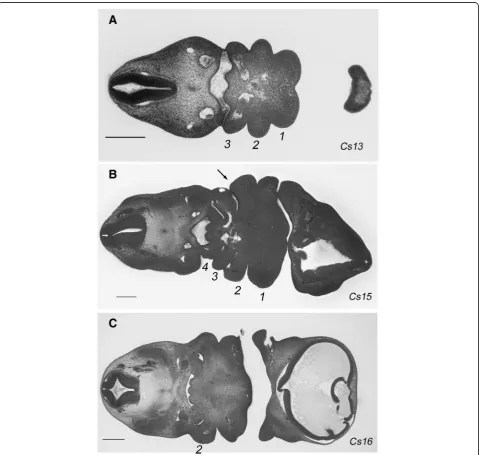
Figure1shows an overview of this process in human em-bryos. At Carnegie stage 13, the three most anterior arches have formed (Fig. 1a), while by stage 15 an add-itional fourth arch has formed (Fig. 1b). Thus, at these stages the segmental nature of the pharynx is readily ap-parent. However, by stage 16 the second arch has ex-panded caudally, covered and subsumed the posterior arches and thus the segmental nature of the pharyngeal region is lost (Fig.1c). Consequently, the segmental nature of the posterior pharynx becomes lost.
In this study, we assessed the early development of the posterior pharyngeal arches and their subsequent envelop-ment by the second arch. We further docuenvelop-ment the rela-tionship between the pharyngeal segments and the formation of the muscular and skeletal derivatives. We find that while there is a clear correspondence between these events and the anterior arches, this is not the case for the posterior segments. Thus, while the processes of myogenesis and chondrogenesis are underway at several sites in the embryo, they are not a feature of the posterior pharyngeal region. We further show that, although the an-terior pharyngeal segments exhibit nested DLX gene ex-pression, the expression of these genes is greatly reduced in the posterior arches, suggesting that the need to region-alise the neural crest along the proximodistal axis in these segments is less in amniotes. We note that the reduction inDLXexpression is more extensive in the mouse than in the chick, and this prompted us to further investigate dif-ferences between the posterior segments in these species. Interestingly, we find that while the 4th pharyngeal pouch contacts the ectoderm in chick and human embryos, it does not do so in mice. Consequently, the posterior pharyngeal segments are not fully delineated in this spe-cies, which further underscores their lack of significance for later events.
Results
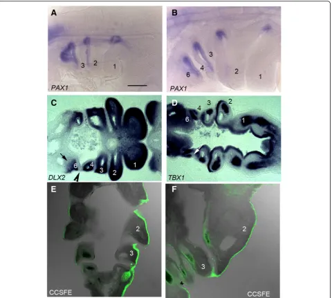
We conducted a detailed analysis of the period covering the formation of the post-otic pharyngeal segments and their subsequent envelopment by the second arch in chick. At stage 17 (HH17), PAX1 staining highlights the formed, and forming, pharyngeal pouches and it is appar-ent that the first three arches are delineated, but that the more posterior segments are not clearly defined (Fig.2a). By HH21, however, the full complement of four pouches and five arches—numbered 1,2,3,4 and 6—have formed (Fig.2b). In amniotes, the most posterior pharyngeal arch is termed the sixth, even though this is numerically the fifth arch, due to the long held, but erroneous, belief that a transient fifth arch formed between this segment and the fourth [11]. We have also used DLX2and TBX1 ex-pression to highlight the distribution of the different em-bryonic populations that contribute to the arches.DLX2is expressed by the neural crest cells and it can be seen in all the arches (Fig. 2c). However, significantly, this staining highlights the fact that while the anterior of the 6th arch has a distinct anterior boundary, the point where the fourth pouch contacts the ectoderm, it does not have a de-fined caudal limit (Fig. 2c). TBX1 expression labels the pharyngeal endoderm, including the pouches, as well as the mesodermal components of the first four arches (Fig. 2d). As development progresses the caudal edge of the second arch expands to cover the more posterior arches. This is shown in embryos in which the ectoderm has been
labelled with a cell tracker, CCSFE. At HH20 the second arch is enlarging and that the point of expansion is at the interface between the ectoderm, which is labelled, and the endoderm (Fig. 2e). This process continues as the 2nd arch increases greatly in size and overhangs the posterior segments (Fig. 2f ). Thus, in amniotes there is an earlier segmental phase of pharyngeal development and a later post-segmental phase.
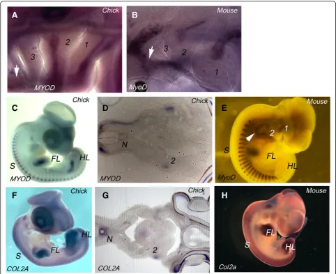
It has been widely documented that in anamniotes myogenesis and skeletogenesis occur within the segmen-tal framework of the pharyngeal arches [12–14]. We therefore sought to determine if there is a relationship between muscular and skeletal differentiation and the segmental organisation of the arches in amniotes, and we used cell type specific markers on both chick and mouse embryos, as representatives of reptiles and mam-mals respectively. To analyse the emerging muscle of the arches, we used MYOD staining. In both HH21 chick and Theiler stage (TS) 16 mouse embryos, where the full cohort of arches have formed, we find that muscle differ-entiation is associated with arches 1 and 2, but not with the more posterior arches (Fig. 3a, b). However, other muscle populations are differentiating in the head, most notably the hypoglossal musculature which migrates from the occipital somites into the ventral pharyngeal region (Fig. 3a, b). We found no expression of collagen
IIA (COL2A), which is a definitive cartilage marker, at any site in the pharyngeal arches at these segmental stages in either chick or mouse (data not shown).
myogenesis or skeletogenesis during their period of definition.
We further assessed the degree to which the pharyngeal arches are regionalised as they develop. The patterning of the neural crest and its skeletal derivatives along the prox-imodistal axis of the arches involves the nested expression of members of the DLX family of transcription factors.
DLXgenes are organised as linked pairs—DLX1/2,DLX3/
DLXpairs in both chick and mouse embryos. In chick, we find that, as for other gnathostomes,DLX2is expressed at high levels throughout the ectomesenchyme all along the proximodistal axis of all the arches (Fig. 4a, b). However, the expression patterns ofDLX6andDLX3 differed from
shows a more restricted expression; again, this gene is strongly expressed in the first two arches but arches 3, 4 and 6 show little expression (Fig.4g, h). The situation in mouse is similar in that Dlx2, Dlx6 and Dlx3 show nested expression in the first two arches, but in this species we find that for all these genes, including
Dlx2, there is little expression in the posterior arches (Figu. 4c, f and i). Similar results have been found from other studies using alternative approaches with
Dlx1/2-cre/rosa 26 and Dlx5–6 cre/rosa26 crosses shows lacZ labelling in the first two arches but not in the posterior arches [19, 20].
Fig. 4DLX expression in the pharyngeal arches in chick and mouse embryos.aSide view of a HH21 chick embryo showingDLX2expression throughout the dorsoventral extent of all the pharyngeal arches.bLongitudinal section through the arches showingDLX2expression in the ectomesenchyme of the arches.cSide view of a TS16 mouse embryo showing pronounced expression ofDlx2in the first two arches, but much reduced expression in the posterior arches.dSide view of a HH21 chick embryo showingDLX6expression in the mid region of the three most anterior arches but not the most posterior arches.eLongitudinal section through the arches showingDLX6expression in the ectomesenchyme of the three most anterior arches.fSide view of a TS16 mouse embryo showing high levels ofDlx6in the mid region of the first two arches, but much reduced expression in the posterior arches.gSide view of a HH21 chick embryo showingDLX3expression in the more distal region of the two most anterior arches but not those lying posteriorly.hLongitudinal section through the arches showingDLX3expression in the
These differences in DLX expression in the posterior arches between chick and mouse prompted us to further analyse the underlying organisation of the arches. Cen-tral to the organisation of the pharyngeal arches is the establishment of the contact between the pharyngeal pouches and the overlying ectoderm. This serves to de-fine the anterior and posterior limits of the arches and to segregate the mesenchymal populations of the differ-ent arches. We have previously documdiffer-ented the forma-tion of the pharyngeal pouches and arches in detail in chick [21] and we therefore assessed if the topology of the mouse pouches and arches were the same. Notably, we find that in mouse the fourth pouch does not contact the ectoderm and thus neither establishes a clearly
mice. We examined HREM data from the four Carnegie stage (CS)15 human embryos catalogued on the DMDD website; this is the stage in humans at which all the arches have formed. In each we noted that the fourth pouch could be seen to contact the ectoderm (Fig.5b). This same topology was also found in histological sections of a CS15 embryo archived on the Virtual Human embryo database (https://www.prenatalorigins.org/virtual-human-embryo/ stage.php?stage=15). Thus, the mouse represents a test of whether the failure to fully establish the posterior pharyngeal segments has any impact on later anatomy; clearly, it does not, and the larynx forms.
Conclusions
A key observation from this study is that the development of the posterior pharyngeal arches in amniotes is markedly different from that of other vertebrate clades. In ana-mniotes, the posterior arches generate muscular and skel-etal structures, exhibit nested expression of DLX linked pairs and these segments form the blueprint for the organ-isation of the branchial apparatus [12, 15–18, 22]. We show here that in chick and mouse the posterior arches do not undergo myogenesis or skeletogenesis while they exist as morphologically discernible entities, and that they exhibit reduced nested expression of theDLXgenes; there has been a suppression of the inner fish here. Conse-quently, the segmental organisation of the posterior em-bryonic pharynx has little significance for the later musculoskeletal anatomy; a point underscored by the fact that, in mouse, the posterior arches fail to fully emerge. Thus, these results lay bare the fact that the correspond-ence between the pharyngeal arches and the components of the larynx, invariably found in anatomy and embryology textbooks, is but supposition [4,6–9].
Finally, it should also be noted that while the skeletal components of the branchial apparatus of anamniotes are generally neural crest derived [23,24], this is not the case for the larynx. Fate mapping studies in birds have demon-strated that the laryngeal cartilages, the cricoid and aryt-enoid, arise from the lateral plate mesoderm [25]. Similarly, a recent study in mouse has also shown a meso-dermal origin for the cricoid and arytenoid cartilages [26]. However, the thyroid cartilage, which is a mammalian novelty, seems to have a more complex origin comprising both a crest derived and a mesodermal component [26]. Thus, the evolution of the larynx cannot have involved a transformation of the developmental programme that would have directed the generation of the branchial appar-atus, but rather the larynx emerges as a new structure.
Materials and methods
Embryo staging
Chick embryos were staged according to Hamburger and Hamilton [27] and mice according to Theiler [28]. The
following mouse lines were used: Wnt1-cre driver: Tg (Wnt1-cre)11Rth [29] and reporter line: R26RmT/mG: GT (Rosa)26Sortm4(ACTB-tdTomato-EGFP) [30]. All animal work was performed in accordance with UK Home Office Regulations. The HREM data from the human em-bryos is from the DMDD website (https://dmdd.org.uk/). Data from Deciphering the Mechanisms of Developmental Disorders (https://dmdd.org.uk/), a programme funded by the Wellcome Trust with support from the Francis Crick Institute, is licensed under a Creative Commons Attribu-tion licence.
In situ hybridisation
Embryos were fixed in in 3.7% (v/v) formaldehyde in PBS and in situ hybridisation carried out as previously described [31]. Gene expression was either examined as whole mounts or embryos were embedded in 20% (w/v) gelatin in PBS and sectioned using a vibratome.
CCSFE labelling
CCSFE labelling was carried out as previously described [10].
Acknowledgments
We would like to thank Dr. Tom Mohun, Crick Institute London, for help with the HREM analysis of the Human and mouse embryos. We thank Caroline Formstone, Clemens Kiecker, Malcolm Logan and Jill Sales for comments on the text.
Funding
This work was funded by a BBSRC grant (BB/R006199/1) to AG.
Availability of data and materials
All data and material are available from the corresponding author. [email protected].
Authors’contributions
The project was conceived by AG. The experimental work was carried out by SP, JR, AS, AD, KL and AG. The paper was written by AG and SP with JR, AS, AD and KL commenting and modifying the draft. All authors read and approved the final manuscript.
Ethics approval and consent to participate
All embryo collection was carried out as prescribed by the UK Animals (Scientific Procedures) Act 1986.
Consent for publication All authors consent to publication.
Competing interests
The authors declare that they have no competing interests.
Publisher’s Note
Springer Nature remains neutral with regard to jurisdictional claims in published maps and institutional affiliations.
Author details
1Centre for Developmental Neurobiology, King’s College London, London,
UK.2Centre for Craniofacial and Regenerative Biology, King’s College London,
Received: 23 November 2018 Accepted: 5 February 2019
References
1. Graham A, Richardson J. Developmental and evolutionary origins of the pharyngeal apparatus. EvoDevo. 2012;3:24.
2. Goodrich E. Studies on the structure and development of vertebrates. London: MacMillan; 1930. p. 387.
3. Romer A. The vertebrate body- shorter version. 4th ed. Philadelphia London Toronto: W. B. Saunders Company; 1971. 452 p.
4. Kardong KV. Vertebrates : comparative anatomy, function, evolution. 6th ed. New York: McGraw-Hill; 2012. xix, 794 p. p.
5. Standring S. Gray's Anatomy. 40th ed: Churchill Livingstone; 2008. 1576 p. 6. Schoenwolf G. Larsen's human Emrbyology: Churchill Livingstone; 2014. 576 p. 7. Standring S. Gray’s anatomy : the anatomical basis of clinical practice
Forty-first edition. ed. xviii, 1562 pages p.
8. Sadler TW, Leland J, Sadler-Redmond SL, Tosney K, Chescheir N, Imseis H, et al. Langman’s medical embryology. 12th ed. Philadelphia, Pa. ; London: Wolters Kluwer/Lippincott Williams & Wilkins; 2012. xiii, 384 p. p. 9. Carlson BM. Human embryology and Dev Biol 4th ed. Philadelphia: Mosby/
Elsevier; 2009. xvi, 541 p. p.
10. Richardson J, Shono T, Okabe M, Graham A. The presence of an embryonic opercular flap in amniotes. Proc R Soc B Biol Sci. 2012;279(1727):224–9. 11. Shone V, Oulion S, Casane D, Laurenti P, Graham A. Mode of reduction in
the number of pharyngeal segments within the sarcopterygians. Zoological Lett. 2016;2:6.
12. Schilling T, Kimmel C. Musculoskeletal patterning in the pharyngeal segments of the zebrafish embryo. Development. 1997;124(15):2945–60. 13. Ziermann JM, Olsson L. Patterns of spatial and temporal cranial muscle
development in the African clawed frog, Xenopus laevis (Anura: Pipidae). J Morphol. 2007;268(9):791–804.
14. Kerney R, Gross JB, Hanken J. Runx2 is essential for larval hyobranchial cartilage formation in Xenopus laevis. Dev Dyn. 2007;236(6):1650–62. 15. Compagnucci C, Debiais-Thibaud M, Coolen M, Fish J, Griffin J, Bertocchini
F, et al. Pattern and polarity in the development and evolution of the gnathostome jaw: both conservation and heterotopy in the branchial arches of the shark, Scyliorhinus canicula. Dev Biol. 2013;377(2):428–48. 16. Borday-Birraux V, Van der Heyden C, Debiais-Thibaud M, Verreijdt L, Stock D,
Huysseune A, et al. Expression of dlx genes during the development of the zebrafish pharyngeal dentition: evolutionary implications. Evolution & Development. 2006;8(2):130–41.
17. Square T, Jandzik D, Cattell M, Coe A, Doherty J, Medeiros D. A gene expression map of the larval Xenopus laevis head reveals developmental changes underlying the evolution of new skeletal elements. Dev Biol. 2015; 397(2):293–304.
18. Gillis J, Modrell M, Baker C. Developmental evidence for serial homology of the vertebrate jaw and gill arch skeleton. Nat Commun. 2013;4.
19. Potter GB, Petryniak MA, Shevchenko E, McKinsey GL, Ekker M, Rubenstein JL. Generation of Cre-transgenic mice using Dlx1/Dlx2 enhancers and their characterization in GABAergic interneurons. Mol Cell Neurosci. 2009;40(2): 167–86.
20. Ruest LB, Hammer RE, Yanagisawa M, Clouthier DE. Dlx5/6-enhancer directed expression of Cre recombinase in the pharyngeal arches and brain. Genesis. 2003;37(4):188–94.
21. Shone V, Graham A. Endodermal/ectodermal interfaces during pharyngeal segmentation in vertebrates. J Anat. 2014;225(5):479–91.
22. Hanken J, Gross J. Evolution of cranial development and the role of neural crest: insights from amphibians. J Anat. 2005;207(5):437–46.
23. Sadaghiani B, Thiebaud C. Neural crest development in the Xenopus-laevis embryo, studied by interspecific transplantation and scanning electron-microscopy. Dev Biol. 1987;124(1):91–110.
24. Kague E, Gallagher M, Burke S, Parsons M, Franz-Odendaal T, Fisher S. Skeletogenic fate of zebrafish cranial and trunk neural crest. PLoS One. 2012;7(11). 25. Evans D, Noden D. Spatial relations between avian craniofacial neural crest
and paraxial mesoderm cells. Dev Dyn. 2006;235(5):1310–25.
26. Tabler J, Rigney M, Berman G, Gopalakrishnan S, Heude E, Al-lami H, et al. Cilia-mediated hedgehog signaling controls form and function in the mammalian larynx. elife. 2017;6.
27. Hamburger V, Hamilton H. A series of normal stages in the development of the chick embryo. J Morphol. 1951;88(1):49.
28. Theiler K. The house mouse - atlas of embryonic development: Springer-Verlag; 1989.
29. Danielian PS, Muccino D, Rowitch DH, Michael SK, McMahon AP. Modification of gene activity in mouse embryos in utero by a tamoxifen-inducible form of Cre recombinase. Curr Biol. 1998;8(24):1323–6. 30. Muzumdar MD, Tasic B, Miyamichi K, Li L, Luo L. A global
double-fluorescent Cre reporter mouse. Genesis. 2007;45(9):593–605. 31. Poopalasundaram S, Chambers D, Graham A, Bouloux PM. Serotonin