R E S E A R C H
Open Access
Newly recorded species of the genus
Synura
(Synurophyceae) from Korea
Bok Yeon Jo and Han Soon Kim
*Abstract
Background:Species in the heterokont genusSynuraare colonial and have silica scales whose ultrastructural characteristics are used for classification. We examined the ultrastructure of silica scales and molecular data (nuclear SSU rDNA and LSU rDNA, and plastidrbcL sequences) to better understand the taxonomy and phylogeny within the sectionPetersenianaeof genusSynura. In addition, we report the first finding of newly recordedSynuraspecies from Korea.
Results:We identified all species by examination of scale ultrastructure using scanning and transmission electron microscopy (SEM and TEM). Three newly recorded species from Korea,Synura americana,Synura conopea, and
Synura truttaewere described based on morphological characters, such as cell size, scale shape, scale size, keel
shape, number of struts, distance between struts, degree of interconnections between struts, size of base plate pores, keel pores, base plate hole, and posterior rim. The scales of the newly recorded species, which belong to the sectionPetersenianae,have a well-developed keel and a characteristic number of struts on the base plate. We performed molecular phylogenetic analyses based on sequence data from three genes in 32 strains (including three outgroup species). The results provided strong statistical support that the sectionPetersenianaewas monophyletic, and that all taxa within this section had well-developed keels and a defined number of struts on the base plate.
Conclusions:The phylogenetic tree based on sequence data of three genes was congruent with the data on scale
ultrastructure. The resulting phylogenetic tree strongly supported the existence of the sectionPetersenianae.In addition, we propose newly recordedSynuraspecies from Korea based on phylogenetic analyses and
morphological characters:S. americana,S. conopea, andS. truttae.
Keywords:Synura americana,S. conopea,S. truttae, Morphology, Ultrastructure, Scale, Molecular phylogeny, Taxonomy
Background
Ehrenberg established the genusSynurain 1834 (Ehrenber 1834), with S. uvella as the type species. Synura is the most common and widespread genus in many phyto-plankton floras (Kristiansen & Preisig 2007). The species in this genus are colonial flagellates with two visible fla-gella and two chloroplasts, and are covered by imbricate silica scales. Several scale morphologies (apical scales, body scales, transition scales, and caudal scales) occur at different locations on the surface of the same cell. These body scales are the most important character for species identification (Kristiansen & Preisig 2007).
Early classification of Synura species using light mi-croscopy (LM) was based largely on features such as cell size and shape, general outline of scales, and the spine or keel (Ehrenber 1834). Previous taxonomical studies of
Synurahave traditionally stressed the distinguishing fea-tures of these scales.
The classification of Synura species using electron mi-croscopy (EM) is based on scale ultrastructure (Korshikov 1929; Petersen & Hansen 1956; Petersen & Hansen 1958; Fott & Ludvík 1957; Asmund 1968; Balonov & Kuzmin 1974; Péterfi & Momeu 1977; Takahashi 1967; Takahashi 1972; Takahashi 1973; Takahashi 1978; Cronberg 1989; Škaloud et al. 2012; Škaloud et al. 2013; Škaloud et al. 2014). In fact, examination of the ultrastructural features of the silica scales has revolutionized Synura taxonomy. The first classification scheme to consider scale ultrastructure * Correspondence:kimhsu@knu.ac.kr
Department of Biology, Kyungpook National University, Daegu 41566, South Korea
Journal of Ecology
and Environment
© The Author(s). 2016Open AccessThis article is distributed under the terms of the Creative Commons Attribution 4.0 International License (http://creativecommons.org/licenses/by/4.0/), which permits unrestricted use, distribution, and reproduction in any medium, provided you give appropriate credit to the original author(s) and the source, provide a link to the Creative Commons license, and indicate if changes were made. The Creative Commons Public Domain Dedication waiver (http://creativecommons.org/publicdomain/zero/1.0/) applies to the data made available in this article, unless otherwise stated.
Jo and KimJournal of Ecology and Environment (2017) 41:1
suggested that the genus Synura is divided into two sections: Petersenianae and Uvellae (Petersen & Hansen 1956). Subsequent classification schemes have made add-itional subgeneric distinctions (Balonov & Kuzmin 1974; Péterfi & Momeu 1977; Takahashi 1967; Takahashi 1972; Takahashi 1973; Takahashi 1978; Cronberg 1989).
The first molecular analyses investigated the genetic vari-ability in 15 individuals ofSynura peterseniiby comparison of nuclear internal transcribed spacer (ITS) sequences (Wee et al. 2001). Subsequent molecular analyses examined ITS sequences from 21 other individuals (Kynčlová et al. 2010). Also, phylogenetic analyses investigated about 100S. peterseniiusing seven-protein gene and confirmed the high degree of cryptic, species-level diversity within this nominal species (Boo et al. 2010). A recent taxonomic assessment of observed cryptic diversity redefined the species concept within the S. petersenii morphotype and recognized six cryptic lineages as separate species: Synura americana, Synura conopea, Synura glabra, Synura macropora, Synura petersenii, and Synura truttae (Škaloud et al. 2012). Most recently, the classification ofSynuradescribed an additional four new species within the S petersenii species com-plex based on scale morphology and sequence data (ITS, rbcL, and cox1) (Škaloud et al. 2014).
Several researchers have studied the genus Synura
from different regions in Korea by the use of EM (Kim 1997). These studies described nine species and provided very short descriptions based on scale ultrastructure (Kim 1997; Kristiansen 1990). Most recently, the first molecular multigene phylogeny of a large number of S. peterseniiconfirmed the high degree of cryptic, species-level diversity (Boo et al. 2010).
The purpose of the present study was to provide a better understanding of the taxonomy and molecular phylogeny within the section Petersenianae of genus
Synura by analysis of the ultrastructure of the silica scales and molecular data (nuclear SSU rDNA and LSU rDNA, and plastidrbcL sequences) and to describe three species ofSynurathat are new to Korea.
Methods
Strains and cultures
The information and accession numbers for the 32 strains (including three outgroup species) examined in this study are in Table 1. Strains were either obtained from culture collections or collected with a 20-μm mesh plankton net (Bokyeong Co., Pusan, Korea) from small ponds in Korea. The details of the culture methods were previously published (Jo et al. 2011; Jo et al. 2013).
Morphological investigations
For field emission scanning electron microscopy (SEM), cells were filtered using nylon membrane filters (Whatman Ltd., Maidstone, UK), rinsed in distilled water, fixed in 1%
OsO4, dehydrated, and then prepared and viewed as
described previously (Jo et al. 2011). Voucher specimens were stored at the Kyungpook National University Herbarium. For field emission transmission electron microscopy (TEM), cells were prepared by air drying onto formvar coated copper grids. The grids were viewed in a JEM 1010 TEM (JEOL Ltd., Tokyo, Japan) at 80 kV. Images were recorded on Kodak EM Film 4489 (Eastman Kodak Co., Rochester, NY, USA) and scanned to digital format using an Epson Perfection V700 Photo scanner (Epson Korea Co., Ltd, Seoul, Korea). The termin-ology used to describe scale ultrastructure follows a previ-ous method (Škaloud et al. 2012).
DNA extraction, amplification, sequence alignment, and phylogenetic analyses
DNA extraction, PCR amplification, PCR product purifi-cation, and sequence alignment were conducted as previously described (Jo et al. 2011; Jo et al. 2013). Phylogenetic analyses were performed using a combined dataset of 5011 characters (nr SSU rDNA = 1638, nr LSU rDNA = 2548, and ptrbcL = 825) by maximum like-lihood (ML) and Bayesian inference (BI). Although nuclear ITS1 and ITS2 sequences were also determined, these sequences were used to examine groups of genetic-ally identical strains and as a barcode to identify species. The sequences of three species of Chrysophyceae (Chromulinasp., Ochromonas danica, andOchromonas
sp.) were used as outgroups to root the tree. Primer regions and ambiguously aligned regions were removed prior to phylogenetic analyses. Prior to ML analysis, the best-fit model for individual and concatenated data sets was traced under Bayesian information criterion (BIC) using Modeltest 3.7 (Posada & Crandall 1998). GTR + I + G model for all the individual and concatenated data sets was selected. We used the GTR + I + G nucleotide model as implemented in RAxML v8 (Stamatakis 2014). Bayesian analyses were run using MrBayes 3.2 (Ronquist et al. 2012) with a random starting tree and ran for 2 × 10⁶ generations, keeping on tree every 1000 generations. The burn-in point was identified graphically by tracking the likelihoods in Tracer v.1.6 (Rambaut et al. 2013). Trees were visualized using the FigTree v.1.4.2 pro-gram (Rambaut A. FigTree v1.4.2 2014). Each analysis was conducted as previously described (Jo et al. 2011; Jo et al. 2013).
Results and discussion Morphological characteristics
We identified all species based on scale ultrastructure from SEM and TEM. This analysis led to identification of three species that are new to Korea:S. americana,S. conopea, and S. truttae (Figs. 1, 2 and 3 and Table 2). The scales of the newly recorded species, all in the section
Table 1List of strains used in the molecular study and GenBank accession number
Taxa/strain GenBank accession
Nuclear ITS Nuclear SSU Nuclear LSU PlastidrbcL
S. americanaKynčlová andŠkaloud
Chimu112407C KP268712 KM590551 KM590617 KM590838
Johae010508F KP268711 JX455151 JX455155 JX455147
CCMP862 GU338124 GU325583 — GU325485
CCMP863 GU338125 GU325584 — GU325486
KNUJO-CM20151226 KX610938 KX610941 KX610944 KX6109447
S. asmundiae(Cronberg and Kristiansen)Škaloud, Kristiansen andŠkaloudová
S90D10 KP268729 KM590553 KM590619 KM590840
S90D11 KP268730 HF549069 — HF549079
S. bjoerkii(Cronberg and Kristiansen)Škaloud, Kristiansen andŠkaloudová
SC57A6 KP268731 HF549070 — HF549080
S. conopeaKynčlová andŠkaloud
Sugyeji041808B KP268690 KM590557 KM590623 KM590844
Yeonseong120807E KP268689 KM590558 KM590624 KM590845
CCMP859 GU338121 GU325580 — GU325482
NIES1007 GU338119 GU325578 — GU325479
KNUJO-YG20160117 KX610939 KX610942 KX610945 KX6109448
S. glabraKorshikov emend. Kynčlová andŠkaloud
Bonggye101407K KP268722 KM590564 KM590630 KM590851
Cheonma041908B KP268716 KM590565 KM590631 KM590852
Dohak111107C KP268721 JX455149 JX455153 JX455145
Geumma020610B KP268718 KM590568 KM590634 KM590855
Hwangsan012508A KP268724 KM590571 KM590637 KM590858
S. macracantha(Petersen and Hansen) Asmund
S90B5 KP268732 HF549064 KM590648 HF549075
S. peterseniiKorshikov emend.Škaloud and Kynčlová
Buje100307A KP268710 KM590586 KM590657 KM590873
Gamgok111107C KP268707 KM590587 KM590658 KM590874
Swaeji103109I KP268705 KM590589 KM590660 KM590876
Yongseong112407A KP268706 KM590590 KM590661 KM590877
Jo
and
Kim
Journal
of
Ecology
and
Environmen
t
(2017) 41:1
Page
3
of
Table 1List of strains used in the molecular study and GenBank accession number(Continued)
Youngji101407A KP268708 JX455150 JX455154 JX455146
S. truttae(Siver)Škaloud and Kynčlová
Hanjeong080611J KP268702 KM590609 KM590680 KM590896
Jangjuk032611J KP268703 KM590610 KM590681 KM590897
CAUP2 GU338138 GU325598 — GU325500
CAUPD5 GU338140 GU325600 — GU325502
KNUJO-HJ20151222 KX610940 KX610943 KX610946 KX610949
Chromulinasp.
SAG 17.97 — EF165103 GU935638 EF165151
Ochromonas danicaPringsheim
SAG 933.7 — JQ281514 GU935636 GU935657
Ochromonassp.
SAG 933.10 — EF165109 GU935637 GU935658
New sequences are indicated in italic type
Jo
and
Kim
Journal
of
Ecology
and
Environmen
t
(2017) 41:1
Page
4
of
Petersenianae, have well-developed keels and a number of struts on the base plate. The terminology used to describe the ultrastructure of these scales follows a previous method (Škaloud et al. 2012). Other studies have described newly recorded species ofSynurafrom Korea based on morpho-logical characters, such as cell size, scale shape, scale size, keel shape, number of struts, distance between struts, de-gree of interconnections between struts, size of the base plate pores, keel pores, base plate hole, and posterior rim (Škaloud et al. 2012). Two of our species (S. americanaand
S. conopea) are morphologically similar toS. petersenii, sug-gesting a close relationship.S. conopeawas most similar to
S. petersenii in terms of cell shape and transverse folds, although these species differ in keel reticulation.S. conopea
is distinguished by its smaller scales and its large and closely arranged keel pores. S. americana is characterized by rounded scales, a near absence of transverse folds, an occasionally triangular keel, and long rear scales.S. truttae
is characterized by small scale size, keel tips, large base plate hole, and short distance between struts.
Taxonomic description
S. americanaKynčlová andŠkaloud 2012 (Fig. 1)
Reference:Škaloud et al. 2012, p. 320, Figs. 62–69.
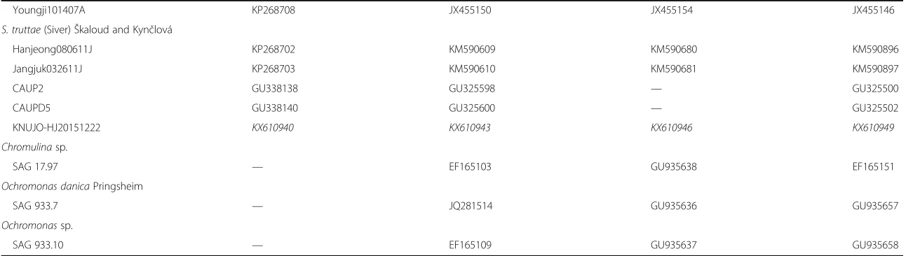
Fig. 2Morphology of the colony and scales ofSynura conopea(a–c: SEM,d: TEM). Allscale bars, 1μm.aSEM image of colony forming cells. bTop surface of a body scale.cBottom surface of a body scale.dTEM image of a body scale
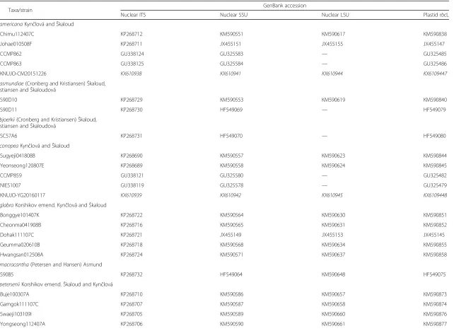
Fig. 1Morphology of the colony and scales ofSynura americana(a–c: SEM,d: TEM). Allscale bars, 1μm.aSEM image of colony forming cells.b Top surface of a body scale.cBottom surface of a body scale.dTEM image of body scale
Specimens examined: KNUJO-CM20151226.
Description: Colonies globular and 22–51 μm in diameter (Fig. 1a). Cells pyriform (22–28 × 8–12 μm) and entirely covered by rounded scales (Fig. 1a). Body scales 3.0–4.2 × 1.7–2.3 μm (Fig. 1b–d). The keel often terminates at an acute tip (Fig. 1b) and is ornamented by medium-sized pores (Fig. 1d). In some cases, the keel is wider in the anterior region, giving it a triangular shape (Fig. 1b). The basal plate, ornamented by numerous small pores, is anteriorly perforated by a rounded base plate hole that is 0.08–0.27μm in diameter (Fig. 1b–d). Numer-ous struts (21–24) extend regularly from the keel to the edge of the scale but almost never interconnect the trans-verse folds (Fig. 1b and d). The spacing between struts is 0.27–0.30μm (Fig. 1b and d).
Site of collection: Chimu, Daesan-myeon, Haman-gun, Gyeongsangnam-do, Korea (35°20′21"N, 128°25′47"E).
Date of collection: 26 Dec 2015.
Distribution: Widely distributed. Canada (Wee et al. 2001), Colombia (Cronberg 1989), Czech Republic (Škaloud et al. 2012; Kynčlová et al. 2010), Denmark (Kristiansen 1988), Germany (Kies & Berndt 1984), Korea (Boo et al. 2010, this study), North America (Kling & Kris-tiansen 1983; KrisKris-tiansen 1975; Wee 1981), and USA (Wee et al. 2001; Boo et al. 2010).
S. conopeaKynčlová andŠkaloud 2012 (Fig. 2)
Reference:Škaloud et al. 2012, p. 324, Figs. 78–85.
Specimens examined: KNUJO-YG20160117, NIBRFL 0000131748, and NIBRFL0000131749.
Description: Colonies globular and 25–47μm in diameter (Fig. 2a). Cells pyriform (20–28 × 8–12 μm) and entirely covered by lanceolate scales (Fig. 2a). Body scales 3.3–4.1 ×
1.4–1.9μm (Fig. 2b–d). The keel terminates at an acute tip (Fig. 2b) and is usually broadened apically and ornamented by medium to large-sized pores (Fig. 2d). The basal plate, ornamented by numerous medium-sized pores, is anteriorly perforated by a round to oblong base plate hole that is 0.19–0.32 μm in diameter (Fig. 2b–d). Numerous struts (24–30) extend regularly from the keel to the edge of the scale but are usually not interconnected by transverse folds (Fig. 2b and d). The spacing between struts is 0.23–0.26μm (Fig. 2b and d).
Site of collection: Yongji, Yongchon-ri, Toseong-myeon, Goseong-gun, Gangwon-do, Korea (38°13′43"N, 128° 33′49"E).
Date of collection: 17 Jan 2016.
Distribution: Widely distributed. Argentina (Vigna & Munari 2001), Brazil (Couté & Franceschini 1988), Czech Republic (Škaloud et al. 2012; Kynčlová et al. 2010), Greenland (Jacobsen 1985), Ireland (Řezáčová & Škaloud 2005), Japan (Boo et al. 2010), and Korea (Boo et al. 2010, this study).
S. truttae(Siver 1987)Škaloud and Kynčlová 2012 (Fig. 3)
Basionym: S. petersenii f. truttae (Siver 1987), p. 111, Figs. 12–14.
Reference:Škaloud et al. 2012, p. 318, Figs. 52–61. Specimens examined: KNUJO-HJ20151222.
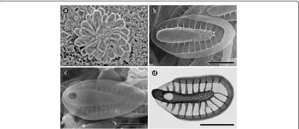
Description: Colonies globular and 35–48μm in diam-eter (Fig. 3a). Cells pyriform (22–31 × 11–13 μm) and entirely covered by lanceolate scales (Fig. 3a). Body scales elongated and 3.3–3.8 × 1.5–1.8 μm (Fig. 3a–d). The keel of the body scales has no apparent tip or a much reduced tip and is ornamented by small pores (Fig. 3b). The keel tip frequently has several (two to Fig. 3Morphology of the colony and scales ofSynura truttae(a–c: SEM,d: TEM). Allscale bars, 1μm.aSEM image of colony forming cells.bTop surface of a body scale.cBottom surface of a body scale.dTEM image of a body scale
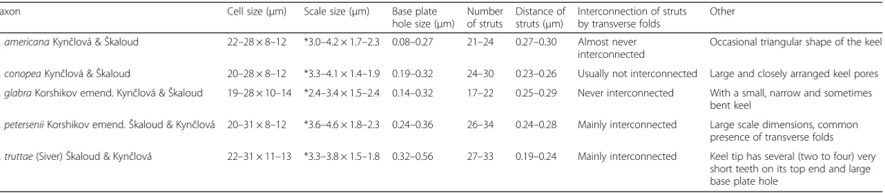
Table 2Summary of the major characteristic features observable with EM used in this study to distinguish between taxa of the sectionPetersenianae
Taxon Cell size (μm) Scale size (μm) Base plate hole size (μm)
Number of struts
Distance of struts (μm)
Interconnection of struts by transverse folds
Other
S. americanaKynčlová &Škaloud 22–28 × 8–12 *3.0–4.2 × 1.7–2.3 0.08–0.27 21–24 0.27–0.30 Almost never interconnected
Occasional triangular shape of the keel
S. conopeaKynčlová &Škaloud 20–28 × 8–12 *3.3–4.1 × 1.4–1.9 0.19–0.32 24–30 0.23–0.26 Usually not interconnected Large and closely arranged keel pores
S. glabraKorshikov emend. Kynčlová &Škaloud 19–28 × 10–14 *2.4–3.4 × 1.5–2.4 0.14–0.32 17–22 0.25–0.29 Never interconnected With a small, narrow and sometimes bent keel
S. peterseniiKorshikov emend.Škaloud & Kynčlová 20–31 × 8–12 *3.6–4.6 × 1.8–2.3 0.24–0.36 26–34 0.24–0.28 Mainly interconnected Large scale dimensions, common presence of transverse folds
S. truttae(Siver)Škaloud & Kynčlová 22–31 × 11–13 *3.3–3.8 × 1.5–1.8 0.32–0.56 27–33 0.19–0.24 Mainly interconnected Keel tip has several (two to four) very short teeth on its top end and large base plate hole
*The dimensions of body scales
Jo
and
Kim
Journal
of
Ecology
and
Environmen
t
(2017) 41:1
Page
7
of
four) very short teeth on its top (Fig. 3d) and is covered by a number of small bumps. The basal plate, ornamented by numerous small pores, is anteriorly perforated by a large, round to oblong base plate hole that is 0.32–0.56 μm in diameter (Fig. 3b–d). Numerous struts (27–33), which are often interconnected, regularly extend from the keel to the edge of the scale (Fig. 3b and d). Scales with nearly absent transverse folds (Fig. 3b–d). The spacing between struts is 0.19–0.24μm (Fig. 3b and d).
Site of collection: Hanjeong, Girin-ri, Soseong-myeon, Jeongeup-si, Jeollabuk-do, Korea (35°33′55"N, 126°46′ 30"E).
Date of collection: 22 Dec 2015.
Distribution: Widely distributed. Czech Republic (Škaloud et al. 2012; Kynčlová et al. 2010), Korea (This study), and USA (Siver 1987; Siver & Wujek 1993; Siver & Lott 2004).
Molecular data
The 5011 nucleotides of the combined data set (nuclear SSU and LSU rDNA, and plastid rbcL) were determined
for 32 strains (Table 1). Although the nuclear ITS1, 5.8S, and ITS2 sequences were also determined, these sequences were only used for to confirm identification, not to assess phylogenetic relationships. The combined sequences had 5011 nucleotides, 4039 variable sites, and 725 parsimoni-ously informative sites. The molecular data contained 12 new sequences (3 new nr SSU rDNA sequences, 3 new nr LSU rDNA sequences, 3 new nr ITS sequences, and 3 new pt rbcL sequences) and 102 published sequences (29 nr SSU rDNA sequences, 20 nr LSU rDNA sequences, 25 nr ITS sequences, and 28 ptrbcL sequences).
Phylogenetic analyses
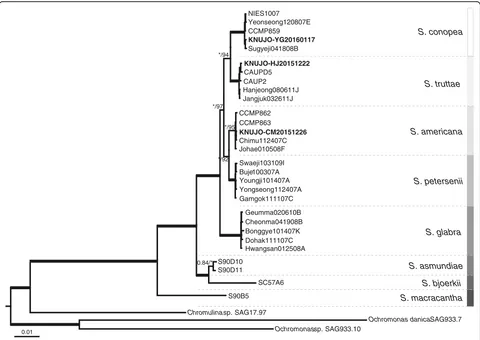
We analyzed nr SSU and LSU rDNA, and pt rbcL se-quences from 32 strains (including three outgroup species). The phylogenetic tree based on the Bayesian analysis was rooted with three species of Chromulinaceae serving as outgroups. The Bayesian and ML analyses recovered a tree with identical topologies (Fig. 4). The phylogenetic tree consisted of species of the section Petersenianae, each of which has a well-developed keel and a number of struts on
0.01
KNUJO-HJ20151222
S. conopea
CAUP2
Youngji101407A
Chromulina sp. SAG17.97 Jangjuk032611J
SC57A6 Johae010508F
Gamgok111107C
Geumma020610B CCMP863
Dohak111107C Hwangsan012508A
Yeonseong120807E NIES1007
Cheonma041908B Hanjeong080611J
CAUPD5 CCMP859
Yongseong112407A
S90D11
S. americana
S90B5
Sugyeji041808B
Ochromonas sp. SAG933.10 Swaeji103109I
S90D10
Bonggye101407K
Ochromonas danica SAG933.7 Buje100307A
*/94
*/95 */97
*/92
0.84/*
CCMP862
Chimu112407C KNUJO-CM20151226
S. glabra
KNUJO-YG20160117
S. petersenii S. truttae
S. asmundiae
S. bjoerkii
S. macracantha
Fig. 4Consensus Bayesian tree of the genusSynurabased on a combined nuclear SSU and LSU rDNA, and plastidrbcL sequences data. Bayesian posterior probability (pp) and maximum-likelihood (ML) bootstrap values are shown above or below the branches. The bold branches indicate strongly supported values (pp = 1.00 and ML = 100).Scale bar, 0.01 substitutions/site
the base plate. The sectionPetersenianaeformed a strongly supported monophyletic lineage (pp = 1.00 and ML = 100). The single strain of Synura macracantha diverged at the base of the tree, followed by Synura bjoerkii and Synura asmundiae. The single strain ofS. bjoerkiiwas closely re-lated to S. asmundiae, which included two strains (pp = 1.00 and ML = 100). Synura glabra formed a sister group withS. americana, S. conopea, S. petersenii, andS. truttae
(pp = 1.00 and ML = 100), andS. americanaandS. peterse-nii diverged at the next S. glabra. The five strains of S. petersenii formed a strongly supported monophyletic lineage (pp = 1.00 and ML = 100) and was a sister group to the five strains ofS. americana, which included KNUJO-CM20151226 (pp = 1.00 and ML = 92). The five strains of
S. americana were monophyletic group (pp = 1.00 and ML = 95), and the intraspecific similarity based on nuclear ITS rDNA sequence data ranged from 99.9% to 100.0%. The five strains of S. truttae (including KNUJO-HJ20151222) were a sister group to the five strains of S. conopea, which included KNUJO-YG20160117 (pp = 1.00 and ML = 94). The five strains of S. conopea formed a monophyletic lineage with strong support values (pp = 1.00 and ML = 100), and the intraspecific similarity based on nuclear ITS rDNA sequence data ranged from 98.5% to 100.0%. The five strains of S. truttaeformed a mono-phyletic lineage with strong support values (pp = 1.00 and ML = 100), and the intraspecific similarity based on nuclear ITS rDNA sequence data was 100.0%.
Conclusions
In summary, we used molecular analysis of three genes and data on the scale ultrastructure to investigate the phylogenetic relationships within Synura, with a focus on the section Petersenianae. The phylogenetic tree based on a combined dataset was well congruent with the ultrastructural characteristics of scales. The phylo-genetic tree was comprised of members of the section
Petersenianae. The sectionPetersenianae was monophy-letic with strong support values and characterized by a well-developed keel and a number of struts on the base plate. In addition, our morphological observa-tions and molecular analyses confirmed unambigu-ously that this is the first report of S. americana, S. conopea, and S. truttae in Korea.
Funding
This work was supported by a grant from the National Institute of Biological Resources (NIBR) funded by the Ministry of Environment (MOE) of the Republic of Korea (NIBR201501209).
Availability of data and materials
The sequence data from this study were deposited in GenBank with the accession codes KX610938-KX610949.
Authors’contributions
Both authors read and approved the final manuscript.
Competing interests
The authors declare that they have no competing interests.
Consent for publication Not applicable.
Ethics approval and consent to participate Not applicable.
Received: 1 August 2016 Accepted: 19 November 2016
References
Asmund, B. (1968). Studies on Chrysophyceae from some ponds and lakes in Alaska. VI. Occurrence ofSynuraspecies.Hydrobiologia, 31, 497–515. Balonov, I. M., & Kuzmin, G. V. (1974). Vidy rodaSynuraEhrenberg (Chrysophyta) v
vodokhranilischchakh Volzhskogo Kaskada.Botanicheskii Zhurnal, 59, 1675–1686. Boo, S. M., Kim, H. S., Shin, W., Boo, G. H., Cho, S. M., Jo, B. Y., Kim, J. H., Kim, J. H.,
Yang, E. C., Siver, P. A., Wolfe, A. P., Bhattacharya, D., Andersen, R. A., & Yoon, H. S. (2010). Complex phylogeographic patterns in the freshwater algaSynura provide new insights into ubiquity vs. endemism in microbial eukaryotes. Molecular Ecology, 19, 4328–4338. doi:10.1111/j.1365-294X.2010.04813.x. Couté, A., & Franceschini, I. M. (1988). Scale-bearing chrysophytes from acid
waters of Florianópolis, Santa Catarina Island, South Brazil.Algological Studies, 88(Suppl 123), 37–66.
Cronberg, G. (1989). Scaled chrysophytes from the tropics.Nova Hedwigia Beiheft, 95, 191–232.
Ehrenber, C. G. (1834). Dritter Beitrag zur Erkenntnis grosser Organisation in der Richtung des kleinsten Raumes.Abhandlungen der Königlichen Akademie der Wissenschaften Berlin, 1833, 145–336.
Fott, B., & Ludvík, J. (1957). Die submikroskopische Struktur der Kieselschuppen bei Synuraund ihre Bedeutung fur die Taxonomie der Gattung.Preslia, 29, 5–16. Jacobsen, B. A. (1985). Scale-bearing Chrysophyceae (Mallomonadaceae and
Paraphysomonadaceae) from West Greenland.Nordic Journal of Botany, 5, 381–398.
Jo, B. Y., Shin, W., Boo, S. M., Kim, H. S., & Siver, P. A. (2011). Studies on ultrastructure and three-gene phylogeny of the genusMallomonas (Synurophyceae).Journal of Phycology, 47, 415–425.
doi:10.1111/j.1529-8817.2010.00953.x.
Jo, B. Y., Shin, W., Kim, H. S., Siver, P. A., & Andersen, R. A. (2013). Phylogeny of the genusMallomonas(Synurophyceae) and descriptions if five new species on the basis of morphological evidence.Phycologia, 52, 266–278. doi:10.2216/12-107.1. Kies, L., & Berndt, H. (1984). DieSynura-Arten (Chrysophyceae) Hamburgs und
seiner nordöstlichen Umgebung.Mitteilungen aus dem Institut für Allgemeine Botanik Hamburg, 19, 99–122.
Kim, H. S. (1997). Silica-scaled chrysophytes (Synurophyceae) in several reservoirs, swamps, and a highland pond from Changnyong County, Korea.Algae, 12, 1–10. Kling, H. J., & Kristiansen, J. (1983). Scale-bearing Chrysophyceae
(Mallomonadaceae) from Central and Northern Canada.Nordic Journal of Botany, 3, 269–290.
Korshikov, A. (1929). Studies on the chrysomonads I.Archiv für Protistenkunde, 67, 253–290.
Kristiansen, J. (1975). Chrysophyceae from Alberta and British Columbia.Syesis, 8, 97–108.
Kristiansen, J. (1988). Seasonal occurrence of silica-scaled chrysophytesunder eutrophic conditions.Hydrobiologia, 161, 171–184.
Kristiansen, J. (1990). Studies on silica-scaled chrysophytes from Central Asia. Archiv für Protistenkunde, 138, 298–303.
Kristiansen, J., & Preisig, H. R. (2007). Chrysophyte and Haptophyte algae. 2. Teil/ Part 2: synurophyceae. In B. Büdel, G. Gärtner, L. Krienitz, H. R. Preisig, & M. Schagerl (Eds.),Süsswasserflora von Mitteleuropa(p. 252). Berlin, Heidelberg: Spektrum Akademischer Verlag.
Kynčlová, A.,Škaloud, P., &Škaloudová, M. (2010). Unveiling hidden diversity in theSynura peterseniispecies complex (Synurophyceae, Heterokontophyta). Nova Hedwigia Beiheft, 136, 283–298. doi:10.1127/1438-9134/2010/0136-0283. Péterfi, L. S., & Momeu, L. (1977). Remarks on the taxonomy of someSynura
species based on the fine structure of scales.Muzeul Brukenthal Studii si Comuicări-Stiinte Naturale, 21, 15–23.
Petersen, J. B., & Hansen, J. B. (1956). On the scales of someSynuraspecies.Biol Medd Kgl. Danske Videnskabernes Selskab., 23(2), 3–27.
Petersen, J. B., & Hansen, J. B. (1958). On the scales of someSynuraspecies. II.Biol Medd Kgl Danske Videnskabernes Selskab, 23(7), 1–13.
Posada, D., & Crandall, K. A. (1998). MODELTEST: testing the model of DNA substitution.Bioinformatics, 14(9), 817–8. doi:10.1093/bioinformatics/14.9.817. Rambaut A. FigTree v1.4.2. 2014. Available online at: http://tree.bio.ed.ac.uk/
software/figtree/
Rambaut A, Suchard MA, Drummond AJ. Tracer v.1.6. 2013. Available online at: http://tree.bio.ed.ac.uk/software/tracer/.
Řezáčová, M., &Škaloud, P. (2005). Silica-scaled chrysophytes of Ireland. With an appendix: geographic variation of scale shape ofMallomonas caudata. Nova Hedwigia Beiheft, 128, 101–124.
Ronquist, F., Teslenko, M., Van Der Mark, P., Ayres, D. L., Darling, A., Höhna, S., Larget, B., Liu, L., Suchard, M. A., & Huelsenbeck, J. P. (2012). MrBayes 3.2: efficient Bayesian phylogenetic inference and model choice a cross a large model space.Systematic Biology, 61(3), 539–42. doi:10.1093/sysbio/sys029. Siver, P. A. (1987). The distribution and variation ofSynuraspecies
(Chrysophyceae) in Connecticut, USA.Nordic Journal of Botany, 7, 107–116. doi:10.1111/j.1756-1051.1987.tb00922.x.
Siver, P. A., & Lott, A. M. (2004). Further observations on the scaled Chrysophycean and Synurophycean flora of the Ocala National Forest, Florida, USA.Nordic Journal of Botany, 24, 211–233. doi:10.1111/j.1756-1051.2004.tb00835.x. Siver, P. A., & Wujek, D. E. (1993). Scaled Chrysophyceae and Synurophyceae from
Florida, USA. IV. The flora of Lower Lake Myakka and Lake Tarpon.Florida scientist (USA), 56, 109–117.
Škaloud, P., Kristiansen, J., &Škaloudová, M. (2013). Developments in the taxonomy of silica-scaled chrysophytes from morphological and ultrastructural to molecular approaches.Nordic Journal of Botany, 31, 385–402. doi:10.1111/j.1756-1051.2013.00119.x.
Škaloud, P., Kynčlová, A., Benada, O., Kofroňová, O., &Škaloudová, M. (2012). Toward a revision of the genusSynura, section Petersenianae (Synurophyceae, Heterokontophyta): morphological characterization of six pseudo-cryptic species.Phycologia, 51, 303–329. http://dx.doi.org/10.2216/11-20.1.
Škaloud, P.,Škaloudová, M., Procházková, A., & Nĕmcová, Y. (2014). Morphological delineation and distribution patterns of four newly described species within the Synura peterseniispecies complex (Chrysophyceae, Stramenopiles).European Journal of Phycology, 49, 213–229. doi:10.1080/09670262.2014.905710. Stamatakis, A. (2014). RAxML version 8: a tool for phylogenetic analysis and
post-analysis of large phylogenies.Bioinformatics, 30(9), 1312–3. doi:10.1093/ bioinformatics/btu033.
Takahashi, E. (1967). Studies on generaMallomonas,Synuraand other plankton in fresh-water with the electron microscope. VI. Morphological and ecological observations on genusSynurain ponds and lakes in Yamagata Prefecture. Bulletin Yamagata University Agricultural Science, 5, 99–118.
Takahashi, E. (1972). Studies on generaMallomonasandSynura, and other plankton in freshwater with electron microscope. VIII. On three new species of Chrysophyceae.The botanical magazine= Shokubutsu-gaku-zasshi, 85, 293–302. Takahashi, E. (1973). Studies on generaMallomonasandSynura, and other
plankton in fresh water with the electron microscope VII. New genus Spiniferomonasof the Synuraceae (Chrysophyceae).The botanical magazine= Shokubutsu-gaku-zasshi, 86, 75–88.
Takahashi, E. (1978).Electron microscopical studies of the Synuraceae (Chrysophyceae) in Japan: taxonomy and ecology. Tokyo: Tokai University Press.
Vigna, M. S., & Munari, C. (2001). Seasonal occurrence of silica scales chrysophytes in a Buenos Aires lake.Nova Hedwigia Beiheft, 122, 195–209.
Wee, J. L. (1981). Studies on silica-scaled chrysophytes from Iowa. II. Common Synuraspecies.Proceedings of the Iowa Academy of Sciences, 88, 70–73. Wee, J. L., Fasone, L. D., Sattler, A., Starks, W. W., & Hurley, D. L. (2001). ITS/5.8S
DNA sequence variation in 15 isolates ofSynura peterseniiKorshikov (Synurophyceae).Nova Hedwigia Beiheft, 122, 245–258.
• We accept pre-submission inquiries
• Our selector tool helps you to find the most relevant journal • We provide round the clock customer support
• Convenient online submission • Thorough peer review
• Inclusion in PubMed and all major indexing services • Maximum visibility for your research
Submit your manuscript at www.biomedcentral.com/submit