University of Pennsylvania
ScholarlyCommons
Publicly Accessible Penn Dissertations
1-1-2014
Development of Spinal Neuronal Hyperexcitability
and Structural Plasticity after Cervical Facet Injury:
Implications for Modulating Persistent Pain
Nathan Crosby
University of Pennsylvania, [email protected]
Follow this and additional works at:http://repository.upenn.edu/edissertations
Part of theBiomedical Commons, and theNeuroscience and Neurobiology Commons
This paper is posted at ScholarlyCommons.http://repository.upenn.edu/edissertations/1246
Recommended Citation
Crosby, Nathan, "Development of Spinal Neuronal Hyperexcitability and Structural Plasticity after Cervical Facet Injury: Implications for Modulating Persistent Pain" (2014).Publicly Accessible Penn Dissertations. 1246.
Development of Spinal Neuronal Hyperexcitability and Structural
Plasticity after Cervical Facet Injury: Implications for Modulating
Persistent Pain
Abstract
Chronic neck pain is a prevalent and costly condition that commonly develops after whiplash injuries. The cervical facet joint and its capsular ligament are frequently identified as a source of pain in patients,
particularly those with whiplash-associated disorders. Spinal neuronal hyperexcitability is a common feature of persistent pain that can be induced by enhanced excitatory signaling in the dorsal horn. Spinal dorsal horn neurons develop hyperexcitability after painful facet joint injury, but the neurophysiological mechanisms that lead to spinal hyperexcitability and persistent pain after facet joint loading remain unclear. This thesis uses a rat model of painful facet joint injury to define the peripheral and spinal signals that promote dorsal horn neuronal hyperexcitability. In particular, the development of spinal hyperexcitability is evaluated by characterizing neuronal activity at multiple times within the first day after painful facet joint injury. To evaluate the role of injury-induced joint afferent activity in the potentiation of excitatory signaling and spinal neuronal excitability, afferent activity is blocked at various times after painful facet joint injury. Excitatory synaptogenesis and the mechanisms inducing synaptogenesis are evaluated to determine whether structural plasticity in the spinal cord contributes to facet-mediated pain. Spinal cord stimulation (SCS) is an effective clinical therapy that attenuates chronic pain by modulating spinal hyperexcitability, but the mechanisms and effectiveness of SCS for persistent neuropathic or joint-mediated pain remain unclear. This thesis evaluates the use of a novel mode of SCS, burst SCS, in rat models of cervical radiculopathy and painful facet joint injury. The role of GABA signaling in the inhibitory effects of burst SCS on dorsal horn neurons is assessed by applying GABA receptor antagonists to the spinal cord during the application of burst SCS. Studies in this thesis demonstrate that afferent discharge induced by injurious loading of the facet joint initiates excitatory synaptic and structural changes in the spinal cord that promote neuronal hyperexcitability, but SCS can attenuate persistent pain by reducing spinal hyperexcitability. This thesis provides a foundation for future investigations into the mechanisms underlying the transduction of mechanical joint injury to centrally-mediated pain and the development of effective therapies for chronic pain.
Degree Type
Dissertation
Degree Name
Doctor of Philosophy (PhD)
Graduate Group
Bioengineering
First Advisor
Subject Categories
DEVELOPMENT OF SPINAL NEURONAL HYPEREXCITABILITY AND STRUCTURAL PLASTICITY AFTER CERVICAL FACET INJURY:
IMPLICATIONS FOR MODULATING PERSISTENT PAIN
Nathan D. Crosby
A DISSERTATION
in
Bioengineering
Presented to the Faculties of the University of Pennsylvania in
Partial Fulfillment of the Requirements for the
Degree of Doctor of Philosophy
2014
Supervisor of Dissertation
________________________________________
Beth A. Winkelstein, Professor of Bioengineering
Graduate Group Chairperson
________________________________________ Jason A. Burdick, Professor of Bioengineering
Dissertation Committee
David F. Meaney, Professor and Chair of Bioengineering
ACKNOWLEDGMENTS
I am immensely grateful to my advisor, Dr. Beth Winkelstein, for her support
throughout my training. Her mentorship and guidance will benefit me throughout the rest
of my professional career, and I hope to continue to gain from her insight and experience
in the years to come. I would also like to thank the members of my committee, Dr. David
Meaney, Dr. Gordon Barr, Dr. Wenqin Luo, and Dr. Iyad Obeid, for their commitment to
my success and their insight during the development and completion of this work.
I would like to thank Dr. Melanie Goodman-Keiser for the training and assistance
she provided during our collaboration. Her experience and advice were instrumental for
designing the experiments, analyzing the data, and interpreting the findings in the studies
in this thesis using spinal cord stimulation.
I am very grateful to the past and present members of the Spine Pain Research
Lab for their support and constant friendship over the years. I would especially like to
thank those lab members that contributed directly to my research, including Kyle Quinn,
Ling Dong, Christine Weisshaar, Martha Zeeman, Jenell Smith, Jeff Kras, Ben Bulka,
and Taylor Gilliland.
I would like to thank my family for their unending encouragement and their
commitment to seeing me achieve my goals. Finally, I would like to thank my wife,
Erika. We leaned on each other frequently as we both navigated graduate school over the
last several years, and I am eternally grateful for her love, patience, perseverance, and
ABSTRACT
DEVELOPMENT OF SPINAL NEURONAL HYPEREXCITABILITY AND STRUCTURAL PLASTICITY AFTER CERVICAL FACET INJURY:
IMPLICATIONS FOR MODULATING PERSISTENT PAIN
Nathan D. Crosby
Beth A. Winkelstein
Chronic neck pain is a prevalent and costly condition that commonly develops
after whiplash injuries. The cervical facet joint and its capsular ligament are frequently
identified as a source of pain in patients, particularly those with whiplash-associated
disorders. Spinal neuronal hyperexcitability is a common feature of persistent pain that
can be induced by enhanced excitatory signaling in the dorsal horn. Spinal dorsal horn
neurons develop hyperexcitability after painful facet joint injury, but the
neurophysiological mechanisms that lead to spinal hyperexcitability and persistent pain
after facet joint loading remain unclear. This thesis uses a rat model of painful facet joint
injury to define the peripheral and spinal signals that promote dorsal horn neuronal
hyperexcitability. In particular, the development of spinal hyperexcitability is evaluated
by characterizing neuronal activity at multiple times within the first day after painful
facet joint injury. To evaluate the role of injury-induced joint afferent activity in the
potentiation of excitatory signaling and spinal neuronal excitability, afferent activity is
blocked at various times after painful facet joint injury. Excitatory synaptogenesis and the
mechanisms inducing synaptogenesis are evaluated to determine whether structural
(SCS) is an effective clinical therapy that attenuates chronic pain by modulating spinal
hyperexcitability, but the mechanisms and effectiveness of SCS for persistent neuropathic
or joint-mediated pain remain unclear. This thesis evaluates the use of a novel mode of
SCS, burst SCS, in rat models of cervical radiculopathy and painful facet joint injury.
The role of GABA signaling in the inhibitory effects of burst SCS on dorsal horn neurons
is assessed by applying GABA receptor antagonists to the spinal cord during the
application of burst SCS. Studies in this thesis demonstrate that afferent discharge
induced by injurious loading of the facet joint initiates excitatory synaptic and structural
changes in the spinal cord that promote neuronal hyperexcitability, but SCS can attenuate
persistent pain by reducing spinal hyperexcitability. This thesis provides a foundation for
future investigations into the mechanisms underlying the transduction of mechanical joint
injury to centrally-mediated pain and the development of effective therapies for chronic
TABLE OF CONTENTS
Page
Acknowledgements ... ii
Abstract ... iii
Table of Contents ...v
List of Tables ... ix
List of Figures ... xi
Chapter 1. Introduction and Background ...1
1.1 Introduction ...1
1.2 Background ...6
1.2.1 Cervical Spine and Facet Joint Anatomy ...6
1.2.2 Neuroanatomy and Neurophysiology of Nociception ...7
1.2.3 Whiplash Kinematics and Facet Joint Injury ...10
1.2.4 Central Sensitization ...14
1.2.5 Spinal Structural Plasticity ...18
1.2.6 In Vivo Animal Models of Facet-Mediated Pain from Joint Trauma ....20
1.2.7 Spinal Cord Stimulation ...23
1.3 Overview ...25
Chapter 2. Rational, Aims, and Hypotheses ...28
2.1 Rationale and Context ...28
2.2 Overall Hypothesis and Specific Aims ...32
Chapter 3. Spinal Neuronal Plasticity is Evident within One Day after a Painful Cervical Facet Joint Injury ...39
3.1 Overview ...39
3.2 Background ...41
3.3 Methods...42
3.3.1 Study Design and Facet Joint Injury ...42
3.3.2 Assessment of Mechanical Hyperalgesia ...44
3.3.3 Spinal Cord Electrophysiological Recordings ...45
3.4 Results ...50
3.5 Discussion ...54
3.6 Conclusions and Integration ...62
Chapter 4. Early Afferent Activity from the Injured Facet Joint Potentiates Spinal Sensitization ...64
4.1 Overview ...64
4.2 Background ...66
4.3 Methods...69
4.3.1 Facet Joint Injury with Immediate or Delayed Intra-articular Bupivacaine...70
4.3.2 Painful Facet Joint Capsule Injury ...71
4.3.3 Intra-articular Bupivacaine Injections...71
4.3.4 Assessment of Mechanical Hyperalgesia ...72
4.3.5 Spinal Cord Electrophysiology and Analysis of Neuronal Excitability .73 4.3.6 Western Blot Analysis of Spinal Cord Tissue ...74
4.3.7 Fluorescent Immunohistochemistry of Spinal Cord Tissue ...76
4.3.8 Painful Facet Joint Injury with Intra-articular Bupivacaine during the Development of Spinal Hyperexcitability ...77
4.4 Results ...78
4.4.1 Immediate, but not Delayed, Intra-articular Bupivacaine Attenuates Mechanical Hyperalgesia after Painful Facet Joint Injury ...78
4.4.2 Dorsal Horn Neuronal Hyperexcitability is Prevented by Immediate Bupivacaine Injection ...80
4.4.3 Excitatory Signaling is Modified by Immediate Bupivacaine Injection .82 4.4.4 Intra-articular Bupivacaine within 8 Hours after Injury Attenuates Hyperalgesia and Spinal Hyperexcitability ...85
4.5 Discussion ...86
4.6 Conclusions and Integration ...96
Chapter 5. Thrombospondin-4 and Excitatory Synaptogenesis Promote Spinal Sensitization after Painful Mechanical Joint Injury ...98
5.1 Overview ...98
5.2 Background ...100
5.3 Methods...104
5.3.2 Assessment of Mechanical Hyperalgesia ...105
5.3.3 Western Blot Analysis of DRG and Spinal Cord Tissue ...106
5.3.4 Immunolabeling of Spinal Cord Tissue ...107
5.3.5 Synapse Quantification in the Dorsal Horn ...108
5.3.6 Intrathecal Injections ...109
5.3.7 Oligonucleotide Treatment ...110
5.3.8 Intrathecal Gabapentin Treatment...111
5.3.9 Intra-articular Bupivacaine Treatment ...111
5.3.10 Recombinant TSP4 Purification and Intrathecal TSP4 Injections ...112
5.3.11 Electrophysiological Recording of Spinal Dorsal Horn Neurons ...114
5.4 Results ...115
5.4.1 Facet Joint Distraction Inducing Mechanical Hyperalgesia Also Increases Excitatory Synapses in the Spinal Dorsal Horn ...115
5.4.2 Altered Expression of TSP4 Parallels the Development of Sustained Mechanical Hyperalgesia ...117
5.4.3 Blocking Spinal TSP4 Expression Prevents Behavioral Sensitivity and Synaptogenesis after Painful Facet Joint Loading ...119
5.4.4 Gabapentin Reduces Injury-Induced Hyperalgesia and Dorsal Horn Excitatory Synaptogenesis ...121
5.4.5 Immediate Intra-articular Bupivacaine Prevents Hyperalgesia and Excitatory Synaptogenesis ...122
5.4.6 Spinal TSP4 Potentiates Hyperalgesia and Neuronal Hyperexcitability Induced by Facet Loading ...124
5.5 Discussion ...130
5.6 Conclusions and Integration ...135
Chapter 6. Optimization of Burst Spinal Cord Stimulation Parameters in a Rat Model of Neuropathic Pain ...137
6.1 Overview ...137
6.2 Background ...139
6.3 Methods...142
6.3.1 Nerve Root Compression Surgery ...142
6.3.2 Assessment of Mechanical Hyperalgesia ...143
6.3.3 Electrophysiological Recordings and Burst Spinal Cord Stimulation ..143
6.4 Results ...148
6.5 Discussion ...154
6.6 Conclusions and Integration ...159
Chapter 7. Evaluating Burst and Tonic Spinal Cord Stimulation for Attenuating Spinal Hyperexcitability ...161
7.1 Overview ...161
7.2 Background ...163
7.3 Burst and Tonic SCS after Painful Nerve Root Compression: The Role of GABA Signaling ...167
7.3.1 Methods...167
7.3.2 Results ...173
7.4 Burst and Tonic SCS after Painful Facet Joint Injury ...179
7.4.1 Methods...179
7.4.2 Results ...181
7.5 Discussion ...184
7.6 Conclusions and Integration ...193
Chapter 8. Synthesis and Future Work ...195
8.1 Introduction ...195
8.2 Summary and Synthesis of Major Findings ...196
8.3 Limitations and Future Work ...209
Appendix A. Protocols for Electrophysiological Recordings and Analysis ...221
Appendix B. Facet Joint Distraction Mechanics and Facet Capsule Strains ...228
Appendix C. Mechanical Hyperalgesia after Facet Joint Injury ...233
Appendix D. Quantification of Dorsal Horn Neuronal Firing ...243
Appendix E. Matlab Codes ...288
Appendix F. Protein Quantification in the DRG and Spinal Cord using Western Blot ...294
Appendix G. Quantification of Immunolabeled Proteins in the Spinal Cord ...298
Appendix H. Protocol for Counting Excitatory Synaptic Puncta ...322
Appendix I. Quantification of Synaptic Puncta in the Spinal Dorsal Horn ...327
Appendix J. Mechanical Hyperalgesia after Nerve Root Compression ...338
Appendix K. Joint Distraction Mechanics, Hyperalgesia, and Synapse Density in the Dorsal Horn at Day 14 after Painful Facet Joint Injury ...340
LIST OF TABLES
Page
Table 3.1 Total number of neurons recorded for each spinal cord level ...51
Table 4.1 Facet joint distraction and capsule mechanical measurements ...78
Table 6.1 Burst SCS conditions and parameter values ...146
Table 7.1 Summary of rat and SCS data for groups receiving painful nerve root
compression or NR compression with GABA receptor antagonists ...174
Table 7.2 Summary of rat and SCS data after a painful facet joint injury ...182
Table B.1 Facet joint distraction mechanics for rats that were tested for hyperalgesia
at 6 hours or 1 day after painful facet joint injury (Chapter 3) ...230
Table B.2 Facet joint distraction mechanics for bupivacaine treatment study
(Chapter 4) ...231
Table B.3 Facet joint distraction mechanics for TSP4 treatment study (Chapter 5) ...232
Table C.1 Paw withdrawal thresholds at 6 hours or day 1 after sham or painful facet
joint injury (Chapter 3) ...235
Table C.2 Paw withdrawal thresholds for bupivacaine treatment study (Chapter 4) ..236
Table C.3 Paw withdrawal thresholds after sham or painful facet joint injury for
characterization of TSP4 expression (Chapter 5) ...237
Table C.4 Paw withdrawal thresholds after painful facet joint injury with
admin-istration of TSP4 antisense or mismatch oligonucleotides (Chapter 5) ...238
Table C.5 Paw withdrawal thresholds for gabapentin treatment study (Chapter 5) ....239
Table C.6 Paw withdrawal thresholds for TSP4 dose-response study (Chapter 5) ...240
Table C.7 Paw withdrawal thresholds for TSP4 treatment study (Chapter 5) ...241
Table C.8 Paw withdrawal thresholds after painful facet joint injury for spinal cord
stimulation at day 7 (Chapter 7)...242
Table D.1 Spike counts at 6 hours or 1 day after sham or painful facet joint injury
(Chapter 3) ...246
Table D.2 Spike counts on day 7 for bupivacaine treatment study (Chapter 4) ...250
Table D.3 Spike counts on day 7 for TSP4 treatment study (Chapter 4)...260
Table D.4 Spike counts for optimization of burst SCS parameters on day 7 after painful
nerve root compression (Chapter 6) ...261
Table D.5 Spike counts after burst or tonic SCS on day 7 following painful nerve root
Table D.6 Spike counts after burst or tonic SCS on day 7 following painful nerve root compression with spinal superfusion of bicuculline (Chapter 7)...274
Table D.7 Spike counts after burst or tonic SCS on day 7 following painful nerve root
compression with spinal superfusion of CGP35348 (Chapter 7)...276
Table D.8 Spike counts after burst or tonic SCS on day 7 following painful facet joint
injury (Chapter 7) ...282
Table F.1 Western blot quantification of glutamatergic signaling proteins and GFAP
on day 7 for bupivacaine treatment study, normalized to sham for each treatment time point (Chapter 4) ...295
Table F.2 Western blot quantification of DRG and spinal TSP4 on day 7 after sham or
painful facet joint injury (Chapter 5) ...296
Table F.3 Western blot quantification of spinal TSP4 on day 7 after painful facet
injury with administration of antisense or mismatch oligonucleotides
(Chapter 5) ...297
Table G.1 Densitometric quantification of pNR1, mGluR5, and GFAP at day 7 for
bupivacaine treatment study (Chapter 4) ...300
Table G.2 Densitometric quantification of TSP4 and GFAP at day 7 after sham or
painful facet joint injury (Chapter 5) ...308
Table G.3 Densitometric quantification of TSP4 and GFAP at day 7 for bupivacaine
treatment study (Chapter 5) ...312
Table I.1 Quantification of synapses at day 7 after sham or painful facet joint injury
(Chapter 5) ...329
Table I.2 Quantification of synapses at day 7 after painful facet injury and prior
treatment with TSP4 antisense or mismatch oligonucleotides (Chapter 5) 331
Table I.3 Quantification of synapses at day 7 for gabapentin treatment study
(Chapter 5) ...333
Table I.4 Quantification of synapses at day 7 for bupivacaine treatment study
(Chapter 5) ...334
Table J.1 Paw withdrawal thresholds after painful nerve root compression for spinal
cord stimulation at day 7 (Chapters 6 and 7) ...339
Table K.1 Facet joint distraction mechanics for rats that were tested for hyperalgesia
through day 14 after painful facet joint injury (Chapter 8) ...342
Table K.2 Paw withdrawal thresholds through day 14 after sham or painful facet joint
injury (Chapter 8) ...342
Table K.3 Quantification of excitatory synapses in the superficial dorsal horn at day 14
after sham or painful facet joint injury (Chapter 8) ...343
Table K.4 Quantification of inhibitory synapses in the superficial dorsal horn at day 14
LIST OF FIGURES
Page
Figure 1.1 Lateral view of the human cervical spinal column ...6
Figure 1.2 Primary afferent connections in the spinal dorsal horn ...9
Figure 1.3 Ascending pathways from the spinal dorsal horn ...11
Figure 1.4 Deformation of the cervical spine during rear-impact ...12
Figure 1.5 The initiation of central sensitization in the dorsal horn ...16
Figure 1.6 Patterns of sensory innervation in the cervical regions of the human and rat...21
Figure 3.1 The right facet joint of the rat with vertebral and capsule bead markers ...44
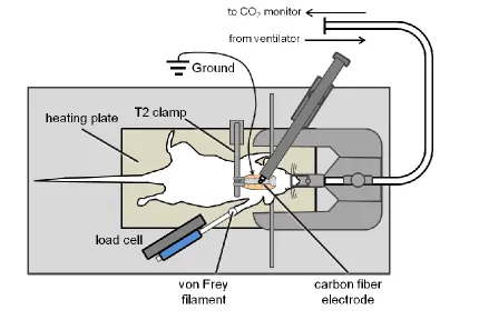
Figure 3.2 Schematic of the instrumentation for extracellular electrophysiological recordings ...46
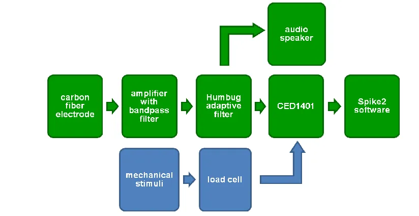
Figure 3.3 General scheme for data collection and processing of electrophysiological recordings ...47
Figure 3.4 Protocol for mechanical stimulation of the forepaw during recording of dorsal horn neuronal firing ...48
Figure 3.5 Mechanical hyperalgesia at 6 hours or 1 day after painful facet joint injury ...51
Figure 3.6 Spontaneous firing in the spinal dorsal horn...52
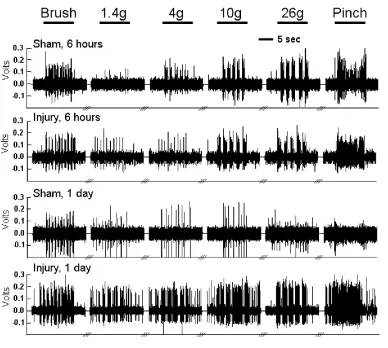
Figure 3.7 Representative firing for sham and injured rats at 6 hours or 1 day ...54
Figure 3.8 Evoked firing in the spinal dorsal horn ...55
Figure 4.1 Behavioral sensitivity after intra-articular bupivacaine given either at injury or 4 days later ...79
Figure 4.2 Extracellular spike activity in the spinal dorsal horn seven days after facet joint injury ...81
Figure 4.3 Western blot of spinal cord at day 7 ...83
Figure 4.4 Immunolabeling of phosphorylated NR1, mGluR5, and GFAP in the spinal dorsal horn ...84
Figure 4.5 Hyperalgesia after bupivacaine administered during spinal sensitization onset ...85
Figure 5.1 Schematic showing the peripheral and spinal treatments used to evaluate the potential relationships between facet joint loading, spinal TSP4 expression,
spinal neuronal sensitization and pain ...104
Figure 5.2 TSP4 antibody specificity ...108
Figure 5.3 Mechanical hyperalgesia after painful facet joint loading ...115
Figure 5.4 Quantification of excitatory synapses in the superficial and deep laminae of the dorsal horn ...116
Figure 5.5 Western blot quantification of DRG and spinal TSP4 ...117
Figure 5.6 Immunolabeling of TSP4 and GFAP in the spinal cord ...118
Figure 5.7 TSP4 antisense oligonucleotides prevent hyperalgesia and spinal excitatory synaptogenesis ...120
Figure 5.8 Gabapentin attenuates mechanical hyperalgesia and excitatory synaptogenesis after painful facet joint loading...121
Figure 5.9 Immediate intra-articular bupivacaine prevents TSP4 upregulation after painful facet joint loading ...123
Figure 5.10 Immediate intra-articular bupivacaine prevents excitatory synaptogenesis ...124
Figure 5.11 Purification of recombinant TSP4 ...125
Figure 5.12 Dose-response of intrathecal TSP4 and mechanical hyperalgesia in the forepaw ...125
Figure 5.13 Mechanical characterization of facet joint loading ...126
Figure 5.14 Spinal TSP4 facilitates hyperalgesia after facet joint loading ...127
Figure 5.15 Spinal TSP4 potentiates dorsal horn neuronal hyperexcitability after facet joint loading ...129
Figure 6.1 Schematic of a burst SCS waveform ...141
Figure 6.2 Stimulation protocol for burst SCS optimization...144
Figure 6.3 Mechanical hyperalgesia after painful cervical nerve root compression ....148
Figure 6.4 Attenuation of neuronal firing by burst SCS ...149
Figure 6.5 Effect of burst parameters on the attenuation of neuronal firing ...150
Figure 6.6 Effect of burst parameters on the percentage of responsive neurons ...151
Figure 6.7 Charge per burst determines the effectiveness of burst SCS ...153
Figure 7.1 Schematic of a C7 nerve root compression and application of SCS ...168
Figure 7.2 Stimulation protocol for recording evoked neuronal activity after burst or tonic SCS ...171
Figure 7.4 Evoked dorsal horn neuronal activity after burst and tonic SCS
application ...175
Figure 7.5 Evoked spike counts for up to 15 minutes after the cessation of burst and tonic SCS application following painful nerve root compression ...176
Figure 7.6 Effects of GABA receptor antagonists on the attenuation of dorsal horn neuronal firing by burst or tonic SCS ...178
Figure 7.7 Mechanical hyperalgesia after painful facet joint injury ...181
Figure 7.8 Representative firing and quantification of neuronal activity after burst and tonic SCS on day 7 following painful facet joint injury ...183
Figure 7.9 Evoked spike counts for up to 15 minutes after the cessation of burst and tonic SCS application following painful facet joint injury ...184
Figure 8.1 Quantification of hyperalgesia through day 14 after painful facet joint injury ...197
Figure 8.2 Temporal development of synaptic and structural modifications in the spinal cord that promote spinal hyperexcitability and behavioral sensitivity after facet joint injury ...199
Figure 8.3 Facet joint injury induces persistent pain through multiple mechanisms ...203
Figure 8.4 Quantification of excitatory and inhibitory synapses at day 14 after painful facet joint injury ...213
Figure A.1 Spike template detection ...225
Figure A.2 Spike template parameter dialogue box ...225
Figure A.3 Quantification of evoked firing during von Frey filament stimulation...227
Figure G.1 Immunolabeling of mGluR5 and GFAP in the spinal dorsal horn at day 7 for the bupivacaine treatment study ...306
Figure G.2 Immunolabeling of pNR1 and GFAP in the spinal dorsal horn at day 7 for the bupivacaine treatment study ...307
Figure G.3 Immunolabeling of TSP4 and GFAP in the spinal cord at day 7 after sham or painful facet joint injury ...311
Figure G.4 Immunolabeling of TSP4 and GFAP in the superficial dorsal horn at day 7 for the bupivacaine treatment study ...319
Figure G.5 Immunolabeling of TSP4 and GFAP in the deep dorsal horn at day 7 for the bupivacaine treatment study ...320
Figure G.6 Immunolabeling of TSP4 and GFAP in the dorsal columns at day 7 for the bupivacaine treatment study ...321
Figure H.1 Puncta Analyzer initialization dialogue box ...323
Figure H.3 Detection threshold for synaptic puncta ...324
Figure H.4 Puncta size dialogue box ...325
CHAPTER 1
Introduction and Background
1.1. Introduction
Chronic pain affects at least one in three adults in the United States, and has an
estimated annual cost of over $635 billion (Roehr, 2011). Costs associated with painful
spine conditions alone increased by 65% to $85.9 billion between 1997 and 2005,
reaching expenditures similar to the costs associated with diabetes and cancer (Martin et
al., 2008). The prevalence of spine pain has been reported at 26-66% in the general
population, with a 30-50% annual incidence of neck or cervical spine pain
(Hogg-Johnson et al., 2008; Linton et al., 1998; Martin et al., 2008). Whiplash injuries are a
primary cause of cervical spine pain, and account for up to 53% of the injuries from
motor vehicle accidents (Freeman et al., 1999). The annual incidence of
whiplash-associated pain has been reported to be at least 300 per 100,000 people in the United
States (Holm et al., 2008). Furthermore, up to 50% of the patients with whiplash injuries
develop chronic pain that is resistant to conservative interventions, like exercise and
physical therapy (Sterling et al., 2012). Recent increases in clinical interventions for
chronic neck pain (Manchikanti et al., 2013), and the fact that motor vehicle collisions
continue to be a leading cause of emergency room visits (Quinlan et al., 2004), suggest a
Many different tissues in the cervical spine have the capacity to generate pain
from injurious mechanical loading owing to their innervation by nerve fibers that detect
and transmit painful signals (Antonacci et al., 1998; Inami et al., 2001; McLain, 1994;
Rhalmi et al., 1993). Among those tissues with the potential to generate pain, the cervical
facet joints are most commonly implicated as the source of chronic pain after whiplash
(Barnsley et al., 1995; Lord et al., 1996; Manchikanti et al., 2004). The facet joints are
bilateral articulations between each pair of vertebrae that guide the motions of the
vertebrae. Each joint is enclosed by a facet capsular ligament, or facet capsule, that is
highly innervated with primary afferent nerve fibers that detect both low-threshold
mechanical stimulation and noxious, painful stimulation (Cavanaugh et al., 1989;
McLain, 1994; Yamashita et al., 1990). Those primary afferent fibers converge in the
dorsal horn of the spinal cord, where they transmit sensory information to second-order
neurons that integrate and project signals supraspinally (Steeds, 2009).
The cervical facet joints are at risk for excessive loading during abnormal motions
of the cervical spine. Biomechanical studies of human volunteers and cadaveric subjects
have determined that the facet joint can undergo injurious motions during simulated
low-velocity rear-impact collisions (Bogduk and Yoganandan, 2001; Deng et al., 2000;
Kaneoka et al., 1999; Panjabi et al., 1998c; Winkelstein et al., 2000). These abnormal
motions of the facet joint can cause tensile and shear loading of the facet capsular
ligament, resulting in strains exceeding the physiologic limit of the capsule (Panjabi et
al., 1998b; Pearson et al., 2004; Siegmund et al., 2001). In a goat model of tensile loading
of the facet joint, facet capsule stretch has also been shown to activate the nerve fibers
has the potential to induce painful signals that are transmitted from the joint to the spinal
cord. Despite evidence of the transduction of mechanical loading to potentially painful
neural signals, the relationship between facet joint loading and the development of
persistent joint-mediated pain is still unclear.
Nociceptive pain is the normal sensory response to stimulation that exceeds the
activation threshold of nociceptors, which are the nerve fibers that specifically respond to
noxious stimuli (Loeser and Treede, 2008). Nociception typically persists only in the
presence of continued suprathreshold stimulation, but pathological pain can develop after
tissue damage and persist even in the absence of noxious stimulation (Coderre et al.,
1993; Steeds, 2009). Chronic pain can be maintained by central sensitization, a state in
which the second-order neurons that integrate sensory information in the spinal dorsal
horn become hyperexcitable (Latremoliere and Woolf, 2009; Loeser and Treede, 2008).
Both nociceptive and nonpainful signals that converge in the spinal cord can be amplified
by increased excitability and decreased activation thresholds of the dorsal horn neurons.
Central sensitization is initiated by a period of sustained afferent discharge (Seltzer et al.,
1991a; Wall et al., 1974), which can occur after excessive loading of the joint that leads
to increased firing of the primary afferents that innervate the facet capsule (Lu et al.,
2005a); despite this notion, no study has defined the role of afferent firing from the facet
joint in the onset of central sensitization.
Central sensitization is associated with widespread changes in the spinal cord that
enhance neuronal excitability and promote persistent pain after tissue injury. For
example, potentiation of excitatory glutamatergic signaling can amplify nociception by
signals (Basbaum et al., 2009; Latremoliere and Woolf, 2009). Structural plasticity, like
the growth of new excitatory synapses, can also promote aberrant nociception in the
spinal cord by increasing the number of excitatory inputs to the dorsal horn neurons
(Jaken et al., 2010; Peng et al., 2010). Noxious loading of the rat facet joint has been
shown to induce dorsal horn neuronal hyperexcitability and spinal modifications that
contribute to the potentiation of glutamatergic signaling (Dong and Winkelstein, 2010;
Lee et al., 2004a; Lee et al., 2008; Quinn et al., 2010b). However, the neurophysiological
mechanisms that underlie the development and maintenance of spinal hyperexcitability
after excessive loading of the facet joint are unknown.
Central sensitization leads to the development of hyperalgesia, defined as a
general increase in pain sensitivity, and allodynia, defined as a painful response to a
typically non-noxious stimulus (Loeser and Treede, 2008). Hyperalgesia can develop
both at the site of injury (primary hyperalgesia) or at remote locations in which there is
no tissue injury (secondary hyperalgesia) because of the amplification of nociceptive
signals as they converge in the spinal cord from widespread anatomical regions (Coderre
et al., 1993). Many of the symptoms exhibited by patients with whiplash-associated pain
are indicative of central sensitization, including decreased pain thresholds in the neck and
back, and at distant sites with no tissue damage, like the head, arms, and legs (Banic et
al., 2004; Curatolo et al., 2001; Curatolo et al., 2004; Herren-Gerber et al., 2004; Jansen
et al., 2008; Lord et al., 1996). In order to prevent the development of persistent pain or
treat the symptoms of pain after facet joint injury, the timeline and factors affecting the
primary and secondary hyperalgesia after whiplash, the relationship between facet joint
injury and the development of centrally-mediated pain has not been defined.
Current treatment strategies for facet joint-mediated pain focus on reducing pain
by eliminating nociceptive input from symptomatic facet joints using peripheral nerve
blocks or neurotomy; however, the effects of those treatments invariably are only
temporary and patients require periodic re-intervention (Barnsley et al., 1995; Lord et al.,
1996; Manchikanti et al., 2008). The inability of peripheral interventions to permanently
reverse central sensitization after its onset is common across many different models of
neuropathic and inflammatory pain (Araujo et al., 2003; Shankarrapa et al., 2012; Xie et
al., 2005). For patients with facet joint-mediated pain after whiplash, conventional
treatment strategies, including physical therapy, pharmacological interventions, or
blocking nerve conduction from the facet joint, fail to target the hyperexcitability of the
neurons in the spinal dorsal horn that contribute to persistent pain (Lord et al., 1996;
Manchikanti et al., 2008; Peeters et al., 2001). Direct modulation of spinal
hyperexcitability, as occurs with spinal cord stimulation, may be more effective for
treating persistent pain after the development of central sensitization (Cameron, 2004;
Compton et al., 2012; Simpson, 1997). Yet, the effectiveness and mechanisms of spinal
cord stimulation for the reduction of spinal hyperexcitability require further investigation.
The next sections of this chapter provide relevant background information about
the cervical spine and facet joint anatomy, neuroanatomy, facet joint injury and whiplash
kinematics, central sensitization, in vivo animal models for the study of facet joint pain,
and spinal cord stimulation. Additional background information related to the specific
1.2. Background
1.2.1. Cervical Spine and Facet Joint Anatomy
The cervical spine is comprised of seven articulating vertebrae that are supported
by soft tissues, including ligaments, musculature, and cartilage. Each pair of bony
vertebrae is connected by the collagenous intervertebral discs ventrally, and by the
bilateral articulating facet (or zygapophyseal) joints posterolaterally (Fig. 1.1a). The facet
joints are formed by contact of the superior and inferior articular facets of two adjacent
vertebrae (Fig. 1.1a). The articulating surfaces of the facet joints are covered by articular
cartilage and lubricated by synovial fluid to allow the vertebral bodies to rotate with
respect to each other during normal head and neck motion (Fig. 1.1b). The joints guide
and limit motions of the head and neck, while protecting the spinal cord from mechanical
loading (Lang, 1993; Watson et al., 2009). The joint space and synovial fluid are
Fig. 1.1. Lateral view of the human cervical spinal column. (a) The facet joints are
formed by the superior and inferior articulating processes of each pair of vertebrae
(adapted from Benzel, 2012). (b) The articulating surfaces of the facet joints are
covered with cartilage (blue), and the facet capsule (red) encloses the joint space.
facet joint
facet capsule
articular cartilage spinous process
b)
inferior articular facet
superior articular facet spinous process
facet capsule
intervertebral disc transverse
process
a) C1
C7
enclosed by the facet capsular ligament, or facet capsule (Fig. 1.1b) (Jaumard et al.,
2014), which is a heterogeneous, fibrous structure composed of dense bundles of collagen
and elastic fibers (Yamashita et al., 1996).
The cervical facet joints are innervated by the medial branches of the dorsal
primary rami immediately superior and inferior to the joint, although the joint can be
innervated to a lesser degree by the dorsal rami of more distant spinal levels (Bogduk and
Marsland, 1988; Kras et al., 2013a; Ohtori et al., 2001). The facet capsule, in particular,
is innervated by mechanosensitive nerve fibers that are activated by capsule stretch
(Cavanaugh et al., 1989; McLain, 1994; Yamashita et al., 1990). Many mechanosensitive
fibers in the capsule exhibit proprioceptive properties, suggesting that the capsule
communicates information about head and neck position and movement (McLain and
Pickar, 1998). The capsule also contains free nerve endings that are typically associated
with nociceptors, the nerve fibers that specifically respond to painful stimuli (Cavanaugh
et al., 1989; McLain, 1994). A subset of the nerve fibers in the capsule express substance
P (SP) and calcitonin gene-related peptide (CGRP), two neuropeptides with demonstrated
roles in nociception, further suggesting that the facet joint is likely capable of signaling
pain under excessive mechanical loading conditions (Beaman et al., 1993; Inami et al.,
2001; Kallakuri et al., 2004; Ohtori et al., 2000; Yamashita et al., 1993).
1.2.2. Neuroanatomy and Neurophysiology of Nociception
Sensory signals are transmitted from the periphery to the spinal cord through
primary afferent nerve fibers. Primary afferents project axons distally from the dorsal
and Robertson, 2004). Primary afferents are classified into several groups (Aα/β, Aδ, C)
based on their structure and function. Aα and Aβ fibers (also called group I and II fibers)
have large, myelinated axons and rapid conduction velocities (Guyton and Hall, 1996).
They generally respond to innocuous mechanical stimuli or movement, and are classified
as low-threshold mechanoreceptors or proprioceptors. The other two types of fibers, Aδ
(group III) and C fibers (group IV), are classified as nociceptors and contribute to the
sensation of pain (Steeds, 2009). Aδ fibers are thinly myelinated, with smaller diameters
and slower conduction velocities than Aβ fibers, and are thought to evoke sharp, pricking
pain and “fast” pain (Basbaum et al., 2009; Guyton and Hall, 1996; Meyer et al., 2006). C
fibers have small-diameter, unmyelinated axons with relatively slow conduction
velocities, and transmit “slow” pain and diffuse pain from heat and pressure (Basbaum et
al., 2009; Meyer et al., 2006). C fibers are further classified as peptidergic fibers, if they
express SP and CGRP, or non-peptidergic fibers, based on the expression of the
purinergic ATP receptor, P2X3, or binding of isolectin B4 (Braz et al., 2005; Snider and
McMahon, 1998).
Nociceptive pain is established in response to stimuli (mechanical, chemical,
thermal, etc.) that exceed the activation thresholds of nociceptive afferent fibers.
Nociception is a protective response designed to warn of impending tissue damage, so it
typically continues only in the presence of noxious stimuli (Costigan et al., 2009).
However, repetitive stimulation of afferent fibers can temporarily sensitize them by
inducing wind-up, which is a progressive increase in dorsal horn neuronal firing in
afterdischarge, which is a continuation of afferent firing after the termination of the
initiating stimulus (Li et al., 1999).
Afferents synapse in the spinal cord with three main classes of second-order
neurons (Fig. 1.2). Nociceptive-specific neurons (NS), or high-threshold (HT) neurons,
receive input only from nociceptive afferents, therefore they are only activated by
noxious stimuli (Dostrovsky and Craig, 2006; Steeds, 2009). Wide dynamic range
(WDR) neurons receive input from non-nociceptive and nociceptive afferents, and
exhibit a graded response to innocuous and noxious stimuli (Baron, 2006; Basbaum et al.,
2009; Dostrovsky and Craig, 2006; Steeds, 2009). Low-threshold mechanoreceptors
(LTM) only process light touch sensation from innocuous stimuli.
Fig. 1.2. Primary afferent connections in the spinal dorsal horn. The superficial
laminae (I-II) contain nociceptive-specific (NS) neurons that receive input from C and Aδ fibers. The deep laminae (III-VI) contain wide dynamic range (WDR) and low-threshold mechanoreceptive (LTM) neurons that receive input from Aδ and Aβ fibers (adapted from Basbaum et al., 2009). Also shown are the dorsal columns, where some Aβ fibers collateralize without first synapsing in the dorsal horn.
dorsal columns
spinal dorsal horn
Aβ fiber
spinal cord
C fiber
Aδ fiber I
II
III
IV
V
NS neurons
WDR/LTM neurons
Primary afferents synapse with second-order neurons in the dorsal horn region of
the spinal cord. The dorsal horn is organized into laminae, or layers, that contain
specialized populations of second-order neurons, and receive input from specific groups
of afferent fibers (Fig. 1.2) (Grant and Koerber, 2004; Molander et al., 1989; Rexed,
1952; Todd and Koerber, 2006; Watson et al., 2009). For example, the most dorsal,
superficial laminae (laminae I and II) almost exclusively contain NS neurons and the
central terminals of nociceptive Aδ and C fibers (Fig. 1.2) (Basbaum et al., 2009; Todd
and Koerber, 2006). In contrast, Aβ mechanoreceptors and some additional Aδ fibers
terminate largely at WDR and LTM neurons in the deeper laminae (laminae III-VI) (Fig.
1.2) (Basbaum et al., 2009; Grant and Robertson, 2004; Todd and Koerber, 2006).
Secondary neurons project supraspinally from the dorsal horn, carrying sensory
information to the brain. Most nociceptive signals are conveyed to the brainstem and
thalamus through the spinothalamic and spinoreticular tracts (Fig. 1.3) (Basbaum et al.,
2009; Watson et al., 2009). For projection of low-threshold mechanical signals, some Aβ
fibers collateralize into the dorsal horn and send mechanosensory information through the
spinothalamic and spinoreticular pathways (Fig. 1.3) (Yaksh and Luo, 2001; Steeds,
2009). However, a large number of Aβ fibers also project directly through the dorsal
columns, carrying proprioceptive and light touch signals to the brainstem without first
synapsing in the dorsal horn (Figs. 1.2 and 1.3) (Steeds, 2009; Watson et al., 2009).
1.2.3. Whiplash Kinematics and Facet Joint Injury
The facet joint and its capsular ligament are among the cervical spinal tissues with
the cervical facet joints in normal subjects by intra-articular injection of fluoroscopic
contrast medium produces neck pain in distributions similar to those observed after
whiplash (Dwyer et al., 1990). Additionally, the cervical facet joints have been
implicated as the source of pain in 54-60% of whiplash patients using diagnostic blocks
of symptomatic joints with intra-articular injection of local anesthetics (Barnsley et al.,
1995; Lord et al., 1996; Manchikanti et al., 2004). The lower cervical levels (C5-C7) are
the most common source of neck and shoulder pain (Barnsley et al., 1995; Bogduk and
Marsland, 1988). Local anesthetic block or radiofrequency ablation of the medial
branches of the dorsal rami that innervate the symptomatic facet joints attenuates pain
(Aprill, 1990; Lord et al., 1996; Manchikanti et al., 2004), further supporting the role of
the facet joints as a primary source of pain following whiplash.
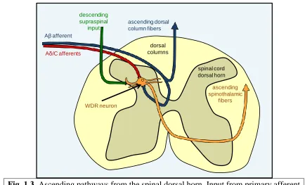
Fig. 1.3. Ascending pathways from the spinal dorsal horn. Input from primary afferent
fibers converges in the spinal dorsal horn, and then continues through spinothalamic tract projections that convey sensory information to the brain. Aβ fibers can also project supraspinally through the dorsal columns (adapted from Baron, 2006).
ascending dorsal column fibers Aβ afferent
Aδ/C afferents
dorsal columns
spinal cord dorsal horn
WDR neuron
ascending spinothalamic
fibers
descending supraspinal
Facet joint injury has been hypothesized to result from excessive loading of the
facet capsular ligament beyond its physiologic limit. Biomechanical studies using human
volunteers and post-mortem human subjects have defined the kinematics of the head and
cervical spine during simulated low-velocity rear-impact collisions (Deng et al., 2000;
Ono et al., 1997; Panjabi et al., 1998a; Pearson et al., 2004; Sundararajan et al., 2004;
Winkelstein et al., 2000; Yoganandan et al., 2001). Based on high-speed X-ray imaging
of the cervical spine during rear impact, the spine forms an S-shaped curvature within the
first 100-120ms after impact, with the upper cervical spine undergoing flexion and the
lower cervical spine being extended (Fig. 1.4) (Bogduk and Yoganandan, 2001; Kaneoka
et al., 1999; Panjabi et al., 1998c). Extension and vertebral retraction at the lower cervical
spinal levels produces tensile and shear loading across the facet joints and capsular
ligaments (Fig. 1.4) (Luan et al., 2000; Panjabi et al., 1998b; Panjabi et al., 1998c;
Siegmund et al., 2001; Winkelstein et al., 2000).
Fig. 1.4. Deformation of the cervical spine during rear-impact. Abnormal motions of
the cervical spine, including excessive extension in the lower cervical levels, can induce excessive strains in the facet capsule that exceed the physiologic levels (adapted from Croft, 2000).
time after rear-end impact (ms)
0 75 150
S-curve C-curve
C7 C2
Facet capsule strains from joint loading during low-speed rear impacts can be as
high as five times the peak strains that are observed for the same cervical levels during
normal neck bending (Panjabi et al., 1998b; Pearson et al., 2004). Capsule strains during
simulated rear-impact collisions are the greatest at the lower cervical facet joints, peaking
at 38.5±24.6% strain at the C5/C6 joint and 39.9±26.3% strain at the C6/C7 joint during
8g accelerations (Pearson et al., 2004), potentially explaining the prevalence of
symptomatic C5-C7 facet joints reported after whiplash (Barnsley et al., 1995; Bogduk
and Marsland, 1988). Capsular strains can be further increased by more complicated
out-of-plane head postures, such as a turned head (Sundararajan et al., 2004; Winkelstein et
al., 2000).
Up to 77% of the nerve fibers that innervate the cervical facet capsule are
activated by physiologic or noxious stretching of the capsule in a goat model of tensile
facet joint loading, and the discharge of nerve fibers is directly related to the magnitude
of facet capsule stretch (Chen et al., 2006; Lu et al., 2005a; Lu et al., 2005b). Although
mechanosensitive fibers are activated by strains as low as 10%, a subpopulation of
high-threshold nociceptors are only activated by noxious strains of 47.2±9.6% (Lu et al.,
2005a). Many activated fibers also exhibit afterdischarge, which is a continuation of
afferent firing after the termination of the initiating stimulus, for up to 30 minutes after
the facet joint is returned to the unloaded configuration (Cavanaugh et al., 1989; Lu et al.,
2005a). Those studies collectively establish the role of nerve fibers in transducing the
magnitude of facet joint loading into proprioceptive and nociceptive signals. However, no
study has investigated the role of discharge and afterdischarge of capsule-innervating
1.2.4. Central Sensitization
Sensory signals are processed in the spinal dorsal horn by the convergence of
excitatory input from afferent fibers, local excitation and inhibition from interneurons,
and descending supraspinal inhibition (Fig. 1.3). Nociception can, therefore, be
augmented or suppressed by changes in those convergent inputs that alter dorsal horn
neuronal excitability. Central sensitization is a state in which nociceptive signaling is
amplified by spinal neuronal hyperexcitability, resulting in persistent pain that outlasts
the inciting injury or tissue damage (Latremoliere and Woolf, 2009). Central sensitization
is characterized by altered neuronal function in the spinal cord that includes increased
spontaneous activity, reduced thresholds for activation, heightened responses to both
non-noxious and non-noxious stimuli, and expansion of receptive fields (Coderre et al., 1993;
Latremoliere and Woolf, 2009; Woolf, 1983; Woolf and Salter, 2000). Those functional
changes in the central nervous system (CNS) result in spontaneous pain, painful
responses to typically non-noxious stimuli like light touch (allodynia), and
hypersensitivity to noxious stimuli at the site of injury (primary hyperalgesia) and at
regions distant from the site of injury (secondary hyperalgesia) (Coderre et al., 1993;
Lamotte et al., 1991).
Central sensitization is initiated by sustained afferent firing that occurs during
noxious stimulation or tissue injury (Seltzer et al., 1991a; Wall et al., 1974). Blocking the
increased afferent discharge that accompanies nerve injury effectively reduces the
subsequent development of central sensitization in models of neuropathic pain
(Dougherty et al., 1992; Gonzales-Darder et al., 1986; Seltzer et al., 1991a). Blocking
analgesia in order to reduce postoperative pain (Coderre et al., 1993; Woolf and Chong,
1983; Woolf and Wall, 1986). Once spinal neuronal hyperexcitability develops, neurons
can return to a baseline state in the absence of continuing afferent discharge, but
hyperexcitability can be maintained by low levels of firing, such as the ectopic discharge
that develops in damaged neural tissue (Devor, 2009; Devor et al., 1992; Djouhri et al.,
2006; Koltzenburg et al., 1992; Xie et al., 2005). Although excessive stretch of the facet
capsule has been shown to induce increased firing of the afferents that innervate the joint
(Chen et al., 2006; Lu et al., 2005a), no study has evaluated the role of injury-induced
afferent discharge from the joint in the development and maintenance of persistent
facet-mediated pain.
Many of the changes in the spinal dorsal horn that are associated with central
sensitization involve altered glutamatergic signaling. Glutamate is the primary excitatory
neurotransmitter in the CNS (Sheng and Lin, 2001; Yaksh, 2006). Glutamate activates
several different ionotropic and metabotropic receptors on the post-synaptic membrane
(Fig. 1.5a). Ionotropic α-amino-3-hydroxy-5-methyl-4-isoxazolepropionic acid receptors
(AMPAR) are permeable to Na+ and K+ currents and mediate rapid excitatory
neurotransmission (Yaksh, 2006). N-methyl-D-aspartate receptors (NMDARs) are a
second class of ionotropic receptor that are activated by glutamate in a voltage-dependent
manner and, once activated, allow calcium influx into the postsynaptic terminal (Fig.
1.5b) (Petrenko et al., 2003; Sheng and Lin, 2002; Yaksh 2006). Metabotropic glutamate
receptors (mGluRs) are a family of G protein-coupled receptors that mediate slower
mGluR1 and mGluR5, can also contribute to calcium influx by releasing calcium from
intracellular stores (Fig. 1.5b) (Sheng and Lin, 2002; Yaksh, 2006).
Central sensitization is induced by increases in intracellular calcium from
NMDAR and group 1 mGluR activation during sustained afferent discharge in the dorsal
horn (Fig. 1.5) (Petrenko et al., 2003; Soliman et al., 2002; Woolf and Thompson, 1991;
Young et al., 1997). Calcium influx activates calcium-dependent kinases, including PKC,
PKA, CamKII, and ERK, that act on numerous targets to directly influence excitatory
signaling (Fig. 1.5c) (Kawasaki et al., 2004; Latremoliere and Woolf, 2009). For
example, phosphorylation of AMPARs and NMDARs at multiple sites increases their
activation kinetics and trafficking to the membrane, enhancing membrane excitability
Fig. 1.5. The initiation of central sensitization in the dorsal horn. (a) Glutamatergic
signaling activates ionotropic (AMPAR, NMDAR) and metabotropic (mGluR5)
receptors. (b) NMDAR and mGluR5 activation cause calcium influx from external
and intracellular sources like the endoplasmic reticulum (ER). (c) Calcium influx
activates kinases (PKC, ERK, and CamKII) and voltage-dependent calcium channels (VDCC) to increase membrane excitability and the strength of excitatory synapses, leading to neuronal hyperexcitability (adapted from Latremoliere and Woolf, 2009).
mGluR5 NMDAR AMPAR
VDCC
mGluR5 NMDAR AMPAR
VDCC
mGluR5 NMDAR AMPAR
VDCC
PKC ERK CaMKII
Ca2+ glutamate
a)
b)
c)
ER
ER
(Daulhac et al., 2011; Liu and Salter, 2010; Petrenko et al., 2003; Ultenius et al., 2006).
Activation of calcium-dependent kinases also leads to transcriptional changes that alter
expression of proteins in primary afferent and dorsal horn neurons to further enhance
spinal excitability (Kawasaki et al., 2004; Latremoliere and Woolf, 2009).
Temporal and spatial management of glutamate concentrations in synapses is
important for preventing aberrant signaling and excitotoxicity. Glutamate signaling is,
therefore, regulated by astrocytic and neuronal transporters that remove glutamate from
the synapse (Danbolt, 2001; Liaw et al., 2005). The excitatory amino acid transporters
(EAAT1-5) are one family of glutamate transporters that are crucial for the regulation of
extracellular glutamate concentrations in the spinal dorsal horn (Queen et al., 2007).
EAAT1 and EAAT2 are homologous to two glutamate transporters in the rat, glutamate
aspartate transporter (GLAST) and glial glutamate transporter 1 (GLT1), and are
expressed primarily on astrocytes. A third member of the transporter family, EAAT3, is
neuronally expressed and is homologous to the rat glutamate transporter EAAC1 (Queen
et al., 2007). In rodent models of neuropathic pain, downregulation of GLAST and GLT1
contributes to persistent behavioral sensitivity (Hu et al., 2009; Sung et al., 2003; Xin et
al., 2009), but dorsal horn expression of those astrocytic transporters has not been
evaluated in the context of facet joint-mediated pain.
Astrocytes can also contribute to central sensitization and pathological pain after
tissue injury. Astrocytes can be activated by excitatory signaling molecules, including
substance P, glutamate, and ATP, that are released by primary afferent nociceptors in
response to tissue damage or noxious stimulation (Milligan and Watkins, 2009). Once
vimentin, and nestin, which are intermediate filament proteins that form part of the
intracellular cytoskeletal network, resulting in hypertrophy of cellular processes
(Benveniste, 1992; Pekny and Nilsson, 2005). Activated astrocytes in the dorsal horn also
release substances that can both enhance neuronal excitability (i.e., excitatory
neurotransmitters, growth factors, and prostaglandins) and promote spinal inflammation
(i.e., pro-inflammatory cytokines) (Benveniste, 1992; Watkins et al., 2001). Astrocytes
may play a particularly important role in facet joint-mediated pain, because spinal GFAP
is upregulated in conjunction with the development of persistent behavioral sensitivity
after injurious facet joint loading (Lee et al., 2004a; Lee et al., 2008). Separately,
anti-inflammatory treatments and spinal treatments that attenuate neuronal excitability reduce
GFAP expression in parallel with attenuation of behavioral sensitivity, further supporting
the contribution of astrocyte activation to facet-mediated pain (Dong et al., 2013a; Dong
et al., 2013b).
1.2.5. Spinal Structural Plasticity
Spinal structural plasticity, which is broadly defined as changes in the number,
distribution, and connectivity of neurons in the spinal cord, has the potential to modify
CNS processing of sensory signals by introducing aberrant synaptic connections between
neurons that do not normally synapse. For example, after peripheral nerve injury,
low-threshold Aβ afferents may sprout from the deep laminae of the dorsal horn into the
superficial laminae and synapse with nociceptive dorsal horn neurons, producing painful
sensation from normally innocuous stimuli (Woolf et al., 1992). Electron microscopy and
dorsal horn in conjunction with the onset of behavioral sensitivity after nerve injury,
supporting the potential role of dorsal horn synaptogenesis in neuropathic pain (Chou et
al., 2002; Chung et al., 1989; Jaken et al., 2010; Lin et al., 2011; Peng et al., 2010).
Although synaptogenesis may contribute to spinal hyperexcitability by altering the
connectivity of nociceptive and non-nociceptive afferents and second-order neurons, the
mechanisms promoting synaptogenesis after injury are unknown.
Astrocytes are important regulators of synapse assembly in the CNS. In the
absence of astrocytes, neurons develop only small numbers of immature synapses in vitro
(Ullian et al., 2001; Christopherson et al., 2005); astrocytes or astrocyte-conditioned cell
culture media significantly increase the number of functional, mature synapses on
cultured CNS neurons (Christopherson et al., 2005). Blocking astrocyte activation after
peripheral nerve injury in the neonatal CNS prevents injury-induced synaptogenesis (Lo
et al., 2011), suggesting that astrocytes mediate synaptogenesis in pathological settings as
well. Studies have identified several astrocyte-derived extracellular matrix proteins with
synaptogenic properties, including hevin, secreted protein acidic and rich in cysteine
(SPARC), and the thrombospondins (TSP1-5) (Christopherson et al., 2005; Kucukdereli
et al., 2011). The thrombospondins in particular have been characterized as
astrocyte-secreted factors necessary for CNS synaptogenesis during development (Christopherson
et al., 2005; Ehlers, 2005; Eroglu et al., 2009; Risher and Eroglu, 2012). One member of
the thrombospondin family, TSP4, is localized to synapses in the CNS and
neuromuscular junctions (Arber and Caroni, 1995). Gene array analysis and subsequent
studies of TSP4 protein expression found upregulation of spinal TSP4 in multiple models
dorsal horn neuronal hyperexcitability (Kim et al., 2009; Kim et al., 2012; Li et al.,
2014a; Zeng et al., 2013). However, despite the synaptogenic properties of TSPs, the
mechanisms by which TSP4 contributes to behavioral sensitivity and neuronal
hyperexcitability remain unclear.
1.2.6. In Vivo Animal Models of Facet-Mediated Pain from Joint Trauma
Whiplash injuries can produce a variety of symptoms that are collectively termed
“whiplash-associated disorders” (Jansen et al., 2008; Spitzer et al., 1995). The symptoms
of whiplash-associated disorders may include neck and back pain, dizziness, tinnitus,
headache, paresthesias, temporomandibular joint pain, and disturbances in concentration,
vision, or memory (Barnsley et al., 1995; Jansen et al., 2008; Lord et al., 1996). Patients
with whiplash-associated disorders demonstrate painful responses to stimuli that are
normally non-noxious (allodynia), increased sensitivity to noxious stimuli (hyperalgesia),
and local and widespread decreases in the activation thresholds of spinal reflexes (Banic
et al., 2004; Curatolo et al., 2001; Moog et al., 2002; Sheather-Reid and Cohen, 1998;
Van Oosterwijck et al., 2013). The body is divided into dermatomes that correspond to
the regions that are innervated by each spinal level (Fig. 1.6). Whiplash-associated pain
symptoms often develop in a dermatome-specific fashion, depending on the spinal level
of the symptomatic facet joints (Dwyer et al., 1990). For example, the C6 and C7 levels
of the spinal cord receive primary afferent innervation from the arms, shoulders, and
back, including the C6/C7 facet joints (Fig. 1.6). Those anatomical regions, collectively
called the C6 and C7 dermatomes, commonly develop decreased pain thresholds after
The cervical spine anatomy, neurophysiology, and dermatomal distribution of the
rat are similar to those of the human (Fig. 1.6), enabling the use of rat models to
investigate the biomechanical and neurophysiological mechanisms underlying persistent
pain. A rat model of facet joint loading has been developed to evaluate the development
of persistent facet-mediated pain. Facet joint loading is induced in rats by distracting the
facet joint to induce capsule stretch and simulate the injurious tensile strains that occur in
the capsule during whiplash (Dong et al., 2012; Lee et al., 2004a; Lee et al., 2008;
Pearson et al., 2004; Siegmund et al., 2001). Behavioral sensitivity develops one day after
injurious C6/C7 facet joint loading and persists for up to six weeks (Rothman et al.,
2008). Pain develops in the shoulders, neck, and forepaws in a dermatome-specific
pattern similar to the symptoms that develop following noxious stimulation of the human
cervical facet capsule (Fig. 1.6) (Crosby et al., 2014a; Curatolo et al., 2001; Dong et al.,
Fig. 1.6. Patterns of sensory innervation in the cervical region of the human and rat.
Pain commonly develops in the C6 and C7 dermatomes after noxious stimulation or
loading of the C6/C7 facet joints. (a) The C6/C7 dermatomes are highlighted on a
human dermatomal map. (b) Corresponding innervation patterns and locations of
behavioral hypersensitivity after painful C6/C7 facet joint injury are shown for the rat (adapted from Takahashi and Nakajima, 1996).
C6/C7 Dermatomes
b)
C2 C3 C4
C5 C6
C7
C8
a)
C5
C6
C7 C8
Front Back
C3 C4
C6
C8 C2
C5
2012; Dwyer et al., 1990; Lee et al., 2008; Van Oosterwijck et al., 2013). However,
physiologic strains in the facet capsule do not induce behavioral sensitivity in the rat, and
capsule transection that alleviates mechanical loading of the capsule prevents the
development sensitivity (Dong et al., 2012; Lee and Winkelstein, 2009; Winkelstein and
Santos, 2008). Together, these findings suggest that capsule strain magnitude plays a key
role in the initiation of persistent facet joint pain.
Injurious C6/C7 facet joint loading that produces pain in rats induces spinal
modifications that are associated with central sensitization. Neurons in the dorsal horn of
the spinal cord develop hyperexcitability by day 7 after painful facet joint injury (Dong et
al., 2013a; Quinn et al., 2010b), including increased responses of WDR neurons to both
non-noxious and noxious mechanical stimulation of the forepaw (Quinn et al., 2010b).
Intrathecal treatment with gabapentin, a neuropathic pain drug that reduces neuronal
excitability, attenuates spinal hyperexcitability and behavioral sensitivity (Dong et al.,
2013a), further supporting that neuronal sensitization contributes to persistent
facet-mediated pain.
Spinal hyperexcitability may be facilitated by increases in excitatory
glutamatergic signaling in the dorsal horn after painful facet joint injury. Metabotropic
glutamate receptor mGluR5 is upregulated in the dorsal horn seven days after painful
facet injury, while neuronal glutamate transporter EAAC1 expression is decreased at that
time point (Dong and Winkelstein, 2010). Increases in mGluR5 and decreases in EAAC1
correlate to the magnitude of capsule strain, suggesting that mechanotransduction of facet
capsule loading alters spinal glutamatergic signaling (Dong and Winkelstein, 2010).
member of one kinase family that contributes to neuronal hyperexcitability in central
sensitization (Chen and Huang, 1992; Kawasaki et al., 2004). Although some
components of glutamate signaling have been characterized after painful facet joint
injury, many glutamate signaling proteins that play important roles in central sensitization
have not been studied after facet joint injury, including the activation of NMDA receptors
and the expression of astrocytic glutamate transporters GLAST and GLT1.
Spinal glial activation also develops after painful facet joint injury in the rat, as
measured by an increase in GFAP expression in spinal astrocytes at day 7 after injury
(Lee et al., 2004a; Weisshaar et al., 2010). Astrocyte activation has been reported to be
modulated by the magnitude of facet capsule stretch, because GFAP expression exhibits a
graded response to sham procedures, physiologic or injurious levels of facet capsule
stretch, and joint distraction that causes rupture of the capsule (Lee et al., 2004a; Lee et
al., 2008). Intrathecal gabapentin treatment that reduces spinal hyperexcitability also
attenuates the injury-induced increase in GFAP in the dorsal horn (Dong et al., 2013a),
suggesting that GFAP expression is modulated by spinal neuronal activity. However, the
relationship between mechanical facet joint injury and the spinal neuronal and glial
activation that may contribute to persistent pain has not been fully defined.
1.2.7. Spinal Cord Stimulation
Spinal cord stimulation (SCS) is used to treat a wide range of chronic neuropathic
pain conditions by directly modulating neuronal activity in the spinal dorsal horn
(Cameron, 2004; Compton et al., 2012; Simpson, 1997). Common clinical indications