Possible mechanisms for oxidative tissue damage in diabetes mellitus e f
and some "anti-diabetic drugs' :^relationship with transftion motalc— /I
PEIMIAN OU
Laboratory o f Toxicology, Division o f Clinical Pharmacology & Toxicology Department o f Medicine, University College London, 5 University Street London W C IE , 6JJ
Submitted for Degree o f D octor o f Philosophy In
The University o f London October, 1995
ProQuest Number: 10016799
All rights reserved
INFORMATION TO ALL USERS
The quality of this reproduction is dependent upon the quality of the copy submitted.
In the unlikely event that the author did not send a complete manuscript and there are missing pages, these will be noted. Also, if material had to be removed,
a note will indicate the deletion.
uest.
ProQuest 10016799
Published by ProQuest LLC(2016). Copyright of the Dissertation is held by the Author.
All rights reserved.
This work is protected against unauthorized copying under Title 17, United States Code. Microform Edition © ProQuest LLC.
ProQuest LLC
789 East Eisenhower Parkway P.O. Box 1346
IN MEMORY OF
ACKNOWLEDGEMENTS
I wish to express my sincere thanks to my supervisor. Dr Simon P Wolff , for his guidance, support and fnendship during the past three years. His rare and generous blend of enthusiasm, stimulating advice and constructive criticism enabled me to undertake the studies and pursue the work developed in this thesis. His patient ajid continuous encouragement in helping me with my English allowed me to finish this thesis.
I am very grateful to the members in the toxicology laboratory for their supports and collaborations, especially. Dr Jafifar Nourooz-Zadeh who gave me useful advice on lipid measurement and assisted me to measure sorbitol of rat lens by GC, Mrs Denise Beales and Dr Martin Frank who assisted me to prepare rat liver slices, Drs Paulo Pereira and Jaffar Nourooz-Zadeh who devoted their time to measure my samples for polyunsaturated fatty acid. My thanks is also due to Dr Charles Stewart’s research group (Scotia Pharmaceuticals) for generously providing rat lenses for this study.
I thank Professor Andre Mclean for his fnendship and constructive criticisms during the period of this course and to Dr John Henry (The National Poison Unit, Guy’s Hospital) for introducing me to his laboratory as my first experience in England and giving me his help in many ways.
I wish to acknowledge the financial award from AST A Medica, Germany, which supported the study and allowed me to present my data in several international conferences.
n
ABBREVIATIONS
AG Aminoguanidine
AGEs Advanced glycosylation products
AH Ascorbic acid
AMT Aminotriazoie
AR Aldose reductase
ARIs Aldose reductase inhibitors BSA Bovine serum albumin
CAT Catalase
CML Carboxymethylated lysine
DETAPAC Diethylenetriamine pentaacetic acid
DM Diabetes mellitus
EDTA Ethylenediamine tetraacetic acid FOX Ferrous oxidation in xylenol orange GSH Glutathione (reduced form)
GSH-Px Glutathione peroxidase GSSG Glutathione (oxidised form) HO" Hydroxyl radical
LP Lipoproteins
LPS Liposomes
MCO Metal ion-catalysed oxidation
MDA Malondialdehyde
NADH Nicotinamide adenine dinucleotide (reduced form)
NADP Nicotinamide adenine dinucleotide phosphate(oxidised form) NADPH Nicotinamide adenine dinucleotide-phosphate (reduced form)
O2- Superoxide
OPT 0-phenanthroline
PBS Phosphate-buSered saline
PC Phosphatidylcholine
PE Phosphatidylethanolamine PUFA Polyunsaturated fatty acid ROS Reactive oxygen species
SBN Sorbinil
SOD Superoxide dismutase T A & L A Thioctic (a-lipoic) acid
TBARS Thiobarbituric acid reactive substances
m
ABSTRACT
Diabetes mellitus and its complications are associated with oxidative stress which might be caused by hyperglycaemia and transition metals. This study examines the role of transition metals in several biochemical pathways associated with hyperglycaemia.
Some experimental methods developed for measurement of the oxidation reactions are discussed: (1) intracellular hydrogen peroxide (H2O2) production in erythrocytes measured by assessing catalase inactivation in the presence o f aminotriazoie; (2) the Ferrous Oxidation in Xylenol orange (FOX) assay applied to measurements o f transition metal (Cu^^-catalysed lipid peroxidation associated with glycation in vitro; (3) assessment o f the ability of drugs to chelate transition metal, particularly copper ion, and (4) transition metal involvement in the sorbitol pathway and activation o f aldose reductase. Results o f the studies presented indicate that:
(1) Erythrocytes exposed to ascorbic acid, but not to glucose in the presence of AMT undergo a dose- and time-dependent inactivation o f endogenous catalase which is proportional to environmental H2O2 concentrations. The production of H2O2 seems to be dependent upon the availability of transition metal chelatable by copper-complexing drugs;
(2) Glucose had little effect upon peroxidation of phosphatidylcholine (PC) liposomes, by contrast with the major role of free copper ions in this process. However pre-autoxidised glucose accelerated the oxidation of lipids and lipoproteins. Furthermore, glucose modified (glycated) forms of protein and lipoprotein were more vulnerable to metal- catalysed oxidative damage compared to native forms o f the proteins.
IV
By contrast, the behaviour of AG was paradoxical. AG slowly generated H2O2 and also inhibited catalase irreversibly. It is possible that such AG-mediated inhibition of catalase might occur in vivo , with presently unpredictable consequences. If irreversible catalase inhibition by AG occurs in vivo, then this would imply that catalase is not involved in oxidative stress regulation in diabetes, or that catalase positively contributes to diabetic tissue damage.
4) An increase in oxidative stress in diabetic patients is also associated with polyol pathway metabolism. Rat lenses incubated with glucose accumulated sorbitol and there was an increase in the level of aldose reductase activity in the lenses as assessed in vitro. Exposure of a lens homogenate to H2O2 was also found to enhance the in vitro AR activity. Exposure of the lenses to glucose in the presence of metal-chelating drugs prevented AR activation. This suggests that activation o f AR may be related to the oxidant production associated with metals and, possibly, glucose autoxidation;
CONTENTS
Section Page
Acknowledgements I
Abbreviations H
Abstract m
Contents V
Lists of figures and table XII
Chapter 1: General Introduction
1.1. Oxidative Stress and H2O2 Metabolism 1
1.1.1. Oxidative stress and H2O2 cytotoxicity 1
1.1.2. Glutathione peroxidase and H2O2 2
1.1.3. Catalase and H2O2 2
1.1.4. Catalase assay and catalase inhibition by aminotriazoie 3 1.2. Characterisation and Assessment of Antioxidant Drugs 4
1.2.1. Definition of antioxidants 6
1.2.2. Antioxidants and prooxidants: Vitamin E and Vitamin C 7
1.2.3. Assessment of antioxidant drugs 7
1.2.4. Metal chelating agents 8
1.2.5. “Antidiabetic Drugs”: Thioctic(a-lipoic) acid and aminoguanidine potentially have both anti- and pro-oxidant activités 9
1.2.5a. Thioctic (a-Lipoic) acid 9
1.2.5b. Aminoguanidine 10
VI
1.3.3. Alteration in tissue oxidative and anti-oxidative systems
in diabetes mellitus 12
1.4. Transition Metals in Diabetes Mellitus 13
1.4.1. Process of metal-induced free radical formation 13 1.4.2. Availability of free metal and increased oxidative stress
in diabetes 15
1.4.3. Copper and diabetes mellitus 16
1.4.4. Iron and diabetes mellitus 17
1.5. Possible Reactions for Increased Oxidative Stress in Diabetes Mellitus 17
1.5.1. “Autoxidation” of Glucose 17
1.5.2. Non-enzymatic glycosylation (glycation) as cause of
tissue damage in diabetes 19
1.5.3. Polyol pathway and oxidative stress 19
1.5.3a. Polyol pathway and diabetic complications 20 1.5.3b Polyol pathway and increase in oxidative stress 20
1.6. Purpose of the present study 25
Chapter 2: General Methods 26
2.1. Materials 26
2.2. Methods for Studying Lipid and Glycated Protein 26
2.2.1. Preparation of artificial liposomes 26
2.2.2. Preparation of artificial lipoprotein 27
2.2.3. Preparation of glycated protein and glycated lipoprotein 27
2.2.4. Protein assay with BCA reagent 28
2.3. Methods for Studying Oxidative Stress 28
2.3.1. H2O2 production with FOX-1 assay 28
2.3.2. Lipid hydroperoxide measurement with FOX-2 assay 29
2.3.3. MDA formation with TEA assay 29
2.3.4. Glutathione (GSH) content in erythrocytes 29
vn
2.4. Methods for Studying Copper Ion and its Catalytic Activity 31 2.4.1. Atomic absorption(AA) spectroscopic method 31 2.4.2. Rate of copper-catalysed ascorbate oxidation 32 2.4.3. Oxygen uptake by copper-catalysed ascorbate oxidation 32 2.4.4. Inhibition of copper-catalysed lipid hydroperoxide production 33 2.5. A Method Modified for Measuring Lipophilicity and Formation
of Drug-copper Complex: n-octanol/water partition 33 2.6. Methods for Studying Aldose Reductase in Rat Lens 34
2.6.1. NADPH oxidation 34
2.6.2. NADP formation 36
2.6.3. Sorbitol formation in rat whole lens incubations 36
2.7. Preparation and Incubation of Tissues 37
2.7.1. Human erythrocytes 37
2.7.2 Rat lens homogenate and incubation 37
2.7.3. Rat liver slices 38
2.8. Statistics 38
Chapter 3. Development of FOX assay for catalase determination andits
application to the study of H2O2 fluxes within erythrocytes 40
3.1. Summary 40
3.2. Introduction 40
3.3. Experimental 41
3.3.1. Discontinuous measurement of erythrocyte catalase 41 3.3.2. Measurement of catalase in the presence of aminotriazoie 42
3.4. Results 42
vni
3.4.5. Discussion 49
Chapter 4. H2O2 production in erythrocytes exposed to ascorbic acid
and glucose: Role of transition metals 50
4.1. Summary 50
4.2. Introduction 51
4.3. Experimental 51
4.4. Results 51
4.4.1. H2O2 production and intracellular catalase inactivation
by ascorbic acid 51
4.4.2. Estimation of intracellular H2O2 production by ascorbic acid 53 4.4.3. Transition metal involvement in erythrocyte catalase inactivation 53 4.4.4. Effect of copper and/or OPT on ascorbate-induced
catalase inactivation 55
4.4.5. Relative rate of ascorbate oxidation in vitro and erythrocytes:
Copper versus iron 58
4.4.6. Relative rate of H2O2 production: Glucose versus ascorbate 58 4.4.7. Oxidative damage and catalase inactivation in erythrocytes 59
4.5. Discussion 63
4.5.1. Hyperglycaemia, ascorbic acid and transition metal: what is a major crucial mechanism in the increase of H2O2 production
in the cell ? 63
4.5.2 Role of glucose and ascorbate in the increase of oxidant
production with relevance to diabetes 63
Chapter 5: Aminoguanidine(AG), a drug proposed for prophylaxis in diabetes, generates H2O2 and inhibits catalase
in erythrocytes and rat liver in vitro 65
IX
5.2. Introduction 65
5.3. Experimental 66
5.4. Results 66
5.4.1. AG increases H2O2 production from glucose during
glycation in vitro 66
5.4.2. H2O2formation by AG 68
5.4.3. Route to H2O2 from AG 68
5.4.4 Inhibition of catalase by AG and aminotriazoie (AMT) 71 5.4.5 Inhibition of catalase by AG in human erythrocytes and rat liver 73
5.5. Discussion 76
5.5.1 AG generation o f H2O2 and its potential as a pro-oxidant 76
5.5.2 Pathway of AG generating H2O2 76
5.5.3 Inhibitory effect of AG with comparison to AMT
with respect to the cytotoxicity 77
5.5.4 The paradox of AG 78
Chapter 6: Glycation and lipid-derived glycoxidation:
role of transition metal 79
6.1. Summary 79
6.2. Introduction 79
6.3. Experimental 81
6.4. Results 81
6.4.1. Effect of glucose on PC peroxidation 81
6.5.2. Possible mechanism of glucose autoxidation in lipid
peroxidation and oxidative glycation 92
Chapter 7 Inhibition of metal-catalysed ascorbate oxidation by thioctic (lipoic) acid (TA) in vitro*, is TA a role of
therapeutic metal-chelating antioxidant ? 93
7.1. Summary 93
7.2. Introduction 93
7.3. Experimental 94
7.4. Results and Discussion 94
7.4.1. TA inhibits ascorbic acid oxidation 94
7.4.2. TA inhibits lipid peroxidation 98
7.4.3. TA facilitates the partition of copper ions into octanol 98 7.4.4 TA inhibits H2O2 production within the erythrocytes 100
Chapter 8 Roles of glucose and transition metal in activation of aldose reductase (AR), and inhibition by aldose
reductase inhibitors (ARIs) in rat lens 103
8.1. Summary 103
8.2. Introduction
8.3. Experimental 104
8.4. Results 104
8.4.1. Increase o f sorbitol accumulation and activity o f AR in
rat lens induced by glucose 105
8.4.2. Inhibition o f glucose activated AR activity by metal
chelating agents 108
XI
8.4.3b. ARIs inhibit copper-catalysed lipid peroxidation and
facilitate copper partition ion into n-octanol 111 8.4.3c. ARIs and metal chelators inhibit H2O2 production by
ascorbate oxidation in human erythrocytes 112
8.5. Discussion 115
8.5.1. Glucose autoxidation and AR activation 115
8.5.2. Possible oxidat - ve mechanism of sorbitol pathway in lens 115
8.5.3. ARIs: metal-chelating antioxidants ? 116
Concluding Remarks 119
References 121
Appendix: Publications from this thesis (1993 -1995) 133
Lists of figures and tables
Chapter 1:
Figure:
1.1. Mechanisms of H2O2 removal in the erythrocyte 5 1.2. Interactions of catalase with H2O2 and aminotriazoie 6 1.3. Interactions of copper(II) with three simple organic legends 9 1.4. Oxidised and reduced forms of lipoic (thioctic) acid 10
1.5. The polyol pathway 20
1.6. Integrative model of polyol pathway and non-enzymatic glycation 22 1.7. NADPH competition for AR and gluthathione reductase 24
XII
Chapter 3:
Figure:
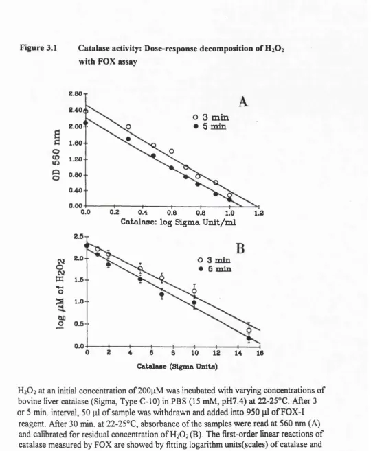
3.1. Catalase activity: Dose-response decomposition of H2O2
with the FOX assay 43
3.2. Measurement of catalase activity in human erythrocyte
and rat liver homogenates with the FOX assay 45 3.3. Kinetics of catalase activity as monitored with the FOX assay 46 3.4. A steady flux of H2O2 generated from the glucose-glucose
oxidase system inactivates catalase in the presence of AMT 47
Chapter 4:
Figure:
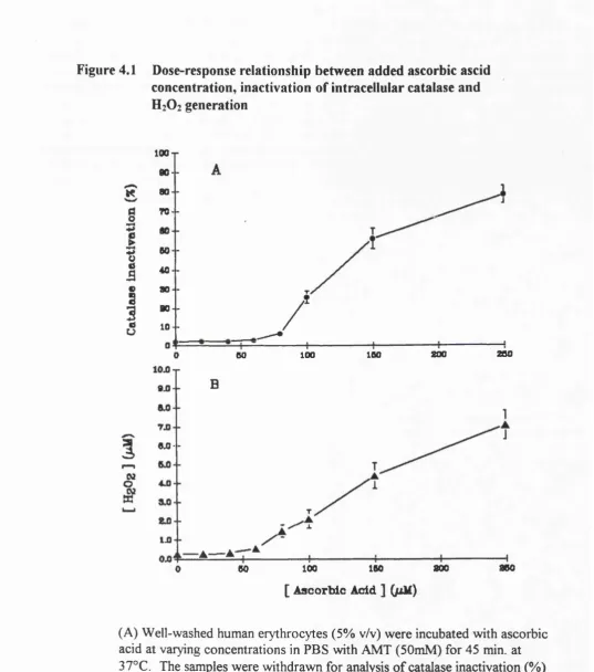
4.1. Dose-response relationship between added ascorbic acid,
inactivation of catalase and H2O2 generation 50 4.2. Inhibition of ascorbate-induced catalase inactivation
requires pre-incubation with metal chelating agent (OPT) 52 4.3. OPT enhances both Cu^^-induced catalase inactivation and H2O2
production during Cu^^ - catalysed ascorbate oxidation but
inhibits Cu^^-catalysed ascorbate oxidation 57
4.4. Comparison of iron and copper in catalysing ascorbate
oxidation in vitro and erythrocytes 60
4.5. Time-course of inactivation of intracellular catalase by ascorbate
and glucose in the presence of AMT 61
4.6. Methemogloblin formation during the incubation of human
erythrocytes with glucose, ascorbate or/and copper 62
Table
4.1. H2O2 generation, catalase inactivation and oxidative damage in erythrocytes exposed to glucose-glucose oxidase in the presence
xm
Chapter 5
Figure:
5.1. The accumulation and Cu^^ - catalysed production of H2O2 from AG 67
5.2. AG increases H2O2 production in vitro 69
5.3. Effect of catalase on hydroperoxide generated by AG 70 5.4. Time-course of inhibition of catalase activity by AG 72 5.5. Dose-response inhibition curve of AMT and AG versus catalase 72 5.6. AG inhibits catalase in liver slices but not in erythrocytes in the
absence of an H2O2 -generating system 74
5.7. AG inhibits erythrocyte catalase in the presence of Cu^Vascorbate 75
Chapter 6
Figure:
6 .1. Effects of glucose and copper on peroxidation of PC liposomes 82 6.2. Effects of glucose on peroxidation of two phospholipid
liposomes: PE and PC 83
6.3. Inhibitory effects of EDTA and DETAPAC on peroxidation
of PE and PC in the presence of glucose 85
6.4. GC profiles of polyunsaturated fatty acid (PUFA) in PC and PE 87 6.5. Oxidation of lipoproteins induced by pre-oxidised glucose
and copper 88
6.6. Susceptibility of glucose modified (glycated) protein and
lipoprotein to Cu^^-catalysed oxidation 89
Table:
6.1. Composition of polyunsaturated fatty acid in PC and PE 86
Chapter 7
XIV
Chapter 8
Figure:
8.1. GC profile of sorbitol accumulation in rat lens caused by glucose 106 8.2. Aldose reductase (AR) in rat lens activated by glucose and
H2O2in vitro 107
8.3. Inhibition of AR activity by OPT, thioctic (lipoic)acid and
sorbinil in rat lens 108
8.4. Aldose reductase inhibitors (ARIs) inhibit Cu^^-catalysed
ascorbate oxidation in vitro 109
8.5. ARIs inhibit Cu^^-catalysed lipid peroxidation 113 8.6. ARIs and OPT facilitate copper partition into n-octanol 113 8.7. ARIs and other metal chelators inhibit ascorbate-metal
induced H2O2 production in erythrocytes 114
8.8. Structures of hydantoin, histidine and aldose reductase inhibitors
used in this study 118
Table:
8.1. Glucose activated AR and sorbitol formation, and the
Chapter 1 GENERAL INTRODUCTION
1.1. Oxidative Stress and H2O2 Metabolism
1.1.1. Oxidative stress and H2O2 cytotoxicity
In the presence of O2, Fe^^ and Cu^^, and an appropriate election donor, a number of enzymatic and nonenzymatic oxygen free radical-generating systems are able to catalyse the oxidative modification of protein, lipid and many biological components to generate hydrogen peroxide (H2O2). H2O2 is a relatively stable and unreactive molecule. Since H2O2 has no unpaired electrons, H2O2 can not be described as a radical. Therefore, the term “reactive oxygen species” (ROS) is now frequently used in description o f not only O and HO*(radicals containing unpaired electrons) as well as H2O2 (which is not a radical) [Halliwell & Gutteridge, 1989]. H2O2 may arise from a number of enzymatic and nonenzymatic pathways, most often through the intermediacy of O2 Superoxide dismutase (SOD) engages in a dismutation reaction, involving oxidation of one O2 to oxygen and reduction o f another O2 to H2O2 :
SOD
O2 + O2 + 2H --- > H2O2 + O2
1.1.2. Glutathione peroxidase (GSH-Px) and H2O2
All aerobic organisms possess enzymatic systems that protect against H2O2 cytotoxicity. These systems include two types of enzymes: the catalases, which catalyse the two step reaction:
2 H 2 O 2 ———^ 2 H 2 O "I" O 2
and the peroxidases, which bring about the general reaction:
SH2 + H2O2 — > S + 2H2O
Glutatione peroxidase [GSH-Px] is a selenium-containing enzyme which acts as a hydroperoxide-decomposing antioxidant in biological systems, often using selenium as a cofactor. GSH-Px requires GSH as co-factor but will act in vivo on a range of peroxides in addition to H2O2 such as fatty acid hydroperoxides, cumene hydroperoxide and other organic hydroperoxides. In each case, the peroxides (ROOH) are reduced to alcohols (ROH). GSH- Px removes H2O2 by catalysing the oxidation of reduced glutathione (GSH) to oxidised glutathione (GSSG):
2GSH + H2O2 ---> 2H2O + GSSG
The relative importance of GSH-Px versus catalase (see next section) may depend on the source and extent of H2O2 exposure [Gaetani et al., 1989]. For many kinds of cells, catalase may be the most important in protection against high H2O2 concentrations because catalases generally have very high turnover numbers [Chance at al.,1979]. However, others have argued that GSH-Px is the more important of the two in removing H2O2, because it is located in the same subcellular compartments(cytosol and mitochondria) as SOD [Cohen & Hochetein,1964; Halliwell, 1991],
Most aerobic cells contain catalase activity. In mammals, catalase exists in all major body organs, being especially concentrated in liver and erythrocytes. Most catalases consist of four protein subunits, each of which contains a haem (Fe^^-protoporphyin) group bound to its active site[Chance, et al.,1979]. Each subunit also usually contains one molecule o f NADPH bound to it, which helps to stabilise the enzyme and sustain it in an active state [Eaton, et al., 1972; Kirkman et al, 1987].
The catalase reaction exhibits dual activities: catalatic(Eqs. 1 and 2) and peroxidatic (Eqs.l and 3) [Chance, et al.,1979]:
ki
Catalase-Fe^^ + H2O2 --- > Compound 1 (1)
k2
Compound 1 + H2O2 > catalase-Fe^^ + H2O + O2 (2)
K3
Compound 1 + AH2 > catalase-Fe^^ + 2H2O + A (3)
The above equations show that the full reaction cycle requires sequential reactions with two molecules of H2O2 Compound 1 is in steady state with H2O2, acting as both oxidising and reducing substrate; in this steady state, only a fraction of the catalase haem is in the form of compound 1. The possible mechanism of the component co-operation in destroying H2O2 is illustrated in the Figure 1.1.
decreased (H2O2) absorbance at 240 nm, or by measuring the release of oxygen using an oxygen electrode. In the present work, we have developed an alternative method in which the disappearance of H2O2 is directly measured by TOX* assay as described in Chapter 3. The specific activity (lU) of catalase is usually expressed as [imoles H2O2 decomposed per min per mg protein.
Catalase activity can be irreversibly inhibited by aminotriazoie (AMT), its inhibitory action being exerted via reaction with the catalase:H2O2 intermediate, compound I. As a result, AMT can only inhibit catalase if H2O2 is present to allow generation of this intermediate. The interactions of catalase with AMT and H2O2 are illustrated in Figure 1.2. This H202-dependent inhibition of catalase by AMT has been used to measure low rates of H2O2 generation in erythrocytes in vitro [Liebowitz & Cohen, 1968; Chapter 4, this thesis] and brain in vivo [Yusa et al., 1987]. Drug-induced H2O2 production in tissues can be measured by assessment of the rate of catalase inhibition [Ou & Wolff, 1993; Giulivi et al., 1994]. Development of the method is described in detail in Chapter 3.
1.2. Characterisation and Measurement of Antioxidant drugs
1.2.1. Definition o f antioxidant
GSH FERRICATALASE
M,0.0,
( • r C M . C H O ) GLUTATHIONE
REDUCTASE
GLUTATHIONE
PEROXIDASE COM POUND II
M , 0 ,
( • > C H , C M , O H )
G S S G COM POUND I
NAD#»' NADP* NAOPtOH*
C O ,
Figure 1.1. Mechanisms o f removal in the ervthrocvte.
Ferricatalase (catalase-Fe^ ) and compound I represent active forms of catalase.
Figure 1.2 Interactions of catalase with HiO? and aminotriazoie fAMT)
Catalase inhibition
Reversible
[Compound II]
|[
6|
Catalase-Fe
[2]
ï
2+ +I%2 Compound I {mtenaedJale]
+AH2 [S]
Catalase-Fe 2HgO + A
Peraxidatie
Iireversihle
[Catalase-AMTi] +AMT
Catalase-Fe^^
2H2O + O2
Caialaüc
Catalase actrvitv
Reactions of catalase with AMT and H2O2
1.2.2. Antioxidants and prooxidants: Vitamin E and Vitamin C
In many ways, vitamins E and C represent the most effective biological antioxidants. Vitamin E (a-tocopherol) is a lipid-soluble chain-breaking antioxidant which reacts with peroxyl and alkoxyl radicals generated during lipid peroxidation. Thus vitamin E protects the membrane by terminating potential peroxidative chain reaction of polyunsaturated fatty acids in cell membranes. Vitamin C has many antioxidant properties and has been called “the most effective aqueous phase antioxidant in human plasma” [Frei et al.,1989]. Ascorbic acid, present in plasma at concentrations of 50-200 jiM, may also scavenge 0% (although this may require the conspiracy of transition metals) and HOCl, and may reduce tocopherol semiquinone, thereby sustaining the antioxidant potency o f vitamin E [reviewed by Winkler et al.,1994].
Under certain circumstances, however, such as in the presence of excess amounts of transition metals, an “antioxidant” may provoke toxic effects on biological systems, through paradoxically acting as a pro-oxidant. In this case, the reducing power of the antioxidant effects, the reduction of transition metals such as Fe^^ and Cu^'*' and the reduced metal may then reduce oxygen or react with oxidants, forming HO* or reactive, higher oxidation states of the metal. This is true of both vitamins C and E.
Thus, for example, ascorbic acid has Janus-like anti- and pro-oxidant properties [Halliwell, 1983]. Metal ions, especially Cu^^, induce ascorbate oxidation producing H2O2 and hydroxyl radicals, and also stimulating lipid peroxidation. These pro-oxidant effects of ascorbic acid in the presence of Cu^^ can lead to damage of both cell membranes and DNA. Ironiascorbate- driven oxidation causes lipid peroxidation and has been indicted in haemoglobin-mediated oxidative damage to the central nervous system [ Sadrzadeh & Eaton, 1988; Prat & Turrens,
sequestration of transition metal ions in forms incapable of participating in free radical reactions.
1.2.3. Assessment o f antioxidant activities
Methods for testing the potency of putative antioxidants often rely on measurements of their reactions with biologically relevant ROS (reviewed by Halliwell, 1990 &1991). Methods exist for measuring the rate of reaction of potential antioxidants with H2O2, O2*’, HO* as well as with other oxidants such as those which form when haem proteins react with H2O2 and ascorbic acid reacts with transition metal ions. Methods also exist to measure the ability of compounds to suppress metal-dependent H2O2 or HO* generation or lipid peroxidation. If an agent acts as a scavenger, the “antioxidant” may itself give rise to damaging radical species. Several of these strategies for determinations of antioxidant actions have been employed in the present studies as described in Chapter 7.8.
1.2.4. M etal chelating agents
Compounds such as ethylenediamine tetraacetic acid (EDTA) and diethylenetriaminepenta- acetic acid (DETAPAC) are commonly used to inhibit radical-generating reactions by chelating iron and copper ions. The chelator either provides a ligand for each of the co-ordination sites on the metal ion, thus excluding oxygen, or it shifts the redox potential of the metal ion so that it is less reactive [Lindenbaum, 1973]. Thus, EDTA is used to prevent oxidative reactions such as the oxidation of plasma lipoproteins which may occur during storage in vitro.
been used as a model of damaging DNA [Dizdaroglu et ai., 1990]. Some examples o f simple copper chelates as shown in Figure 1.3 [Lindenbaum, 1973].
Figure 1.3 Interactions o f Cu (11) with three simple organic ligands
^N. ^ N H 2 . 0
H2C \ H2C \ 0 = C ^ \
Cu""
I
Cu""I
.Cu"" H2ÇCu(II)-ethylenediamine Cu(II)-Glycine Cu(II)-oxalate
1.2.5. Antidiabetic drugs: Thioctic (a-Lipoic) acid and Aminoguanidine potentially have both anti- and pro-oxidant activities.
1.2.5a. Thioctic (a-lipoic) acid
has been proposed that TA may act as a chain-breaking antioxidant in its reduced form, dihydrolipoic acid (DHLA), interacting with vitamin E to block lipid peroxidation. Recently, Handelman et al. (1994) found that normal mammalian cells will reduce TA to dihydrolipoic acid (DHLA), in which the disulphide group of the TA dithiolane ring is reduced to a dithiol (Figure 1.4.).
Figure 1.4. Oxidised and reduced forms o f linoic (Thiotic)acid
S —S
O rlipoic acid
HS SH
d ih y d ro lip o ic acid
COOH
COOH
However, DHLA was also shown to accelerate Fe-dependent HO* generation and lipid peroxidation. By contrast, this pro-oxidant action o f DHLA was inhibited by TA [Scott et al., 1994]. Alternatively, TA may react with oxidants directly [Kagan et al.,1992; Suzuki et al., 1991]. It is currently unclear whether any in vivo antioxidant effect of TA is a direct effect of the compound or depends upon its prior reduction to DHLA. In any case, the antioxidant properties of TA have been proposed to interfere with the pathogenesis o f diabetic polyneuropathy, but the precise mechanism of action o f TA needs further evaluation. In the present studies, we have undertaken analysis of a non radical-scavenging mechanism whereby TA might act as an antioxidant via weak metal-chelating capacity [Sigel & Prijs,1978; Gruner
1960; Ou & Wolff, 1995]. This work is described in detail in Chapter 7.
1.2.5b. Aminoguanidine
reactive carbonyl group o f Amadori products formed by glycation (non-enzymatic glycosylation) of proteins [Brownlee et al., 1986; Lewis & Harding, 1989] and preventing the accumulation of advanced glycation end products (AGE). Furthermore, AG is reported to be a potent inhibitor of NO production [Corbett et al., 1992] and of the activities of aldose reductase [Kumari et al , 1991] and diamine oxidase [Beaven,1982]. Thus, the mechanism(s) whereby AG may moderate diabetic complications are by no means clear.
In fact, AG was recently observed to caus'^ a 60% increase in conjugated dienes in nerves of (streptozotocin-induced) diabetic animals [Kihara et al, 1991]. Thus, possible AG-mediated improvements in nerve function and vascular permeability may not be related to antioxidant effects. In fact, we find that AG actually generates H2O2 and inhibits catalase in liver and erythrocytes [Ou & Wolff, 1993]. AG also increases LDL oxidation [Kortlandt et al., 1994]. In contradiction to this latter report, Bucala et al. (1993) found that AG inhibited lipid- advanced glycosylation, an effect supposedly due to AG-mediated inhibition of lipid peroxidation. Thus, there is much to learn concerning the possible mechanism(s) through which AG might moderate diabetic complications. As reported in Chapter 5, we have experimentally tested the potential toxic and therapeutic effects of this agent in several in vitro
systems.
1.3. Oxidative Stress and Diabetes Mellitus
1.3.1. Diabetes mellitus and oxidative stress
development of diabetic complications. These complications, which are a major threat to both the quality and length of life in diabetic individuals, are a heterogeneous group o f clinical disorders affecting the vascular system, kidneys, retina, peripheral nerves, ocular lens and skin.
There is increasing evidence th a t, regardless of the cause, oxidative damage to tissue proteins is increased in diabetes, and there is a growing suspicion that oxidative stress may be a central mechanism for the development o f diabetic complications [Wol%1987; Baynes, 1991]. The cause of increased oxidative stress in the diabetic patients is still unclear, but may be related to the chronic hyperglycaemic state and gross metabolic abnormalities which include altered lipid metabolism, and an abnormal metal ion homeostasis which may act as a possible source of oxidant stress.
1.3.2. Involvement o f oxygen radicals in the genesis o f diabetes mellitus (DM)
The evidence concerning a role for oxidation in the genesis o f DM comes from the study of two drugs which induce diabetes in experimental animals - alloxan and streptozotocin (STZ)
[Fisher & Harman, 1982; Weiss, 1982]. Both of these diabetogenic agents appear to selectively destroy the islets of Langerhans, which may be uniquely susceptible to oxidant damage by virtue of having limited oxidant defenses. Both SOD and scavengers of HO* (such as vitamin E and BHA) protect against these diabetogenic agents in vivo [Gandy, 1982] lending further support to the idea that drug-mediated oxidation of pancreatic beta cells underlies the diabetogenic effects of these drugs. In fact, similar protective effects of SOD, catalase and HO* scavengers against alloxan toxicity have been shown with isolated islet cells in vitro
[Grankvist et al. 1979]. Alloxan toxicity in vitro and in vivo is also inhibited by metal-chelating agents [Malaisse et al , 1982]. Desferrioxamine also blocks diabetes induced by STZ [Mendola et al , 1989]. These data suggest that transition metal-catalysed free radical reactions may contribute to the toxicity of these two diabetogenic agents.
Although unproven, it is possible that oxidative stress similarly plays a role in the naturally occurring (non-drug induced) destruction of P-cells and the onset of DM and, fiirther, that the sequellae of the diabetic state also involve oxidative processes. Thus, lipid hydroperoxide levels in diabetic plasma, measured with the much-maligned thiobarbituric acid (TEA) assay, are significantly higher than in normal individuals [Kaji et al., 1985]. A further study found that NIDDM patients had higher levels of plasma TBA-reactivity and conjugated diene levels than normal subjects [Collier et al., 1992]. There is also evidence that oxidative events underlie the cataractogenic consequences of diabetes. In diabetic subjects, ocular fluids hive been shown to have increased levels o f H2O2 and lipid hydroperoxides are elevated in the
cataractous lenses of diabetic subjects [Bhuyan & Bhuyan,1984].
Further, albeit indirect, evidence of oxidative stress derives fi'om examination of the antioxidant status of patients with diabetes. Plasma levels o f ascorbic acid are decreased in both humans and animals with diabetes, and the levels o f the oxidation product, dehydroascorbate, are increased [Mclennan et al., 1988; Yue et al , 1989]. Platelet vitamin E level has been observed to be depressed in rats with chemically-induced diabetes [Higuichi,1982].
Rats made diabetic by administraiton of STZ have significantly increased activities of catalase (CAT), glutathione reductase (GR), and CuZn-superoxide dismutase (SOD) in the pancreas and o f CAT and GR in the heart. On the other hand, the livers o f diabetic rats show a generalised decrease in CAT, glutathione peroxidase (GSH-PX), and SOD, as well in the levels of GSH [Wohaieb & Godin, 1987]. Decreased lens antioxidant enzymes and GSH were also observed in lens proteins o f diabetic cataracts [Ready et al., 1976]. The marked alternations in tissue antioxidant enzyme activities may be the result of compensatory increases or oxidant-induced decreases arising from an increased in vivo oxidative stress.
Transition metal ions may catalyse the formation of ROS and promote free radical reactions by accepting and donating single electrons. These transition metals have variable oxidation states (e.g. iron has Fe^^ and Fe^^ and copper has and Cu^^. Changing between oxidation states involves accepting and donating single electrons (e.g. Fe^^ + e' - o Fe^^; Fe^^ + O2 o Fe^^ + O2 ; + e' o ). Thus, in the presence of air, Fe^^ is most stable whereas Fe^^ salts are much less stable and weakly reducing o f oxygen. Several transition metal ions, in vivo primarily Fe^^ and Cu^% also react with H2O2 to form HO*.
The metal ions are remarkably good promoters o f free radical reactions [Hill, 1981] by the so- called Fenton Reaction or through an iron-catalysed Haber-Weiss Reaction. A mixture of hydrogen peroxide and an iron (II) salt reacts with many organic molecules, as was first observed by Fenton in 1894. The reactivity is most likely due to formation of hydroxyl radical :
H2O2 + Fe^^ > GIT + Fe^^+HO* {F entonreaction)
Traces of Fe^^ might be able to react further with H2O2 , although this is very slow at physiological pH;
Fe^^ + H2O2 > Fe^^ + O2 + 2 i T > Fe (IV )0 + OH^ + lT
Also copper (I) salts are thought to react with H2 0 2to make hydroxyl radicals:
Cu^^ + H2O2 --- > Cu^^ + OH- + OH*
Therefore, copper and iron react similarly with H2O2 to produce highly oxidising species (but not necessarily the free OH* shown below):
Fe, Cu
Both iron and copper complexes are able to catalyse HO* formation by this mechanism. For example, the process of iron and copper catalysed lipid peroxidation is believed to involve two metal-dependent reactions: First, these metals participate in the abstraction of hydrogen required for the initial formation of a carbon-centered initiating radical. Second, these metal ions decompose lipid hydroperoxides into peroxyl and alkoxyl radicals that can, in turn, also abstract hydrogen and provoke the chain reaction of lipid peroxidation [Halliwell,1991]. In biological systems, many reductants such as ascorbate, thiol compounds, polyunsaturated fatty acids, proteins and glucose (discussed in following section) are prone to be oxidised by transition metals. In fact, the consequent reduction of the metals (as opposed to O2 dependent reduction) may be most important in the initiation of metal catalyzed oxidative events in vivo
[reviewed by Stadtman, 1990].
1.4.2. Availability o f free metals and increased oxidative stress in diabetes
The endogenous oxidative stress in diabetes - if it exists - may result from reactions between a pool of reactive transition metals and reducing agents such as ascorbic acid and glutathione. Caeruloplasmin is a copper binding and transport protein. It appears to function as an inhibitor of oxidation both by binding copper and by oxidising Fe^""" through its ferroxidase activity [Halliwell & Gutteridge,1989]. However, the copper ion loosely bound to albumin or to histidine can participate in a Fenton-type reaction, reacting with H2O2 to form highly-reactive species with the oxidative potency of OH* and modifying proteins in a “site-specific” manner [Marx & Chevion,1985; Stadtman, 1990]. Similarly, the erythrocyte contains a pool of loosely bound Cu(II)( about 35% of the total ), which catalyses the oxidation of haemoglobin (Rifkind
1974).
peroxidation [Aust,1989]. Recently, haem has been proposed as an important donor o f redox active iron. Added free haem can catalyse the oxidation of LDL and promote oxidant damage of vascular endothelial cells [Vercellotti et al, 1994; Balia, et al., 1993], probably secondary to the oxidative release of free Fe. Fe released from haemoglobin-derived haem also catalyses oxidative injury to neuronal cell membranes [Vercellotti et al. 1994].
There remains, however, the question of whether decompartmentalized transition metal ions exist in vivo in diabetes. The so-called autoxidation o f glucose [Wolff & Dean, 1987] and other oxidation events which seem to occur in diabetes require free transition metal ions. However, the potency of transition metals as catalysts o f such oxidation events is so great that the amounts of metal required may be almost beyond the detection limit of current analytical methods. Furthermore, catalytic amounts of reactive metal may only occur in localised environments in vivo. Of possible relevance in diabetes, carbohydrates have measurable affinities for metal ions and may participate in metal binding reactions in vivo, catalyzing their own 'aut'oxidation [Baynes, 1994]. Occurrence of the oxidation products of glycated proteins such as carboxymethyllysine and fructoselysine in lens and urine of diabetic patients supports the probable availability of redox active transition metals in vivo [ Ahmed et al., 1986].
1.4.3. Copper and Diabetes Mellitus
Increased oxidative stress in diabetes may result from copper-catalysed formation of reactive oxygen species, which induces oxidation o f lipid, protein and many biological components. A generalised theorem can now be developed which argues that diabetes is associated with a redistribution of transition metals, probably copper, into sites where the metal can catalyse the oxidation of susceptible compounds [Wolff, 1987]. Ninety-five percent o f plasma copper is bound tightly to caeruloplasmin, and the remaining copper is associated with histidine groups of albumin and other plasma proteins.
al., 1983]. It is also the case that levels of copper are elevated in diabetic cataractous lenses [Nath et al., 1969], suggesting a possible role of this copper in oxidative damage to the lens. Indeed, crystallins prepared from diabetic rat lenses exhibit greater susceptibility to oxidative insult (H2O2) [Jones & Hothersall,1993]. Comparative studies in vitro showed that copper is more efficient than iron in catalysing autoxidation of sugar and in production of oxidative collagen aggregates in diabetic animal [Chace et al , 1991].
1.4.4. Iron and diabetes mellitus
Patients with idiopathic haemochromatosis have a clear predisposition to the development of adult onset diabetes [Phelps et al., 1989]. A specific link between iron overload and diabetic complications is further suggested by the observation that treatment with the iron-chelating agent desferrioxamine improved fasting glucose (decreases hyperglycaemia) and lowers hypercholesterolaemia and hyperlipidaemia in diabetic individuals with high ferritin but without frank haemochromatosis [Cutler, 1989]. Also consonant with a possible elevation of reactive Fe in diabetes is the observation that levels of plasma and white blood cell ascorbic acid are lower in diabetics (possibly due to increased iron-catalyzed in vivo oxidation of the ascorbic acid) and the in vitro oxidation of vitamin C to dehydroascorbate is higher than in normal individuals [Jennings et al., 1987]. By the same token, patients with iron overload often exhibit low levels of serum and white blood cell ascorbic acid [Nienhuis,1981 & Cohen et al , 1981].
1.5. Possible Mechanisms for Increased Oxidative Stress in Diabetes Mellitus
1.5.1. “Autoxidation** o f Glucose
Simple carbohydrates, particularly glucose, do appear to be a source of metal catalyzed oxidative stress [Wolff & Dean, 1987]. In the presence of metal ions such as Cu^% monosaccharides with a basic alpha-hydroxyaldehyde are able to enolize and thereby reduce oxygen, yielding ketoaldehydes and oxidising intermediates [Wolff et al., 1984]. Glucosone has recently been identified as the oxidised intermediate sugar moiety product during C u ^ - catalysed autoxidation of Amadori compound [Kawakishi,et al.,1991], and the glucose oxidation catalysed by Cu^^ does generate ROS which cause oxidative damage to proteins similar to that caused by the ascorbate-Cu(II) system [Cheng et al., 1992].
Free radicals and hydrogen peroxide slowly produced by glucose “autoxidation” may cause the selective degradation of histidine residues in proteins (to which, as previously discussed, Cu most often binds) [Hunt et al.,1988; Cheng et al.,1992]. A possibly analogous glucose-driven peroxidation of LDL has also been reported [Hunt et al , 1990 & Sakurai et al.,1991]. Nevertheless, caution should be exercised when arguing that diabetes-related increases in oxidative stress may be directly attributed to autoxidation o f glucose in vivo. It is more likely that the increased free radical activity in diabetes is the result o f the glycation process [Hunt et al.,1988, Baynes, 1991] and availability of catalytic metal ions.
1.5.2. Oxidative glycation (glycoxidation) as a cause o f tissue damage in diabetes
Glucose in its straight-chain form is an aldehyde and can slowly condense non-enzymatically with reactive amino groups (a-NH] ) forming, initially, a Schifif base which may rearrange to form the well-known “Amadori adduct” used in the clinical monitoring glycaemic control. This early stage of the reaction is called nonenzymatic glycosylation, or glycation. [Roth, 1983]. The Amadori product may undergo further reactions, known as Maillard or browning reactions, forming more stable products which participate in protein crosslinking/denaturation and formation of Maillard adducts (or Advanced glycation end products: AGEs). The process of formation of glycation (glycosylation) products from glucose is illustrated in the following scheme.
ki k2 kn
Glucose+NH2-R <=> SchifF Amadori > Intermediate -->—>AGE base product glycosylation
products
1.5.3a. Polyol pathway and diabetic complications
Aldose reductase(EC 1.1.1.21; alditol: NAD(P) oxidoreductase), the first enzyme of the polyol pathway, has received special attention because o f its possible role in development o f diabetic complications [Gabbay,1973; Kador & Kinoshita,1985]. Aldose reductase catalyses the reduction of glucose to sorbitol in the polyol pathway (see Figure 1.5), which is normally a minor pathway for metabolism of glucose but may become important during hyperglycaemia. Sorbitol is formed by the reduction of glucose by aldose reductase with the mediation of NADPH by the reaction shown in Figure 1.5 below.
Figure 1.5. The polvol oathwav.
NADPH + H"' NADP""
G lucose "— > Sorbitol Aldose reductase
NAD* NADH + H* _______ X
Sorbitol — ' Fructose
Sorbitol dehydrogenase
Thus, it is not surprising that in the past 20 years numerous aldose reductase inhibitors (ARIs) have been developed, many o f which show promise in preventing or retarding the development o f a wide variety of diabetic complications in experimental animal models o f diabetes (Mayer & Tomlinson, 1983; Robinson et al.,1986]. Some of these ARIs also prevent thickening of the basement membrane. This suggests that there may be a pathogenic role for aldose reductase and the polyol pathway in the development of complications that have generally been ascribed to non-enzymatic glycation.
Vander Jagt and Hunsaker (1993) have recently proposed an integrative model for diabetic complications, combining the polyol pathway theory and the nonezymatic glycocylation theory, but with emphasis also on methylglyoxal/acetol. Methylglyoxal, a toxic 2-oxo-aldehyde produced nonenzymatically from triose phosphates and enzymatically from acetone, is preferred physiological substrate for human aldose reductase, which is also linked with diabetes [Vander Jagt et al.,1992]. This integrative model proposes a central role for aldose reductase in development o f diabetic complications, as postulated in Figure 1.6 [Vander Jagt & Hunsaker, 1993].
1.5.3b Polyol pathway and increases in oxidative stress
Figure 1.6. Central role for aldose reductase in the integrative model o f polvol pathway and nonenzymatic glvcation.
Intagratlve Model of Diabetic Complications (Central Role for Aldose Reductase)
a
Polvol Pathwav/A ldose R e d u c t a s e Theory N onenzym atic Glvcation Theory
G lucose
Sorbitol
Mettiylglyoxal
A M o s a ' X A U O M
R s d u c t i M Rm u c u m
Acetol
1
Altered Myo-inositol
Metabolism HyperosmoticDamage Nonenzymatic Covalent Modification of Proteins
Methylglyoxal, a toxic 2-oxo-aldehyde produced nonenzymatically from triose phosphates and enzymatically from acetone, is preferred physiological substrate for human aldose reductase [Vander Jagt et al.,1992]. Formation o f methylglyoxal is increased during hyperglycaemia [Thomalley, 1992]. Methylglyoxal and its reduction product, acetol, were shown to modify proteins, leading to the formation of fluorescent products with spectral properties similar to those produced by glucose.
NADPH consumption because aldose reductase requires NADPH as a cofactor for the reduction of glucose to sorbitol [Gonzalez, et al., 1986]. The endogenous antioxidant enzymes such as GR also require NADPH for the reduction of GSSG to GSH. Since aldose reductase has a lower Km for NADPH than does GR, severe hyperglycaemia may result in increased flux of glucose through the polyol pathway and diminished availability of NADPH for GR activity as shown in the Figure 1.7. However, one might argue that the molar amount of NADPH which could possibly be oxidized to NADP by AR is so small that it could not have such global metabolic effects [ Eaton, J.,1995, discussion].
Figure 1.7. NADPH com petition for aldose reductase and glutathione reductase
GSSG GLUCOSE
NADPH
Glutathione-reductase
Km = 0.23 mM
Aldose-reductase
Km = 0.02 mM
N A D P
GSH SORBITOL
N A D P H c o m p e t i t i o n f o r a l d o s e r e d u c t a s e a n d g l u t a t h i o n e
r e d u c t a s e . /C^ v a l u e s o f t h e t w o e n z y m e s f o r N A D P H h a v e b e e n
r e p o r t e d b y G o n z a l e z e t al.^’
NADPH competition for aldose reductase (AR) and glutathione reductase (GR). Km values o f two enzymes for NADPH have been reported by Gonzales et al [1986],
1.6. Purpose o f the study
In light of the foregoing data, oxidative stress seems to be a common pathway linking diverse mechanisms for the pathogenesis of complications in diabetes. The processes which may contribute to increased oxidative stress in diabetes are by no means clear, but may involve three major pathways: (1) increases of autoxidation of biological reducing agents, such as ascorbic acid; (2) non-enzymatic glycation as a result of hyperglycaemia and (3) metabolic derangements, especially activition of the sorbitol (polyol) pathway.
The present study focussed on the proposition that transition metals might contribute to all three pro-oxidant processes. Therefore, in vitro experiments have been undertaken to investigate the possible roles of hyperglycaemia (i.e., high levels of glucose in vitro) and of copper ions as initiating factors of oxidative reactions important in the development of diabetic tissue damage resulting from these three pathways.
It is acknowledged that these in vitro experimental studies may not conclusively prove an important role for oxidative processes in the development of diabetic complications. However, the experimental data, if positive, may point the way in development of additional therapeutic strategies for the prevention of diabetic complications.
Chapter 2 GENERAL METHODS
2.1. Materials
Ferrous ammonium sulphate, hydrazine sulphate, 2-hydroxy-1-naphthaIdehyde, 3-amino- 1,2,4-triazole(AMT), ethylenediaminetetra-acetic acid (EDTA), diethylenetriamine penta- acetic acid (DETAPAC), methanol, ethyl acetate, o-phenanthroline (OPT) and cupric sulphate were obtained from the Aldrich Chemical Co. (Poole, U.K.). Bovine serum albumin (BSA)(Bovine: Fraktion V) was obtained from Boehringer (Mannheim,FRG). Xylenol orange, H2O2, sorbitol, catalase (Type C-20), n-octanol, cholesteryl linoleate, trichloroacetic acid (TCA), 2.4-dinitrophenylhydrazine (DNPH), guanidine, L-a-phosphatidylethanol-amine (PE), L-a-phosphatidylcholine(PC), thiobarbituric acid (TBA) semicarbazide, aminogua nidine (AG) bicarbonate, ascorbic acid, glucose, glucose oxidase (from Aspergillus niger) and Chelex (50-100 mesh) metal chelating resin were obtained from Sigma Chemical Co. (Poole, U.K.). Lipoic (thioctic) acid was generously supplied by ASTA Medica, Germany. Aldose reductase inhibitors Sorbinil® and Zopolrestat® were kindly provided by Pfizer (Sandwich, U.K.). Al-1576 was donated by Alcon Inc. All chemicals and reagents were of the highest purity available. All solutions and reagents were prepared with chelex-treated (50-100 dry mesh. Sigma) deionised water.
2.2. Methods of Studying Lipid and Protein Oxidation
2.2.1. Preparation o f artificial liposomes
3 mins on ice. Ethyl acetate was removed by vigorous bubbling under a nitrogen stream. Stock solutions of the small liposomes were then prepared at 25 mg/ml o f PC or PE in phosphate buffer.
2.2 .2. Preparation o f artificial lipoproteins: mixtures o f BSA and cholesteryl linoleate
Artificial lipoproteins, consisting o f BSA and cholesteryl linoleate, were prepared as previously described (Hunt et al. 1993; Werb & Cohn, 1972). The ratio o f lipid to BSA was 1 : 2 (mg/ml). BSA was used at 10 mg/ml in PBS. Cholesteryl linoleate was first dissolved in acetone at 20 mg/ml and then added to 10 mg/ml BSA with vortex mixing. The mixture was ultrasonicated for 2 mins on ice. Acetone was subsequently evaporated by gassing with a nitrogen stream. Lipoproteins were finally prepared at a concentration of 10 mg of cholesteryl linoleate and 20 mg of BSA per ml.
2.2.3. Preparations o f glycated protein and glycated lipoprotein
subsequent procedures were the same as described for liposomes and lipoprotein preparation.
2.2.4. Protein Assay.
Protein assay was performed using a commercial kit (Pierce) based on bicinchoninic acid (BCA)(Smith, et al., 1985). Cu(II) in the reagent reacts with protein, is reduced forming Cu(I) which forms a highly chromophoric BCA:Cu(I) chelate with an absorbance maximum at 562 nm. In the performance of this assay, samples were routinely incubated at 37° C for 30 min and the absorbance at 562 nm was then measured (note that the development o f colour is a cumulative process and has no clear end-point). Protein content was calculated by comparison with results obtained using a BSA standard.
2.3. Methods for Studying Oxidative Stress
2.3.1. Hydrogen peroxide determination
Hydrogen peroxide(H2 0 2) was assayed using the Ferrous Oxidation in Xylenol orange, version 1 (FOX-1) assay. Briefly, 50mL of test sample or H2O2 standard were admixed with 950mL of FOX-1 reagent (composed of 250mM ammonium ferrous sulphate, lOOmM xylenol orange, lOOmM sorbitol in 25mM H2SO4) and incubated for 30 min at room temperature before reading absorbance at 560 nm. Absorbance data were gathered on a Pye Unicam Series 8700 Spectrophotometer. The concentration o f H2O2 dilutions of commercial
-1 -1
2.3.2 Lipid hydroperoxide production with FOX-II assay
Lipid hydroperoxide was measured by the Ferrous Oxidation in Xylenol orange version 2 (FOX-2) assay. One hundred ml of the reaction mixture was mixed with 0.9 ml of methanol and centrifuged at 13,000 g for 5 mins. One hundred ml of the supernatant was then mixed with 0.9 ml of FOX-2 reagent (composed of 100 mM ammonium ferrous sulphate, 250 mM xylenol orange, 4 mM butylated hydroxytoluene (BHT: a lipid-soluble-chain-breaking anti oxidant used for preventing undersirable chain oxidation o f Fe^^ [Jiang et al. 1991] ) and 25 mM H2SO4 in 90 % (v/v) HPLC-grade methanol). The mixture was incubated at room temperature for 30 mins and the absorbance was then read at 560 nm. Where appropriate, flocculated protein was first removed by centrifugation (12,000 g x 5 min). Concentration of lipid hydroperoxide was calculated using an extinction coefiBcient of 4.3 x lO"* M*^ cm*^ [Jiang, et al., 1991].
2.3.3 MDA formation with TBA assay
Erythrocyte GSH was determined by a method based upon the GSH-dependent reduction of S,5-dithiobis-2-nitrobenzoic acid (DTNB) to thio-nitrobenzoic acid (Beutler et al, 1963; Hum et ai., 1987 ). Well-washed human red blood cells (RBC) were lysed by addition of 10 mM potassium phosphate (pH 7.4). One ml of the RBC lysate was mixed vigorously with 2 ml of EDTA/MPA (metaphosphoric acid) reagent. After centrifugation at 3500 rpm for 5 mins, 1 ml of the supernatant was mixed with 4 ml DTNB reagent in tri-potassium citrate buffer (pH = 6.8). After 1 0 mins, the samples were read at 412 nm and GSH content was calculated with reference to a standard curve a^d expressed as mmol GSH/mg haemoglobin content.
2.3.5. Measurement o f Carbonyl content
Carbonyls were determined by the method described by Reznick & Packer (1994) using the reaction of protein carbonyls with 2.4-dinitrophenylhydrazine (DNPH) in HCl, forming protein hydrazones.
Protein (BSA) samples (containing 3 - 5 mg/ml) were precipitated with 5 volumes of 20 % trichloroacetic acid (TCA) ( w/v ) in glass tubes. After centrifugation at 3700 rpm for 15 mins, the protein pellet was resuspended in 1 ml of potassium phosphate buffer (100 mM. pH 7.4 ). Reaction of carbonyls was performed by adding 3 ml o f 10 mM DNPH in 2.5 M HCl to the pellet, mixing by vortexing and incubating for 1 hour at 37° C After the incubation, the mixtures were then mixed with an equal volume of 2 0% TCA solution for 10 mins and then were centrifuged at 3700 rpm for 15 niins. The protein pellets were collected and washed 2 - 3 times with 4 ml o f ethanol - ethyl acetate ( 1 : 1 ) (v/v) by mechanical disruption of the pellets in the washing solution and re-pelleting by centrifiigation at 3700 rpm for 15 mins to remove the free DNPH.
determined on the HCl blank pellets using a BSA standard (EGA assay) as previously described.
2.3.6. Lipid-containing fluorophores o f glycated protein
The process of lipid-centered protein modification can give rise to a organic-soluble fluorescence product formation with excitation at 370nm and emission at 440nm [Monnier & Cerami,1981; Bucala et al., 1993]. After incubation, lOOpl of lipoprotein-glucose sample was diluted into 900pl methanol and mixed. After 10 mins, the samples were centrifuged to remove denatured apoprotein, and then the supernatants were measured for fluorescence with emission at 440 upon excitation at 370nm for estimation of the phospholipid fluorophore using PERKIN-ELMER LS-5 luminescence spectrometer.
2.4. Methods for Assaying Copper Ion and its Catalytic Activity
2.4.1. Atomic absorption (AA) spectrophotometer method
All glassware was soaked in 2% nitric acid for at least 18 hours then thoroughly rinsed in deionised water and allowed to drain dry. Solutions were stored in polythene bottles.
Disposable equipment (syringes, needles, sample tubes ) was also rigorously checked for contamination by filling with deionised water, standing overnight and then analysing for each element.
2.4.2 Rate o f copper-catalysed ascorbate oxidation
Rate of ascorbate oxidation (AOD 265 nm/min) catalysed by exhibits a dose- dependent reaction in vitro, which was monitored spectrophotometrically. Certain drugs which bind copper will inhibit such ascorbate oxidation.
All materials were dissolved in chelex-treated (50-100 dry mesh; Sigma) double distilled water. The mixtures consisted of varying concentrations of test drug and Cu^^ (copper sulphate, 0.1 - 0.5 mM). Ascorbate oxidation was initiated by addition of ascorbic acid (lOOpM ) in 20mM chelex-pretreated potassium phosphate buffer (pH 7.4). This reaction was monitored over an initial 3 min period at 265nm in a Pye unicam 8720 UVATS spectrophotometer thermostatted at 37®C in acid-washed quartz cuvettes. The oxidation rate of ascorbate was calibrated from the known extinction coefficient of ascorbate (Â265=12.5 mM'Vcm'^). All solutions and hardware were shielded as far as possible from contamination by environmental heavy metals. Under these conditions, low baseline rates of ascorbate oxidation were ensured and this reaction yielded very reproducible rates of oxidation (coefficients of variation < 2%).
2.4.3 O2 uptake by copper-catalysed ascorbate oxidation
initial oxygen concentration (air saturated solutions) lost per unit time. In the air-saturated condition, pure water has 250 pM/L of dissolved oxygen at 25® C
2.4.4. Inhibition o f copper-catalysed lipid hydroperoxide production
The ability of drugs to inhibit Cu^^-catalysed lipid peroxidation was assessed by exposure of artificial liposomes to Cu^^ in the presence of test compounds. Lipid hydroperoxide production was measured using the FOX-II assay. L-a-phosphatidylcholine (PC) liposomes (2.5 mg/ml), prepared as described above, were exposed to Cu^^ (0.5mM ) in the presence and absence of test drug at 37®C for 24 hours. At varying time intervals, 0. 1 ml samples were dissolved in 0.9 ml methanol and centrifuged at 13,000 g for 5 mins. The supernatant was assayed for lipid hydroperoxide using the FOX-II assay described previously.
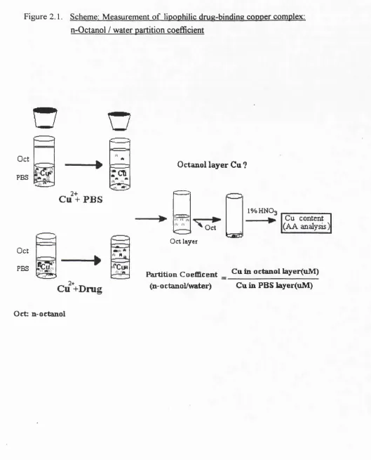
2.5. A Method Modified for Measuring Lipophilicity and Formation of Drug: copper Complex: n-octanol/water partition
N-octanol/water partition is accepted as an index of lipophilicity for drug solubility studies [Cassidy & Henry 1987]. I adapted this method to estimate the ability of drugs to form lipophilic complexes with Cu. The ability o f a compound to cause the partitioning o f Cu(II) into n-octanol is a reflection of the ability to form a lipid-soluble complex with the metal.
(1). Pre-washed n-octanol. n-octanol (Sigma) was washed three times with distilled water, pre-equilibrated with Chelex-treated phosphate buffered saline (PBS: pH 7.4) and stored in the dark until use.
(3). Distribution o f metal-binding drug in n-octanol: 2 ml of n-octanol (pre-equilibrated with PBS) was added to 2 ml of the mixture of drug/copper and drug/PBS (control) and then gently shaken for 3 min. After standing for 20 mins, the samples were further centrifuged at 2 0 0 0 rpm for 1 0 mins to separate octanol from water layer.
(4). Determination o f copper content in octanol. 1 ml of octanol (supernatant) was transferred into a metal-free glass tube. Octanol was evaporated to dryness under N; at
1 0 0* C. The residue was redissolved by addition of 1ml o f 1% HNO3. Cu concentration in the 1% nitric acid was then determined by atomic absorption spectrophotometry (Pye Unicam SP9 Series, England ) at 324.8 nm. The overall process is illustrated in the Figure 2.1. (Scheme).
2.6. Methods for studying Aldose Reductase (AR) in Rat Lens
2.6. 1. Measurement o f aldose reductase-dependent NADPH oxidation.
Figure 2 .1. Scheme: Measurement o f lipophilic drug-binding copper complex: n-Qctanol / water partition coefficient
O
n
... CL- ZZ
Oct A
PBS
---► i c T
Cu + PBS
Oct
PBS
&
lU *
C u + D n ig
Octanol layer Cu ?
"— ' C = ^
r. 3 !•: !•: ,1,
^ Oct
Saif’S
l% H N O .
Cu content (AA analysis )
Oct layer
Partition Coefficent _ Cum octanol byer(tiM) (n-octanol/water) Cu in PBS layer(uM)
2.6.2 Measurement o f aldose reductase-dependent NADP formation
The assay of aldose reductase activity was also performed by determining NADP formation measured by the change of fluorescence at excitation 360nm and emission 460nm [Song et al, 1987]. The reaction system (in 0.2 ml reaction volumes) contained a 50ml aliquot of an appropriate amount of enzyme (lens homogenate), 0.4 M lithium sulphate, (used for stabilising the enzyme activity o f lens homogenate [Song et al, 1987].) 5 mM
2-mercaptoethanol in 50 mM potassium phosphare, pH 6.0, and was incubated for 3 minute at 37°C. Subsequently, 0.1 mM NADPH was added and the reaction mixture was incubated for an additional minute. The reaction was started by the addition o f 10 mM D-glucose and stopped after 5 min by the addition of 0.2 ml 0.5 N HCl. After 10 min, 2 ml of 6N NaOH/10 mM imidazole was added to the samples. Fluorescence of the mixture, after cooling, was determined using a PERKIN-ELMER LS-5 Luminescence Spectrometer. A standard of NADP (100 - 1,000 picomoles) was prepared for the calibration.
2.6.3. Aldose reductase-dependent sorbitol formation.
Sorbitol formation by rat lens homogenates was determined by capillary gas chromatography (GC) as described in Jansen et al (1986) and Petchey & Crabbe (1984). D-mannitol, D- glucose, D-sorbitol and P-D(-) fructose were used as standards and prepared as 5 mM stock solutions in methanol. D-mannitol was used as an internal standard. 50ml of 5mM mannitol was added to 0.2ml of samples of lens homogenate (20mg) making a total volume of 250 ml and a final mannitol concentration of ImM. One ml o f methanol was added to the tissue samples which were held at -20°C for 2 0 min. The samples were centrifuged at 1 0 0 0 x g for 10 min, supernatants were collected and extracted with 1ml of hexane for two times. The upper layer (hexane) was finally discarded and methanol layer (containing polar substances) was dried under a stream of nitrogen.